Foehring - Sensory Spinal Pathways Flashcards
(41 cards)
What are the two systems for somesthesis? Fiber types and ascending tracts?
- PROTOPATHIC: anterolateral -> pain, crude touch, and temperature
1. Low spatial/temporal resolution
2. Sm, slowly conducting, lightly myelinated (A-delta) and unmyelinated (C) fibers
3. Lateral spinothalamic tract - EPICRITIC: lemniscal -> form, texture, touch, pressure, slippage, vibration, position (info about internal body: muscles, joints, internal organs)
1. High temporal/spatial resolution
2. Lg, rapidly conducting, myelinated fibers
3. Posterior/posterolateral columns
Which sensory fibers are chemonociceptive? Be specific.
C fibers
What will happen in the case of a lesion to the spinothalamic tract?
- Loss of pain and temp contralaterally, beginning several levels below the spinal cord lesion
What kinds of sensory info do the dorsal columns conduct?
- Vibration
- Joint position sense
- Discriminative touch
Woman presents with bilateral, cape-like loss of sensation in upper extremities, and forehead.
Where would you place this lesion? Dx?
- Midline of the cervical spinal cord —> C2 dermatome extends up to the top of the head (almost forehead)
- Syringomyelia: hole in the center of the spinal cord with LMN findings
Pt comes in for AAA repair. When he awoke from anesthesia post-op, he was unable to move his legs. There were no reported cxs during surgery. On exam, 0-1/5 strength in all leg mm. Lost sensation to pin in both legs up to level of umbilicus. Vibratory sensation and ability to sense joint mvmt in both legs normal.
Where is the lesion? What happened?
- Bilateral lesion in the thoracic cord
- When repairing this, can get thrombus in great radicular artery of Adamkiewicz, resulting in bilateral, anterior lesion of the spinal cord
74-y/o man wakes from sleep with numbness on R side of body. PMH of HTN and diabetes. No other neuro complaints. Elevated threshold to pin, temp, and proprioceptive sensation over right head and body. When touched with a safety pin that was moved from right thorax to his left, he quickly noted the sensation of a sharp object precisely at midline. Vibratory sensation over forehead was felt bilaterally, and only minimally diminished when crossing the midline.
Where was the lesion? What may have caused it? How could you confirm this?
- Left thalamus. Not much of a difference b/t left and right forehead vibration due to radicular nerve fibers. Thalamic lesion classically affects the epicritic and protopathic pathways; hard to affect only these 2 in other parts of the spinal cord/brain stem without affecting other structures
- This could be caused by an ischemic stroke, which tend to occur at night —> do neural imaging (MRI superior to CT in terms of resolution for anatomical structures). Diffusion weighted imaging (MRI) good for detecting ischemia. CT only better for hemorrhage detection.
What is the general arrangement of spinal cord and nerve roots (sensory and motor)?
- MOTOR: motoneuron cell bodies in spinal cord ventral horn, and axons exit ventral horn via ventral roots, traveling in spinal NN to targe mm -> final comm path for motor activation/efferent spinal reflexes
- SENSORY: receptors in skin (cutaneous), mm and joints, and visceral organs, and communicate w/CNS via peripheral processes of pseudo-unipolar sensory neurons with cell bodies outside the spinal cord in the dorsal root ganglia (DRGs)
1. AP’s in DRG cell peripheral process continue in dorsal roots along central process to dorsal horn, then may synapse there, or project up spinal cord to brainstem (depending on type of receptor, and which somesthetic system is involved)

Briefly, describe the dermatomes (image).

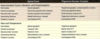
What are the classifications of peripheral nerve fibers, incl. motor (table; 8)?
- Motor: A-alpha, A-gamma
- Muscle spindle: Ia, II, A-gamma
- Golgi tendon organ (and Ruffini): Ib
- Posterior/medial lemniscus: A-beta
- Aneterolateral/spinothalamic: A-delta, C
- Autonomic: preganglionic B, postganglionic C

What are some of the different types of somatosensory receptors? At a basic level, how do these work?
- Exteroreceptors: external events
- Proprioceptors: position of joints/muscles in space
- Enteroreceptors: state of internal organs
- Also: chemo, photo, thermo, mechano, and nocireceptors
- Energy from these sources that exist in nature is transduced by sensory receptors into graded electrical signals that are integrated -> when threshold is reached, AP’s are generated
What five types of receptors are found in glabrous and hairy skin?
- PACINIAN CORPUSCLE: lg, lamellar, rapidly adapting mechanoreceptor that detects gross pressure and vibratory skin stimuli (optimal freq 250 Hz)
1. Found in subcu skin, joints, muscle, mesentery - MEISSNER CORPUSCLE: rapidly adapting mechano-receptor sensitive to light touch and vibration <50 Hz
1. Located in glabrous skin, right below epidermis - MERKEL DISKS: slowly adapting mechanoreceptors in skin and mucosa; in glabrous skin, clustered under ridges of fingertips, and clustered into specialized epi structures called “touch domes/hair disks” in hairy skin
- RUFFINI ORGANS: slowly adapting mechanoreceptor in deep layers of glabrous skin that respond to sustained pressure and skin stretch
1. Responsible for detecting objects slipping along skin, and contribute to position sense - FREE NN ENDINGS: no accessory structures; typically responsive to temp and nociceptive stimuli

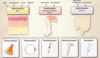
What is receptor adaptation?
- ADAPTATION: reduced response in face of constant, continued stimulus (see attached image)
1. TOP = no adaptation: AP firing maintained at constant rate as long as stimulus is applied
2. MIDDLE: firing maintained for some time, then freq of AP’s slows, despite maintained stimulus -> slowly adapting receptor
3. BOTTOM: rapid adaptation; brief/transient response

Describe the mechanism of the Pacinian corpuscle sensory receptor.
- Rapidly adapting receptor consisting of free NN ending encapsulated by series of modified Schwann cells (lamellae)
- Pressure applied to lamellae causes lateral mvmt, and corpuscle is distorted; mechanical stimulus transduced to AP at onset of stimulus, followed by adaptation while pressure is maintained
- When pressure released, second AP occurs, so organ signals onset and offset of stimulus, but makes no signals during maintained stimulus
- Transduction via stretch-sensitive ion channels in NN ending, and depolarizations transduced to AP’s at first Node of Ranvier of afferent NN
- Lamellae are accessory structure that determine adaptation in Pacinian corpuscle; w/o lamellae, the corpuscle responds to different stimuli and in a different way

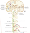
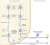
Describe the lemniscal pathway.
- Subcu Pacinian corpuscle (esp. sensitive to vibration) in leg that is rapidly-adapting, connected to lg afferent axon (Ab = T2) that projects via dorsal root into dorsal horn of spinal cord
- Axon continues into ipsilateral, posterior (dorsal) column of spinal cord, and ascends entire spinal cord to ipsilateral medulla (brainstem)
-
First synapse = posterior column nuclei in medulla: nucleus gracilis (L-S), nucleus cuneatus (C-T)
1. NG axons travel in fasciculus gracilis (thin; most medial post column) in spinal cord
2. NC axons travel in more lateral (wedge-shaped), fasciculus cuneatus (T6 is the cut-off) - Axon of 2o neuron (w/cell body in post column nucleus) crosses mid-line of brainstem (decussates) and runs in contralateral MEDIAL LEMNISCUS to part of thalamus involved in somesthesis (synapse #2), the ventral posterolateral nucleus (VPL)
- 3rd order neuron sends axon to ipsilateral 1o somato-sensory cortex, in postcentral gyrus (3rd synapse)

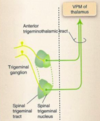
Describe the spinothalamic pathway.
- Free NN ending in skin (cutaneous) of leg (pain, temp receptors usually relatively unspecialized free NN endings) with small, slowly conducting axon (A-delta or C fibers; T3, T4)
- Afferents enter spinal cord via dorsal root, and synapse in ipsilateral dorsal horn (first synapse), unlike lemniscal pathway
- 2nd order neuron (soma in dorsal horn) sends axon across mid-line at same level of spinal cord to contralateral anterolateral funiculus (aka, fasciculus: surface feature indicating underlying axon tract), then turns and ascends through brainstem to VPL in thalamus (synapse #2, contralateral to receptor)
1. NOTE: decussation in anterior commissure of the spinal cord - 3rd order neuron projects ipsilaterally to somato-sensory cortex (post-central gyrus), synapse #3
Briefly compare the cells, synapses, and decussations for the epicritic and protopathic pathways.
- EPICRITIC (via medial lemniscus): enters ipsilateral dorsal horn and ascends in ipsilateral post column to ipsilateral posterior column nuclei (synapse #1), then 2nd order neuron decussates in medulla and ascends as contralateral medial lemniscus to contralateral (to receptor) VPL nucleus of thalamus (synapse #2), then 3rd order neuron sends axon (thalamic radiations) to 1o somatosensory cortex (postcentral gyrus; synapse #3)
- PROTOPATHIC: enters ipsilateral dorsal horn and synapses there (synapse #1), 2nd order neuron decussates at that level of spinal cord and travels in contralateral anterolateral spinal cord in spinothalamic tract, then synapses (#2) in thalamic VPL (contralateral to receptor), and 3rd order neuron projects to 1o somatosensory cortex (postcentral gyrus; synapse #3)
- NOTE: in both pathways, the VPL and somatosensory cortex involved is contralateral to peripheral receptor, and the VPL always projects to its ipsilateral cortex
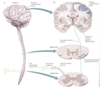
Describe the anatomical pathway of the lemniscal pathway from the brainstem on. Be specific.
- Posterior column nuclei (gracilis and cuneatus) are in the caudal medulla; this is where DECUSSATION of axons from these nuclei occur -> crossing fibers also called internal arcuate fibers
- Medulla = metencephalon
- In rostral medulla, medial lemniscus (axons of contralateral post column nuclei) runs near midline, towards anterior medulla -> as it travels rostrally, its path takes it gradually away from the midline, through the pons (metencephalon) and midbrain (mesencephalon)
- Projections from VPL to cortex travel through posterior limb of the internal capsule
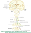
Anatomically, how do afferents add to the sensory pathways as they ascend the spinal cord? What other types of processing may happen along this path?
- Ascending the spinal cord, afferents add to the pathway lateral to lower regions (cuneatus is lateral to gracilis; this is visible anatomically)
- Collaterals of axons from peripheral receptors also synapse with the ipsilateral dorsal and ventral horn, so in addition to conveying peripheral info for processing in brainstem, thalamus, cortex, additional processing occurs in both the dorsal and ventral horn
- NOTE: also indicated in this image is pathway from VPL to Cortex via posterior limb of internal capsule

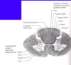
What are Rexed’s laminae? How do they apply to the sensory pathways?
- I-V = dorsal horn; VI, VII = intermediate zone; VIII, IX = ventral horn
- EPICRITIC:
1. Proprioceptive (purple) afferents send collaterals to deep layers of dorsal horn (V-VII) and ventral horn (incl. some type 1a afferents from spindles; monosynaptic on motoneurons)
1. Touch and pressure (blue) afferents send collaterals most to dorsal horn, with few in ventral
a. A-alpha (Ia) from mm receptors, spindles and GTO’s, project to ventral/deep layers of dorsal
b. A-beta (Ib) incl. many cutaneous and joint receptors (2o spindle organs) that project mostly to dorsal horn) - PROTOPATHIC (red): synapse immediately in ipsilateral dorsal horn (mostly I-V) and 2nd order projection is to contralateral spinothalamic tract
1. Some axons travel short way (2-3 segments) in Lissauer’s tract before synapsing in dorsal horn; it takes 2-3 spinal cord segments for all afferents entering at given level to cross to contralateral cord -> lesions at a given spinal cord level result in loss of inputs from 2-3 segments below lesion

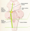
What are the 4 spinocerebellar tracts? Describe their role, and courses.
- Convey info about limb/joint position to ipsilateral cerebellum (input mainly from mm spindles/GTOs)
- DORSAL (leg/body): afferents pass via fasiculus gracilis and synapse in Clarke’s Nucleus (column of relay neuron cell bodies in medial gray matter, T2-L2), then these neurons project to cerebellum via INFERIOR cerebellar peduncle
- VENTRAL (leg/body): afferents synapse in layer VII of spinal cord (L3-S3), and project to contralateral lateral funiculus to synapse with neurons in region of SUPERIOR cerebellar peduncle -> some cross midline again, ending on ipsilateral cerebellum (relative to original afferents); others stay on contralateral side
- CUNEO (arm): afferents synapse in accessory cuneate nucleus (lateral to cuneate nucleus in medulla), then 2nd order neurons project to ipsilateral cerebellum via INFERIOR cerebellar peduncle
- ROSTRAL (arm): afferents synapse in layer VII of spinal cord (above L3) at dorsal horn, and ascend ipsilaterally to cerebellum via INFERIOR cerebellar peduncle

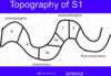
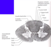
Briefly describe the somatotopy of the posterior and anterolateral columns.
- POSTERIOR COLUMNS: cervical cord is lateral to lumbar cord
- ANTEROLATERAL: neck/occiput to arm to upper/lower trunk to leg
- NOTE: image shows tracts in fixed/stained (real) human spinal cord -> stained for myelin, so fiber tracts dark (axons myelinated; funiculi), and cell bodies (dorsal/ventral gray matter) light
Describe the evolution of the somatotopy of the posterior columns/medial lemniscus as they ascend the spinal cord/brainstem/cortex.
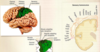
- Fibers add laterally moving up the SPINAL CORD: leg afferents enter in lumbar cord, thoracic cord (lower trunk) added next, upper limb added at cervical levels
- At decussation (MEDULLA), what was medial in spinal cord (lumbar) arcs to become lateral in brainstem (e.g., legs lateral)
- As medial lemniscus ascends in brainstem, lateral representation becomes more posterior (dorsal) too, so legs are lateral and dorsal -> this orientation is preserved through THALAMUS AND CORTEX
- In CORTEX, representation basically standing on its head; superior cortex more midline and inferior cortex more lateral (from the shape of the brain)

What is the Substantia gelatinosa? Lissauer’s Tract?
- SUBSTANTIA GELATINOSA: lamina I and II of spinal cord gray matter; site of first modulation of pain and temperature info
- LISSAUER’S TRACT (aka, posterolateral fasiculus): composed of sensory fibers carrying pain/temp that ascend or descend several spinal cord levels before synapsing in dorsal horn; also contains short axons of projections of neurons from laminae I and II