Foehring - Cortex Flashcards
(45 cards)
Describe the prenatal devo of the cerebral cortex.
- Develops as outpocketings of Prosencephalon (most anterior/rostral part of neural tube), but is specifically a Telencephalic structure
1. Two cerebral hemispheres form laterally on either side of the Telencephalon - ~100d gestation: cerebral hemispheres grown over most of the rest of the brain -> at this age (until about 6 months), cortical surface is smooth or lissencephalic
- 9 mos (birth): surface of cortex covered in pattern of ridges and valles (gyri and sulci) -> gyrencephalic
1. Reflects solution to packing problem bc larger head would pose problems during birth - Cortex is of similar thickness over much of its extent

What is the significance of brain size?
- Human brain about 3-4 lbs
- While brain size to body weight differences between species correlate overall with our perceptions of animal intelligence, clearly something else must be going on as well
- There has never been any credible correlation bt brain size and intelligence within humans or any animal species
- In other words, we don’t really know…

What are the 4 cortical lobes?
- FRONTAL: from frontal pole to central sulcus
- PARIETAL: from central sulcus to imaginary line connecting preoccipital notch to parietooccipital sulcus
- OCCIPITAL: aforementioned line to occipital pole
- TEMPORAL

What are the 3 histological kinds of cortex?
- Defined on the basis of histo:
1. ALLOCORTEX: incl hippocampal formation, olfactory complex (3 layers)
2. ISOCORTEX: 6 layers, at least some pt in devo
3. MESOCORTEX: less regular, and may have 3-5 layers - NOTE: in mature brain, some cortical areas emphasize particular layers, and o/layers may be DEC or absent

What does this figure illustrate?

- Nissl (cell body) stain of the 6 layers of ISOCORTEX (aka, neocortex)
- Layers are named I to VI, going from pia surface to deep white matter
1. LAYER I is cell poor, and different layers differ in size and density of cells - NOTE: this example is from 1o somatosensory cortex of a rat
Describe the cellular composition of the 6 layers of isocortex.
- Layer I = molecular layer, and is poor in cells (in mature brain, only GABAergic interneurons)
- Layers II and III are continuous and hard to tell apart from e/o -> collectively, the superficial pyramidal cell layer (most common cell type)
- Layer IV has many small cells (looks like grains of sand) = granular layer
1. Since layer IV is granular layer, I-III are known as supragranular layers (above granular), and layers V-VI subgranular layers (or infragranular) - Layer V = deep pyramidal cell layer, and largest pyramidal cells are found here
- Layer VI contains multiple cell types, and is known as the polymorphic layer

What are the 3 evolutionary types of cortex? From what are they formed?
- Defined in terms of evolutionary/embryo origin:
1. PALEOCORTEX: incl olfactory cortex -> formed from lateral pallium
2. ARCHICORTEX: incl hippocampal formation -> formed from medial pallium
3. NEOCORTEX: synonymous w/isocortex -> formed from dorsal pallium
What are the 3 major classes of neurons in the neocortex (soma shape scheme)?
- Based on soma shape + configuration of dendrites
- PYRAMIDAL: pear-shaped soma and single dominant apical dendrite (+ basal rosette of dendrites) -> send their axon deep to white matter (projection neurons)
1. Project locally, to o/cortical, & subcortical areas
2. Excitatory: glutamate or aspartate as 1o NT - NON-PYRAMIDAL: mostly GABAergic interneurons (local circuit neurons that only project locally in given area of cortex) -> typically multipolar (several similar sized dendrites radiating from soma) or bipolar (2 similar sized dendrites on opposites sides of soma)
- SPINY STELLATE CELL: another type of non-pyramidal neuron in layer IV of 1o sensory cortex uses glutamate as its transmitter -> may be a subtype of pyramidal cell, but only project locally (local circuit interneuron)
- NOTE: as many as 45+ different cell types in neocortex, but only need to focus on 2 for this course

What is the second classification system for neocortex neurons?
- Divides cells into spiny and aspiny (sparsely spiny)
- Dendritic spines are sites of excitatory synapses on dendrites that isolate individual synapses electrically and biochemically
- SPINY: pyramidal cell dendrites are covered with dendritic spines (spiny), as are dendrites of spiny stellate cells in layer IV (spiny)
- ASPINY: GABAergic interneurons are aspiny or very sparsely spiny
Describe the cortical cell types based on soma shape, apical dendrite, spines, axonal projection, and transmitter.
- PYRAMIDAL: pear-pyramid shape, apical dendrite, spines, axonal projection to white matter, glutamate, excitatory
- SPINY STELLATE: pear-pyramid shape, NO apical dendrite, spines, local axonal projection, glutamate, excitatory (distinguishing features from pyramidal underlined)
- NON-PYRAMIDAL: variable shape, NO apical dendrite (multi- or bipolar), no or sparse spines, local axonal projection, GABA, INH
- Non-pyramidal cell types: chandelier, basket, neuroglia form, bipolar
What is this? Describe its key characteristics.

- Typical neocortical PYRAMIDAL CELL: single, dominant apical dendrite, basal rosette of dendrites, and axon that leaves soma and projects deep to white matter, giving off collaterals on the way
- Variable density of spines along dendrites (highest density intermediate distance from soma)
- ATTACHED IMAGE: examples of human pyramidal cells (and dendrite with spines at higher power) filled w/dye (biocytin)

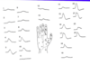
What cells do you see here? Describe their key characteristics.

- NON-PYRAMIDAL cells: diverse, Golgi-filled neurons (>40 types in cortex)
- A, B = basket cells: in layers II, III, and V, and vary in size -> multipolar (many similar-sized dendrites), and axons have basket-shaped terminations surrounding somas of pyramidal cells
- D = chandelier cells: also named for their axonal terminations, whose cassettes contact initial segments of pyramidal neurons and collectively make the cell look like a chandelier
- F-G = various bipolar and bi-tufted cells -> have long dendrites and axons organized vertically as opposed to the more horizontal organization of basket and chandelier cells, and tend to innervate more distal dendrites of pyramidal neurons (compared to chandelier or basket cells)

Where do the main inputs to the cortex come from/go?
- Dominant input to most cortical neurons from other cortical neurons -> excitatory pyramidal neurons are highly interconnected
- Main extrsinsic input to cortex = THALAMUS
1. Specific: from thalamic nuclei that project to single cortical area, and typically concern a single modality -> ex. incl VL to motor cortex, VPL to somatosensory cortex, lateral geniculate to visual cortex, and medial geniculate to auditory
a. Input from specific thalamic nuclei centered on layer IV (in 1o sensory areas, synapse is on spines of spiny stellate cells)
2. Non-specific: thalamic nuclei that integrate info from many sources that is important for general brain states and arousal -> ex. incl intralaminar and midline thalamic nuclei
a. Projection is primarily to layer I (local interneurons and apical tuft of pyramidal cell apical dendrites) - Another source of extrinsic input = widely projecting brainstem nuclei, which serve modulatory functions
1. Incl: locus ceruleus (NE), raphe nuclei (5-HT), ventral tegmental area (DA), basal forebrain nuclei (Ach) - NOTE: all of extrinsic inputs enter the cortex from the deep white matter and travel vertically

Describe the cortical outputs.
- Pyramidal cells = principal cortical projection neurons
- LAYERS II-III: main sources of cortico-cortical connections, incl. association fibers that project ipsilaterally (local and long distance) and callosal projections (cross to equivalent areas of contralateral cortex via CC)
1. Send some axons to subcortical telencephalon (esp. basal ganglia) - LAYER V: project to various subcortical regions, incl spinal cord (corticospinal tract), pons (corticopontine), tectum (corticotectal), basal ganglia (corticostriatal)
- LAYER VI: primarily project to thalamus (same areas from whence afferent signals came) -> feedback loop is basis for thalamocortical rhythms observed in EEG
1. Rhythms important in regulation of sleep-wake cycle, consciousness, and several pathological conditions (e.g., absence epilepsy)

What makes up the cortical white matter? Describe some of the terminal projections.
- Made up of axons of cortical projection (pyramidal) neurons, which have several different targets.
1. LAYERS II-III: primarily project to contralateral cortex (COMMISSURAL) or o/cortical areas on same side of brain (ASSOCIATIONAL); some also project to striatum (subcortical; in telencephalon)
2. LAYER V: more hetero in their projections; those in more superficial part (5A) tend to be thinner, w/less robust apical dendrite, project to contralateral cortex and subcortical telencephalic targets like striatum (like layers II, III), meanwhile those in deeper parts (5B) more robust, and tend to project beyond telencephalon (e.g., spinal cord, tectum, pons, brain stem) - IMAGE: subcortical projections

What percentage of cells in most areas of the cortex are pyramidal?
80% (rest non-pyramidal)
Briefly, how are pyramidal and non-pyramidal cells different?
- Pyramidal: excitatory (glutamate, aspartate), projection
- Non-pyramidal: GABAergic, INH, local circuit
Most inputs to a given pyramidal cell are from…? What are the implications of this?
- Other pyramidal cells (excitatory) -> this system of mutual excitation would lead to unstable network if not for the less numerous, but very important INH interneurons
- Many neural diseases may reflect relatively subtle changes in the balance of excitation to INH in local cortical circuits (EX: epilepsy, which is characterized by abnormal excitability and synchrony between cells)
1. More recently, alteration of this balance has been implicated in Autism, Alzheimer s disease, and various forms of mental retardation
Briefly describe the interactions b/t cells in the cortex.
- Numerous local circuits (synaptically connected neurons) that work together on a particular task
- Influenced by extrinsic inputs from thalamus and brainstem modulatory systems + more distal cortical circuits -> white matter tracts are anatomical substrate for these interactions bt local circuits
- ATTACHED IMAGE: 2 human layer III pyramidal cells (leftmost 2) and 1 multipolar basket cell (rightmost) in close proximity in human temporal cortex
1. Note the vertical arrangement of the pyramidal cells (pia is at top of photo) and more horizontal arrangement of the basket cell

What is going on in this image? Describe the key features of these cells.

- Typical layer V pyramidal cell: apical and basal rosette of dendrites -> different orientation of these 2 types of dendrite allows sampling of different inputs
- Chandelier cell: terminal cassettes selectively contact basal dendrite and esp. axon initial segment of pyramidal cell -> allows GABAergic (INH) chandelier cell to powerfully INH output of layer V pyramidal cell (and thus the output of this local circuit) bc initial segment/first node of Ranvier is site of AP initiation
- Basket cell: axon terminals terminate as baskets that surround soma of pyramidal cell, allowing INH control at final summing point for synaptiv input from whole dendritic tree at soma -> particularly powerful INH influence on pyramidal cell and cortical output
- Bipolar (double bouquet) cell: also GABAergic and INH, but its primary axonal termination is on more distal braches of apical and basal dendrites, making these cells more influential on local signal processing in dendrites (where most excitatory input to pyramidal cells occurs on spines)
- NOTE: non-pyramidal cells show axonal arborization

What is a cortical column?
- Cortex is composed of repeated modules called cortical columns (not exactly a column, but grouping and repeatability)
- All cells in a mini column (~30 microns diameter: 100-200 cells) encode similar features -> associations bt cells in mini columns may be dynamic and depend upon particular tasks
- Idea of a microcolumn or hyper-column (~0.5-1 mm diameter) would encompass all of the cells (several microcolumns) allied together for a particular function
- Current idea is that a macro column (~10,000 cells) is the basic functional unit

Describe the anatomical nature of a cortical column.
- Specific (excitatory) thalamic input is to a small group of layer IV spiny stellate cells -> these excitatory layer IV cells in turn project to layer II, III pyramidal cells
- Layer II, III pyramidal cells project to layer V, VI pyramidal cells (excitatory), which are the excitatory output of the column
- Axons of layer V pyramidal cells project to o/columns locally, other areas of cortex, and subcortically)
- The anatomy of pyramidal cell axons, together with lateral inhibition by interneurons, set the dimensions of the column

What is the radial unit hypothesis?
- All pyramidal cell members of developmental mini-column are ancestors of a single precursor cell in the ventricular zone of embryonic cerebral vesicles
- Precursors give rise to pyramidal cells and radial glia; radial glia migrate to layer I, and nuerons migrate along radial glia to their mature laminar location, where they fully differentiate
- Interneurons are NOT generated in the ventricular zone of cerebral vesicles, but rather derive from cells from medial ganglionic eminence (and caudal gang eminence), and migrate to neocortex to give rise to majority of cortical GABAergic interneurons
- According to this theory, a single functional column that responds to same receptive field in the periphery might consist of several ontogenetic radial columns originating from the adjacent proliferative units in the ventricular zone
- NOTE: columns can be defined based on anatomy or function, but anatomical and physiological columns do not always match

Can functions be localized within the cortex?
Yes