The Ear/ Integumentary System Flashcards

(92 cards)

1
Q

what are the three parts of the ear?

A

1) outer
2) middle
3) inner

2
Q

what is the function of the outer and middle ear?

A

hearing

3
Q

what is the function of the inner ear?

A

hearing and balance

4
Q

what is the outer ear composed of?

A

1) The auricle (pinna)
2) External auditory canal
3) Tympanic membrane (eardrum)

5
Q

The pinna is…

A
  • shell shaped
  • cartilage covered by thick skin
6
Q

the external auditory canal is..

A
  • short, curved tube in temporal lobe
  • filled with ceruminous glands
  • accumulates earwax
7
Q

the Tympanic membrane (eardrum) is..

A
  • Thin connective tissue membrane that vibrates in response to sound
  • Transfers sound energy to the middle ear ossicles
  • Boundary between outer and middle ears
8
Q

what are the functions of the external ear?

A
  • •Pinna collects and transmits sound waves to middle ear, causing tympanic membrane to vibrate
  • Hairs and ear wax in external auditory canal prevent foreign materials entering ear
9
Q

what is the structure of the middle ear?

A
  • Small, air-filled cavity in the temporal bone
  • Tympanic membrane separates middle ear from external ear
  • Oval and round windows separate middle ear from inner ear
10
Q

what is the structure of the middle ear?

A
  • Tympanic membrane
  • Oval window
  • Round window
  • Pharyngotympanic tube to pharynx
  • Mastoid cavity of temporal bone
11
Q

what are the three ear ossicles and where are they located?

A

1) malleus (hammer)
2) incus (anvil)
3) stapes (stirrup)

Located in the tympanic cavity

12
Q

what is the function of the ear ossicles?

A

Transmit vibratory motion of the eardrum to the oval window

13
Q

what are the functions of the middle ear?

A
  • ossicles transmits vibrations from tympanic membrane to cochlea (inner ear)
  • Equalizes pressures on both sides of tympanic membrane
  • Pharyngotympanic tube revents membrane from rupturing
  • opens when yawning or swallowing
14
Q

how does the middle ear provide protection?

A
  • reducing the motion of the ossicles resulting from very large sounds
  • is done by contracting two little muscles attached to malleus (tensor tympani and stapes)
15
Q

what is the stapedius muscle?

A

the smallest skeletal muscle in the body

16
Q

what happens when there is a sudden loud noise?

A

the muscles needs time to contract, so they do not always provied protection fot sudden noises (ex: gun shot)

17
Q

how can a throat infection cause an infection to the middle ear?

A
  • the throat is involved with the pharyngotympanic tube which is connected to the middle ear.
  • pathogens in the throat can travel up the tube and cause otitis media
18
Q

what does the inner ear contain?

A

1) bone labyrinth
2) Membranous labyrinth
3) Vestibule
4) Cochlea

19
Q

what is the bony labyrinth and what does it contain?

A
  • canals hollowed out of the temporal bone
  • Contains 3 areas:
    1) semicircular canals
    2) vestibule
    3) cochlea
  • Filled with perilymph
20
Q

what is the membranous labyrinth and what does it contain?

A
  • series of membranous sacs within bony labyrinth
  • Contains potassium-rich fluid called endolymph
21
Q

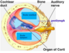
what is the cochlea and what does it contain?

A
  • a spiral, conical, bony chamber that extends from the anterior vestibule
  • contains:
  • the cochlear duct, which ends at the cochlear apex
  • the organ of corti (hearing receptor)
22
Q

what are the three chambers that the cochlea is divided into?

A

1) scala vestibuli
2) scala media
3) scala tympani

**image cut in cross section**

23
Q

what is the helicotrema?

A

the part that the two points meet

24
Q

what must be moved to get hearing receptor to act?

A

scala media

25
scala vestibuli and scala tympani facts
- filled with perilymph - continuous with each other by the helicotrema - scala tympani terminates at the round window
26
what is the scala media filled with?
endolymph
27
what is the "floor" (scala media) composed of?
the basilar membrane supports the organ of corti
28
what runs from the organ or corti to the brain?
the cochlear branch of nerve VIII
29
what is sound and how is it tansmitted?
- a pressure disturbance originating from a vibrating object - represented by a wave - described by wavelength, frequency, and amplitude - short wave= high frequency= high pitch
30
what is the transmission of sound to the inner ear?
1) outer ear (pinna) 2) auditory canal 3) eardrum 4) middle ear (malleus, incus, stapes) 5) oval window 6) inner ear (scala vestibuli/tympani to cochlear duct) 7) the cochlear branch of nerve VIII 8) brain
31
what is the pathway of sound waves?
- pressure waves created by the stapes, pushing the oval window moves through fluid in the scala vestibuli
32
what are short/stiff and long/floppy fibres and where are they located?
short/stiff fibres: - for loud sounds - found near base of cochlea (surrounding long/floppy fibres) Long/floppy fibres: - for soft, quiet sounds - found in the centre of cochlea (inner part)
33
what is sound dependent on?
the fibres moving
34
what is the organ of the corti composed of?
- supporting cells and outer/inner har cells - afferent fibres of the cochlear nerve attached to the base of hair cells
35
where is the stereocila (hairs) located?
- protrude into the endolymph - touch the tectorial membrane - pushes against membrane when sound comes in
36
how does the excitation of hair cells int he organ of corti work?
- bending cilia: - opens machanically gated ion channels (potassium/calcium) - causes depolarization hair cells that cause the release of neurotransmitters to cochlear nerve - neurotransmitters causes cochlear fibres to transmit impulses to the brain
37
what is the vistibular apparatus?
Equilibrium receptors in the semicircular ducts and vestibule - maintain out orientation and balanc in space
38
what are the two different types of balance?
1) static balance 2) dynamic balance
39
what is static gravity?
maintenance of position of the bosy relative to the force of gravity - forward, backward, side-to-side head movements
40
what is responsible for static balance?
Vestibule
41
what is dynamic balance?
maintenance of body position in response to sudden movements ex: rotation, acceleration, deceleration sensed by the head
42
what is the vestibule ?
the central egg shaped cavity of the bony labyrinth
43
what are the two sacs suspended in the perilymph of the vestibule and their functions?
1) utricle (superior) 2) saccule (inferior) Functions: - house equilibrium receptors in their maculae - respond to gravilty and changes in the position of the head
44
what is in the walls of the utricle and saccule?
maculae
45
what is the anatomy of maculae?
the sensory receptors for static equilibrium, in walls of utricle and saccule - contain supporting cells/hair cells - each hair cell has stereocilia embedded in the otolithic membrane
46
what is the otholithic membrane?
jelly-like mass, studded with tiny CaCO3 stones called otoliths
47
what responds to horizontal movement and what responds to veticle movements?
- utricular hairs = horizontal movements - saccular hair = vertical movement
48
what happens when the head bends forward?
- otolithic membrane slides forward, bending the stereocila attached to the hair cells - hair cells move forward, opening mechanical gates which rushes in potassium, depolarizing hair cells, causing action potential - rate of fire neurons fires more forcefully
49
what happens when the head tilts backwards?
- potassium gates close and less neurotransmitter is released, causing less action potentials in the associated neuron - head bends back, neurons stop producing neurotransmitters (rate of fire neurons fire less)
50
what is used for dynamic balance?
the ampulla in the semicircular canals
51
what are the semicircular canals?
three bony canals that lie in the three plans of space - anterior - posterior - lateral
52
what is the crista ampullaris (crista)?
- the receptor for dynamic equilibrium - located in the ampulla of each semicircular duct - responds to angular movements
53
what happens with dynamic movements?
- the semicircular ducts and hair cells move with it - endolymph (fluid in membrane) lags behind, pulling on the cupula - pulling of cupula causes the hair bundles to bend - bending of the stereocilia opens/closes the mechanical postassium gates, which causes hair cells to release more or less neurotransmitter, changin frequency of action potentials
54
what is the equilibrium pathway to brain?
- impulses travel along the vestibular brance of cranial nerve VIII, going straight to refelx centres in the brainstem and cerebellum - information is put togehter by information from the eyes and receptors that give information on body positioning to allow for reflex movement that maintains body position
55
what is transmission deafness?
- something covers sound conduction to the fluids of the inner ear ex: ear wax, ruptured ear drum
56
what is osteosclerosis of the ossicles?
excessive hardening of bone which results in bone not being able to vibrate properly
57
what is perceptive (sensorineural) deafness?
results from damage to the neural structures at any point of the choclear hair cells to the auditory cortical cells
58
hearing aids only helps what form of deafness?
conductory deafness
59
what is tinnitus and what causes it?
ringing of clinking sound in the ears in the absence of auditory stimuli causes: - wax build up - drugs - perforated tympanic membrane - disturbance of auditory nerve or cerebral cortex
60
what are components of the integumentary system?
- skin - skin derivatives: - hair - glands - nails - sensory receptors
61
why is skin considered an organ system?
- conprises different tissues and organs - all 4 tissue type - hair, nails, lips, glands one of the largest organs in the body
62
what are the 7 major functions of skin?
1) protection 2) tempurature regulation 3) sensation 4) excretion 5) immunity to disease (defence) 6) vitamin D production 7) Behavioural
63
what are the 2 major layers to the skin?
1) epidermis 2) dermis
64
what is the epidermis made up of?
epithelial cells
65
what is the dermis made up of?
connective tissue containing elastin and collagen
66
what is the hypodermis?
layer between skin and muscles - fat is present here - not part of the skin
67
what are the 5 layers of the epidermis?
1) stratum basale 2) stratum spinosum 3) stratum granulosum 4) stratum lucidum 5) stratum corneum (order from bottom to top)
68
stratum basale facts
- single layer of cuboidal/columnar keratinocytes - cells are alive and always dividing - when cells divide, one cell stays, other pushes up to next layer - has cells that produce melanin
69
stratum spinosum facts
- contains many filaments (spines) to join cells together - produces protein keratohyalin (protein granules)
70
stratum granulosum facts
- above stratum spinosum - cells start to die
71
stratum lucidum facts
- above stratum granulosum - only in palms of hands and soles of feet - cells are dead - filled with keratin
72
stratum corneum facts
- top layer of epidermis - outer layer of dead cells filled with keratin - can cause calluses from friction
73
what is the difference of epidermis and dermis?
Epidermis: - avascular - innervated - epithelial cells - sensation but no blood supply Dermis: - vascular - innervated - hair follicles - glands (oil/sweat)
74
what are the two layers of the dermis?
1) Papillary region 2) reticular region
75
what is in the papillary region?
- finger like projections going into epidermis - firm and flexible junction - loose connective tissues for easier access of immune cells - has some sensory structures (innervated) - contins small blood vessels (vascular)
76
what is in the reticular region?
below papillary region - dense connective tissue - elastin and collagen (flexible/strength) - contains various glands, nerve endings, blood vessels, hair follicles (vascular/innervated)
77
what is the difference of the epidermis and dermis?
Epidermis: - epithial cells - Avascular - Innerveated - sensation, but no blood supply Dermis: - vascular - innervated - hair follicles - glands (oil/sweat)
78
what are the two cells found in the epidermis?
1) keratinocytes 2) melanocytes
79
what are keratinocytes?
produce keratin, which protects the skin and keeps it waterproof cell with keratin in it
80
what are melanocytes?
produce melanin, one of the major pigments responsible for skin colour protects skin from UV rays
81
how do we get skin pigment?
from melanosomes - other cells takes up melanosomes, then breaks it which distributes the pigment - we have the same amount of melanocytes, the type of melanin produced distinguishes the different colours
82
hair facts and functions
- occurs on most skin surfaces - amount/distribution depends on age, gender, genetics - each hair made of dead keratinized cells function: sensation protection tempurature regulation
83
what is th eimportance of sebaceous glands for hair?
as the hair grows, the sebaceous glands helps prevent hair from drying out/breaking
84
where is the hair follicle found?
dermis
85
what are the three types of glands of the skin?
1) sebaceous glands 2) ceruminous (wax) glands 3) sudoriferous (sweat) glands
86
what is the use of sebaceous glands?
- prevents hair from drying out and becomming brittle - keeps the skin pliable and hydrated - has some anti-microbial properties
87
what is the use of ceruminous glands?
- a wax located in ear canal - protects ear against dust, insects - has some anti-microbial properties
88
what are the two types of sudoriferous glands?
1) apocrine 2) eccrine
89
where is apocrine located and facts?
- located around armpits and groin area - produces sweat but also has some fat/protein - gives out the yellowish colour in sweat - contains pheromones - not active before puberty
90
where is eccrine located?
throughout the skin in most regions of the body produces sweat
91
what are some nail facts and functions?
- from the epidermis - made of compacted, keratin filled, dead epithelial cells Fucntions: - protect the tips of fingers and toes - aid to grasp/manipulate objects - sratching/digging - defense
92