Patients may find it difficult to articulate their symptoms of visual loss, so need to ask direct questions. What 3 questions can you start with?
- Do things look distorted/crooked?
- Is there a shadow?
- Does the shadow move?
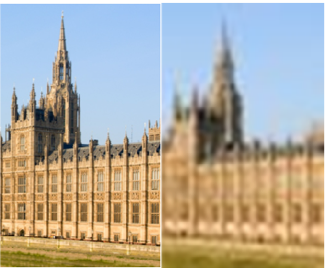
How would ‘blurred vision/fuzziness’ present?
- Out of focus/not sharp
- No distortion or shadowed (no bent/nothing missing)
What are 2 potential causes of blurred vision?
- a refractive problem (cornea, lens, shape of eye)
- a macular problem
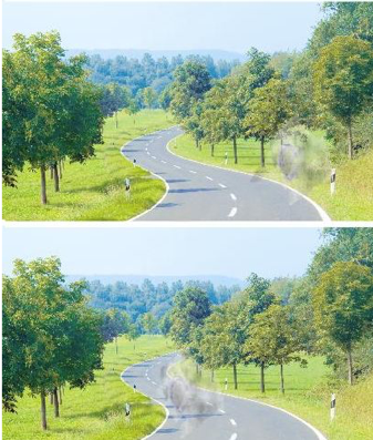
How would ‘glare’ typically present?
- Difficulty seeing in bright light
- Low sun
- Driving at night
- Fluorescent light in supermarkets
- Similar effect produced by having dusty car windscreen
- Vision good otherwise
What is ‘glare’ often due to?
Cataract
How would ‘distorted vision’ typically present?
- Things look wavy, jumbled up. Lines not straight/ bent. Kink in lines
- Effect could be produced by having crumpled film in camera (or crumpled photo)

Typical causes of distorted vision?
- Condition affecting retina:
- Wet macular degeneration
- Macular hole
- Macular pucker
- Retinal detachment
What could cause a patient’s vision to be described as ‘pale’?
- Optic nerve disease:
- Optic neuritis e.g. MS
- Compressive optic nerve disease e.g. pituitary tumour
- Condition affecting retina:
- Wet macular degeneration
- Central serous retinopathy (fluid beneath the retina)

What is a ‘floater’?
Floaters are tiny pieces of debris in the eye’s fluid, known as the vitreous humour –> important feature is movement
What is a ‘floater’ caused by?
Degeneration in vitreous humour:
- Vitreous syneresis
- Posterior vitreous detachment
- Vitreous haemorrhage
A visual field defect (e.g. part of vision missing/ missing things/ shadow/ thumb print) is always a sign of what?
A serious problem
If the visual field is homonymous, what does this indicate?
same visual field both eyes –> a defect of visual pathways
If the visual field is not homonymous, what does this indicate?
Retinal or optic nerve problem
What is confrontation testing?
Confrontation visual field testing involves having the patient looking directly at your eye or nose and testing each quadrant in the patient’s visual field by having them count the number of fingers that you are showing. This is a test of one eye at a time
Label the types of visual defects:
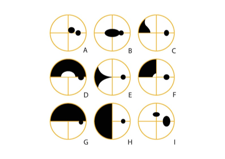
A. Central B. Cecocentral C. Nasal
D. Arcuate E. Nasal wedge F. Quadrantic
G. Altitudinal H. Hemianopia
I. Paracentral + enlarged blind spot
What is a Glaucoma?
A common eye condition where the optic nerve becomes damaged. Usually caused by abnormally high pressure in eye e.g. due to blocked or restricted drainage in your eye or high blood pressure.
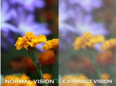
What is ‘cataract’?
- WHAT: Opacity of the lens
- Common aging change
Symptoms of cataract?
- Gradual onset
- Blurred vision, glare, change in refraction
Treatment of cataract?
phacoextraction with lens implant –> great improvement of symptoms
There are 2 blood supplies to the retina. What are they?
1) Choroid – underneath the sclera, supplies the outer 2/3 of retina
2) Vascular supply – supplies inner 1/3 of retina
A lot of supply to the retina therefore requires diffusion from the choroid.
How does retinal pigment epithelium maintain the environment of photoreceptors?
RPE removes waste product from cones and rods
What can reduced function of retinal pigment epithelium lead to?
Drusen
What are drusen?
Drusen are small yellow deposits of lipids that accumulate under the retina
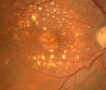
What are the signs of dry age related macular degeneration (ARMD)?
- Drusen
- RPE pigmentation
- RPE atrophy
- Gradual deterioration
- Particularly affects reading vision
- Loss of small area leads to severe visual loss as it is central vision that is impaired, not peripheral
- Early stages often symptomatic

-
Development of CNS53
-
Intro to Human Brain78
-
The Skull146
-
Head and Neck 1: Front of Neck SDL124
-
Meninges and Ventricles106
-
Blood Supply128
-
Head and Neck Practical 1111
-
Head and Neck 2: Face and Skull93
-
Head and Neck Practical 284
-
Braindeath29
-
Brainstem and Cerebellum118
-
Neuroanatomy 1 SDL59
-
Neuroanatomy Practical 1113
-
Cranial Nerves83
-
Forebrain (Cerebrum)61
-
Control Week 1 MCQs16
-
Neuroanatomy Practical 2112
-
Neuroanatomy 2 SDL: Brainstem, Cerebellum and Cranial Nerves130
-
Localisation91
-
AFL 522
-
Muscles of Mastication12
-
Facial Nerve Revision30
-
Arteries of the Head and Neck29
-
Vertebral Column89
-
Spinal Cord and Nerves90
-
Backpain32
-
Ascending Sensory Pathways122
-
Descending Motor Pathways91
-
Contents of the Vertebral Canal SDL45
-
Vertebral Column and Contents Practical71
-
Revision - Different Regions of the Spine23
-
Control Week 2 MCQs11
-
AFL 624
-
Neuroanatomy 3 SDL: Forebrain, Ventricles and CSF40
-
Neuroanatomy Practical 3107
-
Autonomic Nervous System76
-
Skin Histology0
-
Somatosensory Disorders - Sensory Loss1
-
Headache60
-
Skin SDL44
-
Somatosensation Revision49
-
Epilepsy - Clinical Lecture85
-
Clinical - Tumours of the CNS44
-
Spinal Reflexes54
-
Muscle Weakness37
-
Trigeminal Nerve Revision27
-
Control Week 3 MCQs17
-
Pain & Somatosensory Disorders45
-
Peripheral Somatosensory System17
-
Cerebellum90
-
Head Injuries SDL33
-
AFL 726
-
Pain49
-
Local Anaethesia85
-
General Anaesthesia60
-
Visual Pathway112
-
Visual Defects119
-
Visual Defects40
-
SDL - Eye and Ear70
-
Auditory Pathway95
-
Clinical - Multiple Sclerosis0
-
Control Week 5 MCQs15
-
Control Week 6 MCQs32
-
NeuroAnatomy Revision57
-
ANS Revision97
-
Skin and Sensory Receptors MCQs10
-
Basal Ganglia73
-
Limbic System62
-
AFL 825
-
Physiology of Vision27
-
AFL 925
