Congenital Lesions of the Face and Neck Flashcards
Development of the Face
- The external human face develops between the … and … week of embryonic development.
- Many opportunities for abnormal development
- Critical periods of development
- The external human face develops between the 4th and 6th week of embryonic development.
- Many opportunities for abnormal development
- Critical periods of development

Development of the Face
- These developments take place in week … (left) and week … (right)

at 4 weeks and 5 weeks
Development of the Face
- These developments take place in week … (left) and week … (right)

at 6 weeks and 7 weeks

Development of the Face
- These developments take place in week … (left) and week … (right)

At 8 Weeks and 9 Weeks

Electron Micrograph showing development of the …

face
Basic Facial Structure
- 1 x f…
- 2 x …medial
- 2 x …lateral
- 2 x m….
- 2 x m…
- m… and m… 1st … arch
- m… arch
- 1 x frontonasal
- 2 x nasomedial
- 2 x nasolateral
- 2 x maxillary
- 2 x mandibular
- maxillary and mandibular 1st pharyngeal arch
- mandibular arch

Formation of the Palate
- The maxillary process develops … processes
- Should fuse to form … palate in midline
- Anteriorly, … palate formed
- Part of that becomes … palate
- Any problem in formation of these and fusion of these - give rise to conditions of … …
- The maxillary process develops palatine processes
- Should fuse to form hard palate in midline
- Anteriorly, primary palate formed
- Part of that becomes soft palate
- Any problem in formation of these and fusion of these - give rise to conditions of cleft palate

Electron Micrograph showing what happens during formation of the …

Palate
Abnormalities in Development of Face
- A-C =
- D-E =

- A-C = cleft lip
- D-E = cleft palate
The Pharyngeal Arches
- How many?
- What are they called?
- They all have m.., car…, n… and a branch of the … arch
- Mandibular, hyoid, third and fourth
- All have mesoderm, cartilage, a nerve, and a branch of the aortic arch

The Pharyngeal Apparatus

Pharyngeal Pouches - Embryology

Pharyngeal Apparatus

Embryology - Pharyngeal Apparatus
Pharyngeal Arch 1 is the … arch
Pharyngeal Arch 1 is the Mandibular arch
Pharyngeal Arch 1 - Mandibular
- Cranial nerve …
- Muscles of …
- What arises from it?
- Cranial nerve 5 - trigeminal
- Muscles of mastication
- Meckel’s cartilage, sphenomandibular ligament, malleus, incus
(From Teach me anatomy)
- the first pharyngeal arch is the largest and forms a dorsal maxillary process and a ventral mandibular process, which contains Meckel’s cartilage. It contributes to the development of the face and several facial bones as well as the temporal bone
- cranial nerve: Cn Vc (Trigeminal)
- mesodermal derivatives: tensor tympani, muscles of mastication, mylohyoid, anterior belly of digastric, tensor veli palatini
- artery: first aortic arch (temporary)
- neural crest cell derivatives: incus, malleus, anterior ligament of malleus, sphenoid spine, sphenomandibular ligament and the genial tubercle of the mandible

Pharyngeal Arch 2 is …
Hyoid

Pharyngeal Arch 2: Hyoid
- Cranial nerve …
- Muscles of …
- What arises from it?
- Cranial nerve - 7 (Facial)
- Muscles of facial expression
- Part body and lesser horn hyoid, stapes, styloid process, stylohoid ligament, thyroid cartilage, cricoid cartilage arise from it
(From Teach me anatomy)
- the second pharyngeal arch contains Reichert’s cartilage
- cranial nerve: CN VII (facial)
- mesodermal derivatives: stapedius, stylohyoid, muscles of facial expression, posterior belly of digastric
- artery: stapedial artery (temporary)
- neural crest cell derivatives: stapes, styloid process of the temporal bone, stylohyoid ligament, lesser horn and upper part of the body of the hyoid bone

Derivates of First and Second Arches (Pharyngeal arches)

Skeleton of Pharyngeal Arches 3 and 4
- Give rise to part body + greater horn … (3) and cartilages of the … (4)
- Supplied by cranial nerves … and …
- Give rise to part body + greater horn hyoid (3) and cartilages of the larynx (4)
- Supplied by cranial nerves 9 and 10 (glossopharyngeal and vagus)

Muscles Pharyngeal Arches 3 and 4
- 3 = cranial nerve …, muscle = …
- 4 = cranial nerve …, muscle = …
- 3 = cranial nerve 9 (glossopharyngeal), muscle = stylopharyngeus
- 4 = cranial nerve 10 (vagus), muscle = pharyngeal muscles

Embryonic Pharynx

Pharyngeal Grooves and Pouches
- a number of outpocketings appear on the lateral wall of the pharynx – the pharyngeal pouches.
- The pouches separate the arches on the internal (…) surface whilst the clefts separate the arches on the external (…) surface.
- a number of outpocketings appear on the lateral wall of the pharynx – the pharyngeal pouches.
- The pouches separate the arches on the internal (endodermal) surface whilst the clefts separate the arches on the external (ectodermal) surface.

Pharyngeal Grooves

- Note - thyroglossal duct
- cyst or fistula after birth
- While a baby is developing in the womb, the thyroid gland begins at the base of the tongue. Before birth the thyroid gland moves in the neck to its usual position below the thyroid cartilage and above the sternum. A portion of this path in which the thyroid gland moves may fill with mucus-like fluid, creating a thyroglossal duct cyst.
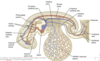
Tongue and Thyroid Gland


Electron micrograph of … and … gland
tongue and thyroid gland
