Physiology of Skeletal Muscle Contraction Flashcards
The basic unit of skeletal muscle is the multinucleated …
The basic unit of skeletal muscle is the multinucleated myofiber
… are long filaments that run parallel to each other to form muscle (myo) fibers
Myofibrils are long filaments that run parallel to each other to form muscle (myo) fibers
… are long filaments that run parallel to each other to form muscle (myo) fibers
Myofibrils are long filaments that run parallel to each other to form muscle (myo) fibers
The … are the contractile proteins in the myofibers that are arranged into groups that cause the cytoplasm to appear repetitively banded (or striated).
The myofilaments are the contractile proteins in the myofibers that are arranged into groups that cause the cytoplasm to appear repetitively banded (or striated).
Sarcomere
- Each sarcomere consists of a central …-band (thick filaments) and two halves of the …-band (thin filaments).
- The …-band from two adjacent sarcomeres meets at the Z-line. The central portion of the …-band is the M-line, which does not contain actin.
- Each sarcomere consists of a central A-band (thick filaments) and two halves of the I-band (thin filaments).
- The I-band from two adjacent sarcomeres meets at the Z-line. The central portion of the A-band is the M-line, which does not contain actin.

Sarcomere
- Each sarcomere consists of a central A-band (… filaments) and two halves of the I-band (… filaments).
- The I-band from two adjacent sarcomeres meets at the …-line. The central portion of the A-band is the M-line, which does not contain …
- Each sarcomere consists of a central A-band (thick filaments) and two halves of the I-band (thin filaments).
- The I-band from two adjacent sarcomeres meets at the Z-line. The central portion of the A-band is the M-line, which does not contain actin.

The myofilaments include thick filaments, composed mainly of …, and thin filaments composed mainly of …
The myofilaments include thick filaments, composed mainly of myosin, and thin filaments composed mainly of actin.
Skeletal muscles are tissues comprising … and … filaments, which require adenosine triphosphate (ATP) to produce muscle contractions
Skeletal muscles are tissues comprising actin and myosin filaments, which require adenosine triphosphate (ATP) to produce muscle contractions
Tropomyosin (TPM)
- Controls whether … and … interact
- Sits on … filament in groove of double helix
- At rest, TPM covers … binding site for …
- Thus, TPM blocks …-… interaction
- Pulled out of the way when muscle is active
- Controls whether actin and myosin interact
- Sits on thin filament in groove of double helix
- At rest, TPM covers actin’s binding site for myosin
- Thus, TPM blocks actin-myosin interaction
- Pulled out of the way when muscle is active
Tropomyosin (TPM)
- Controls whether actin and myosin interact
- Sits on thin filament in groove of double helix
- At rest, TPM covers actin’s binding site for myosin
- Thus, TPM blocks actin-myosin interaction
- Pulled out of the way when muscle is active
- Controls whether actin and myosin interact
- Sits on thin filament in groove of double helix
- At rest, TPM covers actin’s binding site for myosin
- Thus, TPM blocks actin-myosin interaction
- Pulled out of the way when muscle is active
Cross bridge cycling
- … pulling …, consuming …, and resetting
- Controlled by calcium
- … pulled during … power stroke
- Myosin pulling actin, consuming ATP, and resetting
- Controlled by calcium
- Actin pulled during myosin power stroke
Troponin (Tn): 3 subunits
- Troponin proteins control whether
- tropomyosin allows or blocks …-… interaction
- Troponin is made up of 3 subunits (in this order):
- troponin T (T = tropomyosin-binding)
- troponin C (C = calcium-binding)
- troponin I (I = inhibitory / binds to actin)
- Troponin proteins control whether
- tropomyosin allows or blocks actin-myosin interaction
- Troponin is made up of 3 subunits (in this order):
- troponin T (T = tropomyosin-binding)
- troponin C (C = calcium-binding)
- troponin I (I = inhibitory / binds to actin)
Troponin (Tn): 3 subunits
- Troponin proteins control whether
- tropomyosin allows or blocks actin-myosin interaction
- Troponin is made up of 3 subunits (in this order):
- troponin …
- troponin …
- troponin …
- Troponin proteins control whether
- tropomyosin allows or blocks actin-myosin interaction
- Troponin is made up of 3 subunits (in this order):
- troponin T (T = tropomyosin-binding)
- troponin C (C = calcium-binding)
- troponin I (I = inhibitory / binds to actin)
Cross Bridge Cycle (illustration)
- REACTIONS
- 1 Myosin releases …
- 2 Myosin head … …
- 3 Myosin binds actin
- 4 Power stroke
- REACTIONS
- 1 Myosin releases actin
- 2 Myosin head cleaves ATP
- 3 Myosin binds actin
- 4 Power stroke

Cross Bridge Cycle (illustration)
- REACTIONS
- 1 Myosin releases actin
- 2 Myosin head cleaves ATP
- 3 Myosin … actin
- 4 … stroke
- REACTIONS
- 1 Myosin releases actin
- 2 Myosin head cleaves ATP
- 3 Myosin binds actin
- 4 Power stroke

Excitation-Contraction Coupling
- describes the rapid communication between electrical events occurring in the plasma membrane of skeletal muscle fibres and …+ release from the …, which leads to contraction.
- describes the rapid communication between electrical events occurring in the plasma membrane of skeletal muscle fibres and Ca2+ release from the SR, which leads to contraction.
Excitation-Contraction Coupling
- describes the rapid communication between electrical events occurring in the plasma membrane of skeletal muscle fibres and Ca2+ release from the SR, which leads to contraction.
- In skeletal muscle there is a direct … connection between calcium channels of membrane and calcium release channels of sarcoplasmic reticulum
- membrane … -> membrane calcium channels undergo a … change -> SR calcium release channels undergo a conformational change that opens them -> calcium flows from SR to cytosol
- describes the rapid communication between electrical events occurring in the plasma membrane of skeletal muscle fibres and Ca2+ release from the SR, which leads to contraction.
- In skeletal muscle there is a direct physical connection between calcium channels of membrane and calcium release channels of sarcoplasmic reticulum
- membrane depolarises -> membrane calcium channels undergo a conformational change -> SR calcium release channels undergo a conformational change that opens them -> calcium flows from SR to cytosol
Excitation-Contraction Coupling
- describes the rapid communication between electrical events occurring in the plasma membrane of skeletal muscle fibres and Ca2+ release from the SR, which leads to contraction.
- In skeletal muscle there is a direct physical connection between calcium channels of membrane and calcium release channels of sarcoplasmic reticulum
- membrane depolarises -> membrane calcium channels undergo a conformational change -> SR calcium … channels undergo a conformational change that … them -> calcium flows from SR to …
- describes the rapid communication between electrical events occurring in the plasma membrane of skeletal muscle fibres and Ca2+ release from the SR, which leads to contraction.
- In skeletal muscle there is a direct physical connection between calcium channels of membrane and calcium release channels of sarcoplasmic reticulum
- membrane depolarises -> membrane calcium channels undergo a conformational change -> SR calcium release channels undergo a conformational change that opens them -> calcium flows from SR to cytosol
Excitation-Contraction Coupling
- describes the rapid communication between electrical events occurring in the plasma membrane of skeletal muscle fibres and Ca2+ release from the SR, which leads to contraction.
- In skeletal muscle there is a direct physical connection between calcium channels of membrane and calcium … channels of sarcoplasmic reticulum
- membrane depolarises -> membrane calcium channels undergo a … change -> SR calcium release channels undergo a … change that opens them -> calcium flows from SR to cytosol
- describes the rapid communication between electrical events occurring in the plasma membrane of skeletal muscle fibres and Ca2+ release from the SR, which leads to contraction.
- In skeletal muscle there is a direct physical connection between calcium channels of membrane and calcium release channels of sarcoplasmic reticulum
- membrane depolarises -> membrane calcium channels undergo a conformational change -> SR calcium release channels undergo a conformational change that opens them -> calcium flows from SR to cytosol
… = the link (molecular process) between the depolarisation of the membrane (with a tiny influx of calcium) and the consequent huge increase in cytosolic calcium that then leads to contraction
E-C coupling = the link (molecular process) between the depolarisation of the membrane (with a tiny influx of calcium) and the consequent huge increase in cytosolic calcium that then leads to contraction
… = one long multi-nucleate muscle cell
Myofibre = one long multi-nucleate muscle cell
… = organelle, string of sarcomeres
Myofibril = organelle, string of sarcomeres
… = thick or thin filament (molecules)
Myofilament = thick or thin filament (molecules)
Contraction = when ends of sarcomere (… lines) are pulled toward each other by … filament pulling … filaments
Contraction = when ends of sarcomere (z lines) are pulled toward each other by myosin filament pulling actin filaments
Troponin: how it works
- … Ca2+ bind to troponin C (C = calcium binding),
- In heart TnC only binds to … Ca2+ ions
- TnC changes …
- … change in TnC “shuts off” TnI
- tropomyosin-troponin leaves F-actin groove
- unmasks the myosin binding site on actin
- next myosin heads make cross bridges (cycling) to actin
- Myosin breaks down ATP
- Myosin pulls thin filaments
-
4 Ca2+ bind to troponin C (C = calcium binding),
- In heart TnC only binds to 3 Ca2+ ions
- TnC changes conformation
- conformational change in TnC “shuts off” TnI
- tropomyosin-troponin leaves F-actin groove
- unmasks the myosin binding site on actin
- next myosin heads make cross bridges (cycling) to actin
- Myosin breaks down ATP
- Myosin pulls thin filaments
Troponin: how it works
- 4 Ca2+ bind to troponin C (C = calcium binding),
- In heart TnC only binds to 3 Ca2+ ions
- TnC changes conformation
- conformational change in TnC “shuts off” Tn…
- tropomyosin-troponin leaves …-actin groove
- unmasks the myosin … site on actin
- next myosin heads make cross bridges (cycling) to actin
- Myosin breaks down ATP
- Myosin pulls thin filaments
- 4 Ca2+ bind to troponin C (C = calcium binding),
- In heart TnC only binds to 3 Ca2+ ions
- TnC changes conformation
- conformational change in TnC “shuts off” TnI
- tropomyosin-troponin leaves F-actin groove
- unmasks the myosin binding site on actin
- next myosin heads make cross bridges (cycling) to actin
- Myosin breaks down ATP
- Myosin pulls thin filaments
Troponin: how it works
- 4 Ca2+ bind to troponin C (C = calcium binding),
- In heart TnC only binds to 3 Ca2+ ions
- TnC changes conformation
- conformational change in TnC “shuts off” TnI
- tropomyosin-troponin leaves …-actin groove
- unmasks the myosin binding site on actin
- next myosin heads make … bridges (cycling) to actin
- Myosin breaks down ATP
- Myosin pulls thin filaments
- 4 Ca2+ bind to troponin C (C = calcium binding),
- In heart TnC only binds to 3 Ca2+ ions
- TnC changes conformation
- conformational change in TnC “shuts off” TnI
- tropomyosin-troponin leaves F-actin groove
- unmasks the myosin binding site on actin
- next myosin heads make cross bridges (cycling) to actin
- Myosin breaks down ATP
- Myosin pulls thin filaments
Troponin: how it works
- … Ca2+ bind to troponin C (C = calcium binding),
- In heart TnC only binds to 3 Ca2+ ions
- TnC changes conformation
- conformational change in TnC “shuts off” TnI
- tropomyosin-troponin leaves F-actin groove
- unmasks the myosin binding site on actin
- next myosin heads make cross bridges (cycling) to actin
- Myosin breaks down …
- Myosin pulls … filaments
-
4 Ca2+ bind to troponin C (C = calcium binding),
- In heart TnC only binds to 3 Ca2+ ions
- TnC changes conformation
- conformational change in TnC “shuts off” TnI
- tropomyosin-troponin leaves F-actin groove
- unmasks the myosin binding site on actin
- next myosin heads make cross bridges (cycling) to actin
- Myosin breaks down ATP
- Myosin pulls thin filaments
Total TnI = marker for total muscle …
Total TnI = marker for total muscle breakdown
Cardiac TnI = marker for … …
Cardiac TnI = marker for myocardial infarct
Cardiac Tn… = marker for myocardial infarct
Cardiac TnI = marker for myocardial infarct
Cross bridge cycling
- Molecular cycle of actin-myosin interaction
- Mechanism of Contraction at … level
- Contraction depends on binding of … heads to … filaments (actin) at specific binding sites
- In resting state of sarcomere, myosin heads are blocked from binding to actin by tropomyosin, which occupies the specific binding sites (in F-actin double helical groove)
- Molecular cycle of actin-myosin interaction
- Mechanism of Contraction at Molecular level
- Contraction depends on binding of myosin heads to thin filaments (actin) at specific binding sites
- In resting state of sarcomere, myosin heads are blocked from binding to actin by tropomyosin, which occupies the specific binding sites (in F-actin double helical groove)
Cross bridge cycling
- Molecular cycle of actin-myosin interaction
- Mechanism of Contraction at Molecular level
- Contraction depends on binding of myosin heads to thin filaments (actin) at specific binding sites
- In … state of sarcomere, myosin heads are … from binding to actin by …, which occupies the specific binding sites (in F-actin double helical groove)
- Molecular cycle of actin-myosin interaction
- Mechanism of Contraction at Molecular level
- Contraction depends on binding of myosin heads to thin filaments (actin) at specific binding sites
- In resting state of sarcomere, myosin heads are blocked from binding to actin by tropomyosin, which occupies the specific binding sites (in F-actin double helical groove)
Force generation vs. sarcomere length
- Fill in blanks

- Between A & B: the degree of filament overlap is directly proportional to force muscle can generate.
- When the effect of small shortening of one sarcomere is multiplied by many sarcomeres in a myofibril and the shortening of many myofibrils together is summed, the whole muscle fibre shortens.
- Shortening of many muscle fibres is manifest as contraction of the entire muscle with change in muscle length

Cross Bridge Cycle (illustration)
- REACTIONS
- 1 - Myosin releases actin
- 2 - Myosin head … ATP
- 3 - Myosin binds actin
- 4 - …. …

- REACTIONS
- 1.Myosin releases actin
- 2.Myosin head cleaves ATP
- 3.Myosin binds actin
- 4.Power stroke
Tropomyosin: The important thing to notice in the animation (you can animate this by running this powerpoint in slide show mode (shift-F5) is that the tropomyosin (long blue string) rolls up and down. It turns out that myosin (top and left) can only interact with specific bits of the actin molecule. In the absence of calcium, this active site of actin is covered by tropomyosin. In the presence of calcium (yellow balls that fly in to cartoon), tropomyosin is rolled away from actin’s active site because the calcium causes troponin (3 purple beads connected to one another, one of which is connected to the blue tropomyosin) to drag tropomyosin down (i.e. away from the active site of actin). This allows myosin to interact with actin at its active site.
Cross Bridge Cycle - Explanation

Tropomyosin: The important thing to notice in the animation (you can animate this by running this powerpoint in slide show mode (shift-F5) is that the tropomyosin (long blue string) rolls up and down. It turns out that myosin (top and left) can only interact with specific bits of the actin molecule. In the absence of calcium, this active site of actin is covered by tropomyosin. In the presence of calcium (yellow balls that fly in to cartoon), tropomyosin is rolled away from actin’s active site because the calcium causes troponin (3 purple beads connected to one another, one of which is connected to the blue tropomyosin) to drag tropomyosin down (i.e. away from the active site of actin). This allows myosin to interact with actin at its active site.
Rigor Mortis
- After death, … ceases to occur, depleting the corpse of oxygen used in the making of Adenosine triphosphate (ATP).
- ATP depleted after death
- Muscle cell does not resequester Ca2+ into …
- Increase in … Ca2+
- Ca2+ allows crossbridge cycle contraction
- Until ATP & creatine-P run out
- W/o ATP -> myosin stops just after power stroke
- With myosin still bound to actin
- Rigor mortis ends when muscle tissue degrades after 3 days
- After death, respiration ceases to occur, depleting the corpse of oxygen used in the making of Adenosine triphosphate (ATP).
- ATP depleted after death
- Muscle cell does not resequester Ca2+ into SR
- Increase in Cytosolic Ca2+
- Ca2+ allows crossbridge cycle contraction
- Until ATP & creatine-P run out
- W/o ATP -> myosin stops just after power stroke
- With myosin still bound to actin
- Rigor mortis ends when muscle tissue degrades after 3 days
Rigor Mortis
- After death, respiration ceases to occur, depleting the corpse of oxygen used in the making of Adenosine triphosphate (ATP).
- ATP depleted after death
- Muscle cell does not resequester Ca2+ into SR
- Increase in Cytosolic Ca2+
- Ca2+ allows … cycle contraction
- Until … & …-P run out
- W/o ATP -> myosin stops just after power stroke
- With myosin still bound to actin
- Rigor mortis ends when muscle tissue degrades after 3 days
- After death, respiration ceases to occur, depleting the corpse of oxygen used in the making of Adenosine triphosphate (ATP).
- ATP depleted after death
- Muscle cell does not resequester Ca2+ into SR
- Increase in Cytosolic Ca2+
- Ca2+ allows crossbridge cycle contraction
- Until ATP & creatine-P run out
- W/o ATP -> myosin stops just after power stroke
- With myosin still bound to actin
- Rigor mortis ends when muscle tissue degrades after 3 days
Rigor Mortis
- After death, respiration ceases to occur, depleting the corpse of oxygen used in the making of Adenosine triphosphate (ATP).
- ATP depleted after death
- Muscle cell does not resequester Ca2+ into SR
- Increase in Cytosolic Ca2+
- Ca2+ allows crossbridge cycle contraction
- Until ATP & creatine-P run out
- W/o ATP -> myosin stops just after … …
- With myosin still bound to …
- Rigor mortis ends when muscle tissue degrades after 3 days
- After death, respiration ceases to occur, depleting the corpse of oxygen used in the making of Adenosine triphosphate (ATP).
- ATP depleted after death
- Muscle cell does not resequester Ca2+ into SR
- Increase in Cytosolic Ca2+
- Ca2+ allows crossbridge cycle contraction
- Until ATP & creatine-P run out
- W/o ATP -> myosin stops just after power stroke
- With myosin still bound to actin
- Rigor mortis ends when muscle tissue degrades after 3 days
Rigor Mortis
- After death, respiration ceases to occur, depleting the corpse of oxygen used in the making of Adenosine triphosphate (ATP).
- ATP depleted after death
- Muscle cell does not resequester Ca2+ into SR
- Increase in Cytosolic Ca2+
- Ca2+ allows crossbridge cycle contraction
- Until ATP & creatine-P run out
- W/o ATP -> myosin stops just after power stroke
- With myosin still bound to actin
- Rigor mortis ends when muscle tissue degrades after … days
- After death, respiration ceases to occur, depleting the corpse of oxygen used in the making of Adenosine triphosphate (ATP).
- ATP depleted after death
- Muscle cell does not resequester Ca2+ into SR
- Increase in Cytosolic Ca2+
- Ca2+ allows crossbridge cycle contraction
- Until ATP & creatine-P run out
- W/o ATP -> myosin stops just after power stroke
- With myosin still bound to actin
- Rigor mortis ends when muscle tissue degrades after 3 days
ATP, creatine phosphate and creatine phosphokinase
- Creatine found in muscle …
- Phosphorylated to creatine …
- This is how energy is stored in muscle
- When cross bridge cycling hydrolyses ATP to ADP + Pi, creatine phosphate donates a high energy phosphate to ADP restoring it to ATP
- ATP levels must be kept stable – buffering & regeneration
- The reaction is catalysed in both directions by the enzyme creatine phosphokinase (a/k/a CK, CPK)
- Creatine found in muscle fibres
- Phosphorylated to creatine phosphate
- This is how energy is stored in muscle
- When cross bridge cycling hydrolyses ATP to ADP + Pi, creatine phosphate donates a high energy phosphate to ADP restoring it to ATP
- ATP levels must be kept stable – buffering & regeneration
- The reaction is catalysed in both directions by the enzyme creatine phosphokinase (a/k/a CK, CPK)

ATP, creatine phosphate and creatine phosphokinase
- Creatine found in muscle fibres
- Phosphorylated to creatine phosphate
- This is how energy is stored in muscle
- When cross bridge cycling … ATP to ADP + Pi, creatine phosphate donates a high energy phosphate to ADP restoring it to …
- ATP levels must be kept stable – buffering & regeneration
- The reaction is catalysed in both directions by the enzyme creatine … (a/k/a CK, CPK)
- Creatine found in muscle fibres
- Phosphorylated to creatine phosphate
- This is how energy is stored in muscle
- When cross bridge cycling hydrolyses ATP to ADP + Pi, creatine phosphate donates a high energy phosphate to ADP restoring it to ATP
- ATP levels must be kept stable – buffering & regeneration
- The reaction is catalysed in both directions by the enzyme creatine phosphokinase (a/k/a CK, CPK)

Creatine vs creatinine
- Creatine is a small molecule that can accept high energy phosphate bonds from …
- Creatine-phosphate is the above molecule after phosphate has been added to it
- Creatine-… (CPK) is the enzyme the adds phosphate to creatine
- This is a plasma marker of muscle destruction
- It is a large molecule detected by antibodies
- Creatine-kinase (CK) is just another name for creatine phosphokinase (above). They are the same thing.
- Creatinine is a diagnostic marker of kidney function. It is a breakdown product of creatine.
- Creatine is a small molecule that can accept high energy phosphate bonds from ATP
- Creatine-phosphate is the above molecule after phosphate has been added to it
- Creatine-phosphokinase (CPK) is the enzyme the adds phosphate to creatine
- This is a plasma marker of muscle destruction
- It is a large molecule detected by antibodies
- Creatine-kinase (CK) is just another name for creatine phosphokinase (above). They are the same thing.
- Creatinine is a diagnostic marker of kidney function. It is a breakdown product of creatine.
Creatine vs creatinine
- Creatine is a small molecule that can accept high energy phosphate bonds from …
- Creatine-phosphate is the above molecule after phosphate has been added to it
- Creatine-phosphokinase (CPK) is the enzyme the adds phosphate to creatine
- This is a plasma marker of muscle …
- It is a large molecule detected by …
- Creatine-kinase (CK) is just another name for creatine phosphokinase (above). They are the same thing.
- Creatinine is a diagnostic marker of kidney function. It is a breakdown product of creatine.
- Creatine is a small molecule that can accept high energy phosphate bonds from ATP
- Creatine-phosphate is the above molecule after phosphate has been added to it
- Creatine-phosphokinase (CPK) is the enzyme the adds phosphate to creatine
- This is a plasma marker of muscle destruction
- It is a large molecule detected by antibodies
- Creatine-kinase (CK) is just another name for creatine phosphokinase (above). They are the same thing.
- Creatinine is a diagnostic marker of kidney function. It is a breakdown product of creatine.
Creatinine is a diagnostic marker of … function. It is a breakdown product of creatine.
Creatinine is a diagnostic marker of kidney function. It is a breakdown product of creatine.
Creatine-kinase (CK) is just another name for creatine … - They are the same thing.
Creatine-kinase (CK) is just another name for creatine phosphokinase - They are the same thing.
Calcium triggers contraction
- There are … Ca2+ gradients
- … vs. … free Ca2+
- … vs. … free Ca2+
- Efflux of Ca2+ from sarcoplasmic reticulum to cytoplasm provides most of calcium
- Calcium entering cell from outside provides only small fraction of calcium needed for contraction
- There are two Ca2+ gradients
- Extracellular vs. cytosolic free Ca2+
- SR vs. cytosolic free Ca2+
- Efflux of Ca2+ from sarcoplasmic reticulum to cytoplasm provides most of calcium
- Calcium entering cell from outside provides only small fraction of calcium needed for contraction
Calcium triggers contraction
- There are two Ca2+ gradients
- Extracellular vs. cytosolic free Ca2+
- SR vs. cytosolic free Ca2+
- … of Ca2+ from … … to cytoplasm provides most of calcium
- Calcium entering cell from outside provides only small fraction of calcium needed for contraction
- There are two Ca2+ gradients
- Extracellular vs. cytosolic free Ca2+
- SR vs. cytosolic free Ca2+
-
Efflux of Ca2+ from sarcoplasmic reticulum to cytoplasm provides most of calcium
- Calcium entering cell from outside provides only small fraction of calcium needed for contraction
Excitation-Contraction (EC) Coupling
- Excitation-contraction coupling = the molecular mechanism for how the depolarisation of the plasma membrane leads to the release of Ca2+ into the cytoplasm followed by contraction.
- … Receptor (RyR)
- In SR membrane
- Releases Ca2+
- From SR
- Triggered by voltage sensor on Ca2+ channel
- …
- In SR membrane
- Pumps Ca2+ Back into SR
- Needs ATP
- Excitation-contraction coupling = the molecular mechanism for how the depolarisation of the plasma membrane leads to the release of Ca2+ into the cytoplasm followed by contraction.
-
Ryanodine Receptor (RyR)
- In SR membrane
- Releases Ca2+
- From SR
- Triggered by voltage sensor on Ca2+ channel
-
SERCA
- In SR membrane
- Pumps Ca2+ Back into SR
- Needs ATP

Tetany: molecular basis
- A … AP -> Ca2+ release from SR -> …
- Ca2+ ions are rapidly pumped back into SR -> end of …
- Frequent APs -> insufficient Ca2+ resequestration -> summation of contraction
- A single AP -> Ca2+ release from SR -> twitch
- Ca2+ ions are rapidly pumped back into SR -> end of twitch
- Frequent APs -> insufficient Ca2+ resequestration -> summation of contraction

Tetany: molecular basis
- A single AP -> Ca2+ release from SR -> twitch
- Ca2+ ions are rapidly pumped back into SR -> end of twitch
- … APs -> insufficient Ca2+ … -> … of contraction
- A single AP -> Ca2+ release from SR -> twitch
- Ca2+ ions are rapidly pumped back into SR -> end of twitch
- Frequent APs -> insufficient Ca2+ resequestration -> summation of contraction
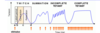
Sarcomere length
- Below is a graph of sarcomere spacing (i.e. sarcomere length) vs. tension.
- For each segment (e.g. between A-B or between B-C), what is the relative length of the sarcomeric A band to the sarcomeric I band?


Basis of muscle fibre types

Exercise and adaptation
- Normal gastrocnemius - T1 and T2 Fibres mixed together, some dark (T1) some light (T2)
- … runner - Very little T1 (slow) most T2 (fast)
- … … runner - Very little T2 (fast) a lot more T1 (slow)
- Normal gastrocnemius - T1 and T2 Fibres mixed together, some dark (T1) some light (T2)
- Sprint runner - Very little T1 (slow) most T2 (fast)
- Long Distance runner - Very little T2 (fast) a lot more T1 (slow)

Exercise and adaptation
- Normal gastrocnemius - T1 and T2 Fibres mixed together, some dark (T1) some light (T2)
- Sprint runner - Very little T1 (… twitch) most T2 (… twitch)
- Long Distance runner - Very little T2 (… twitch) a lot more T1 (… twitch)
- Normal gastrocnemius - T1 and T2 Fibres mixed together, some dark (T1) some light (T2)
- Sprint runner - Very little T1 (slow) most T2 (fast)
- Long Distance runner - Very little T2 (fast) a lot more T1 (slow)

Motor units
- Definition: A … alpha motor … and all muscle fibres it …
- Functions as a single … unit of skeletal muscle
- all muscle fibres in a single motor unit are of the same type
- (e.g. slow oxidative, fast oxidative, fast glycolytic).

- Definition: A single alpha motor neuron and all muscle fibres it innervates.
- Functions as a single contractile unit of skeletal muscle
- all muscle fibres in a single motor unit are of the same type
- (e.g. slow oxidative, fast oxidative, fast glycolytic).
Motor units
- Definition: A single … motor neuron and all muscle fibres it innervates
- Functions as a single contractile unit of skeletal muscle
- all muscle fibres in a single motor unit are of the … …

- Definition: A single alpha motor neuron and all muscle fibres it innervates.
- Functions as a single contractile unit of skeletal muscle
- all muscle fibres in a single motor unit are of the same type
- (e.g. slow oxidative, fast oxidative, fast glycolytic).
Types of muscular force generation
- Muscle contraction ≠ (necessarily) muscle …
- … – force during contraction – tossing a ball into air
- … (negatives) – force during muscle elongation
- e.g. when “braking” or when the weight of the object is overwhelming – catching a ball
- Both types of force generation can occur in one behaviour
- Proprioception controls force gen. based on length and stretch
- Muscle contraction ≠ (necessarily) muscle shortening
- Concentric – force during contraction – tossing a ball into air
-
Eccentric (negatives) – force during muscle elongation
- e.g. when “braking” or when the weight of the object is overwhelming – catching a ball
- Both types of force generation can occur in one behaviour
- Proprioception controls force gen. based on length and stretch

Types of muscular force generation
- Muscle contraction ≠ (necessarily) muscle shortening
- Concentric – force during … – tossing a ball into air
- Eccentric (negatives) – force during muscle …
- e.g. when “braking” or when the weight of the object is overwhelming – catching a ball
- Both types of force generation can occur in one behaviour
- … controls force gen. based on length and stretch
- Muscle contraction ≠ (necessarily) muscle shortening
- Concentric – force during contraction – tossing a ball into air
- Eccentric (negatives) – force during muscle elongation
- e.g. when “braking” or when the weight of the object is overwhelming – catching a ball
- Both types of force generation can occur in one behaviour
- Proprioception controls force gen. based on length and stretch

Types of muscular force generation
- Fill in the blanks


Upper vs Lower Motor Neurons
- Lower motor neuron disease
- Leads to …
- Leads to Muscle …
- Upper motor neurone disease
- Spasticity, hypertonia
- Lower motor neuron disease
- Weakness
- Muscle atrophy
- Upper motor neurone disease
- Spasticity, hypertonia

Upper vs Lower Motor Neurons
- Lower motor neuron disease
- Weakness
- Muscle atrophy
- Upper motor neurone disease
- S…, H…
- Lower motor neuron disease
- Weakness
- Muscle atrophy
- Upper motor neurone disease
- Spasticity, hypertonia

Stretch Reflex
- Controls Muscle Length
- … Muscle Force
- Lack of patellar reflex = … sign
- Controls Muscle Length
- Increases Muscle Force
- Lack of patellar reflex = Westphal’s sign

Stretch Reflex controls muscle … and increases muscle …
Stretch Reflex controls muscle length and increases muscle force (lack of patellar reflex - westphal’s sign)

Patellar Reflex
- Sensory spindle fibre = Muscle Spindle Fibre
- Detects …, i.e. Length
- Proprioception
- Spindle is … to other muscle fibres
- Ipsilateral Spinal reflex
- Monosynaptic

- Sensory spindle fibre = Muscle Spindle Fibre
- Detects Stretch, i.e. Length
- Proprioception
- Spindle is Parallel to other muscle fibres
- Ipsilateral Spinal reflex
- Monosynaptic
Patellar Reflex
- Sensory spindle fibre = Muscle Spindle Fibre
- Detects Stretch, i.e. Length
- P…
- Spindle is Parallel to other muscle fibres
- Ipsilateral … reflex
- Monosynaptic

- Sensory spindle fibre = Muscle Spindle Fibre
- Detects Stretch, i.e. Length
- Proprioception
- Spindle is Parallel to other muscle fibres
- Ipsilateral Spinal reflex
- Monosynaptic
Tendon Reflex
- Protects from …
- … Muscle Force -> dropping the load
- Sensor firing -> decreased contraction
- Protects from overloading
-
Decreases Muscle Force -> dropping the load
- Sensor firing -> decreased contraction

Tendon Reflex
- Protects from overloading
- Decreases Muscle Force -> dropping the load
- Sensor firing -> … contraction
- Protects from overloading
- Decreases Muscle Force -> dropping the load
- Sensor firing -> decreased contraction

Tendon Reflex
- Sensor to Spinal Cord
- Interneuron to motor neuron
- Motor neuron …
- Motor neuron to muscle
- Sensor to Spinal Cord
- Interneuron to motor neuron
- Motor neuron inhibited
- Motor neuron to muscle

Tendon Reflex
- Sensor = … Tendon Organ
- Detects …
- In series with muscle
- In tendon
- Near border with muscle
- Disynaptic
- Ipsilateral Spinal reflex
- Sensor = Golgi Tendon Organ
- Detects Tension
- In series with muscle
- In tendon
- Near border with muscle
- Disynaptic
- Ipsilateral Spinal reflex

Tendon Reflex
- Sensor = Golgi Tendon Organ
- Detects Tension
- In … with muscle
- In tendon
- Near … with muscle
- Disynaptic
- Ipsilateral Spinal reflex
- Sensor = Golgi Tendon Organ
- Detects Tension
- In series with muscle
- In tendon
- Near border with muscle
- Disynaptic
- Ipsilateral Spinal reflex

Tendon Reflex
- Sensor = Golgi Tendon Organ
- Detects Tension
- In series with muscle
- In tendon
- Near border with muscle
- …synaptic
- Ipsilateral Spinal reflex
- Sensor = Golgi Tendon Organ
- Detects Tension
- In series with muscle
- In tendon
- Near border with muscle
- Disynaptic
- Ipsilateral Spinal reflex

Tendon Reflex
- Sensor = Golgi Tendon Organ
- Detects Tension
- In series with muscle
- In tendon
- Near border with muscle
- Disynaptic
- … Spinal reflex
- Sensor = Golgi Tendon Organ
- Detects Tension
- In series with muscle
- In tendon
- Near border with muscle
- Disynaptic
- Ipsilateral Spinal reflex
