Anterior Thigh, Leg, & Foot Flashcards
(15 cards)
Anterior compartment of the thigh
- Actions?
- Innverated by?
- Emerges? Passes?
- Innervates muscles where?
- What part innerves skin where?
- Muscles of the anterior compartment of the thigh?
Anterior compartment of the thigh
- Action:
- Flex hip joint
- Extend knee joint
- Innervation:
- Femoral nerve (L2-L4)
- Emerges lateral to psoas major
- Passes deep to the inguinal ligament
- Innervates muscles in the anterior compartment of the thigh and skin on the anterior medial thigh
- Terminal branch, the saphenous nerve, innervates skin on the anterior medial aspect of the knee, leg, and foot
- Muscles of the anterior compartment of the thigh:
- Quadriceps femoris
- Rectus femoris
- Vastus lateralis
- Vastus medialis
- Vastus intermedius
- Iliopsoas
- Satorius
- Pectineus
- Quadriceps femoris
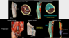
Quadriceps femoris (of the anterior compartment of the thigh)
- What are the four muscles that make up the quadriceps femoris?
- Origin and action for each of the main two?
- The four parts unite to form?
- What is the patella and what does it do?
- Where are the two places bursitis can develop?
- What is the patellar reflex?
Quadriceps femoris (of the anterior compartment of the thigh)
-
Rectus femoris
- Orgin: anterior inferior iliac spine (AIIS)
- Action: flex hip joint and extend knee joint
-
Vastus
- Lateralis
- Medialis
- Intermedius (deep to rectus femoris)
- Orgin: femur
- Action: extend knee joint
- 4 parts unite distally to form the quadriceps tendon, which continues as the patellar ligament and inserts on the tibial tuberosity
- Patella: large sesamoid bone, embedded in the quadriceps tendon, increases leverage of the quadriceps and increases power of knee extension
- Bursitis: can develop in the prepatellar bursa (between skin and patella) or the suprapatellar bursa (deep to quadriceps tendon)
- Patellar tendon reflex: tests the integrity of the femoral nerve and the L2-L4 spinal nerves. With knees flexed and relaxed, the patella ligament is tapped to elicit rapid extension of the knee joint (exploiting proprioception)

(Of the anterior compartment of the thigh)
Iliopsoas
- Composed of what two muscles?
- Origin, insertion, action?
Sartorius
- Origin, insertion, action?
Pectineus
- Origin, insertion, action?
- Innervation
(Of the anterior compartment of the thigh)
Iliopsoas
- Iliopsoas = iliacus + psoas major
- Origin:
- Iliac fossa (iliacus)
- Lumbar vertebrae (psoas major)
- Insertion:
- Lesser trochanter of femur
- Actions:
- Flex hip joint (if lower extremity is free)
- Stabilize the trunk (if lower extremity is fixed)
Sartorius
- Origin:
- ASIS
- Insertion:
- Medial aspect of proximal tibia
- Actions:
- Flex (b/c it is posterior)
- Abduct (b/c it is lateral to hip joint)
- Laterally rotate hip joint
- Flex knee joint (sit cross legged)
Pectineus
- Orgin:
- Pubis
- Insertion:
- Proximal femur
- Actions:
- Weakly flex hip joint
- Adduct hip joint (b/c it is medial to hip joint)
- Innervation:
- Femoral nerve (similar to rest of the compartment)
- Branch of obturator nerve
Medial compartment of the thigh
- Origin, action, and innervation?
- Courses along? Exits?
- Innervates what?
- What muscles make up the medial compartment of the thigh?
Medial compartment of the thigh
- Origin: pubis
- Action: adduct hip joint (medial to hip joint)
- Innervation: obturator nerve (L2-L4)
- Courses along lateral walls of pelvis and exits via the obturator canal
- Innervates muscles of medial compartment of thigh and skin on medial thigh
- Muscles that make up the medial compartment of the thigh:
- Adductor longus
- Adductor brevis
- Adductor magnus
- Gracilis
- Obturator externus

(Of medial compartment of the thigh)
- Adductor longus vs brevis?
- Adductor magnus
- Adductor vs hamstring part?
- Hamstring origin, insertion, action, and innervation?
- Gracilis insert and action?
- Obturator externus origin, insertion, and action?
(Of medial compartment of the thigh)
Adductor longus
- Longer and more superficial
Adductor brevis
- Shorter and deeper
Adductor magnus
-
Adductor part
- Follows compartment rules for action and innervation
-
Hamstring part
- Origin: ischial tuberosity
- Insertion: adductor tubercle of femur
- Action: extend hip joint
- Innervation: tibial nerve of sciatic nerve
Gracilis
- Insertion: medial aspect of proximal tibia
- Actions:
- Weakly adduct hip joint
- Flex knee joint
Obturator externus
(remember: obturator internus is inside the pelvis and obturator externus is outside)
- Origin:
- External aspect of obturator membrane
- Margin of obturator foramen
- Insertion:
- Proximal femur
- Actions:
- Laterally rotate hip joint
- Stabilize hip joint

Crural fascia and compartments
- What are the three fascial compartments in the leg?
- The crural fascia thickens where to form what?
- How are tendons protected under the retinaculum? Function of this?
Crural fascia and compartments
- Three fascial compartments in the leg:
- Anterior
- Lateral
- Posterior
- Superficial
- Deep
- The crural fascia thickens distally to form:
- Flexor retinacula
- Extensor retinacula
- Fibular retinacula
- As tendons pass under a retinaculum, they are protected by synovial tendon sheaths, which facilitate sliding by reducing friction and can become inflamed causing pain

Anterior compartment of the leg
- Action and innervation?
- Sciatic nerve divides into?
- Common fibular nerve winds around?
- Common fibular nerve divides into?
- Deep fibular nerve innervates what?
- What are the muscles?
Anterior compartment of the leg
- Action:
- Dorsiflex ankle joint
- Extend toes
- Innervation:
- Deep fibular nerve (L4-S1)
- Sciatic nerve divides into:
- Tibial nerve
- Common fibular (peroneal) nerves
- Common fibular nerve winds around the neck of the fibula
- Common fibular nerve divides into superficial fibular and deep fibular
- Deep fibular (peroneal) nerve
- Innervates the muscles of the anterior compartment of the leg and dorsal foot
- Innervates the skin of the dorsal foot between the hallux and second digit
- Deep fibular (peroneal) nerve
- Sciatic nerve divides into:
- Deep fibular nerve (L4-S1)
- Muscles:
- Tibialis anterior
- Extensor hallucis longus
- Extensor digitorum longus
- Fibularis (peroneus) teritus

(Of the anterior compartment of the leg)
Tibialis anterior
- Insertion and action?
- What are shin splints?
Extensor hallucis longus
- Insertion and action?
Extensor digitorum longus
- Insertion and action?
- What forms on dorsal surface of the phalanges?
(Of the anterior compartment of the leg)
Tibialis anterior
- Insertion:
- Medial cuneiform
- Base of first metatarsal
- Action:
- Dorsiflex ankle joint
- Invert foot
- Shin splints: anterior tibialis strain, results from repeated microtrauma or overexertion, or small tears in the periosteum (tissue enclosing bone), inflammation can cause compartment syndrome
Extensor hallucis longus
- Insertion:
- Distal phalanx of the hallux
- Action:
- Extend hallux (MTP and IP joints)
- Weakly dorisflex ankle joint
Extensor digitorum longus
- Insertion:
- Distal phalanges of digits 2-5
- Action:
- Extend digits 2-5 (MTP, PIP, and DIP joints)
- Dorsiflex ankle joint
- Note: the extensor digitorum longus and extensor hallucis longus tendons form extensor expansions on the dorsal surface of the phalanges

(Of the anterior compartment of the leg)
Fibularis (peroneus) tertius
- Insertion and action?
- Possible abnormalities of this?
Common fibular nerve
- Why is this the most frequently injured lower extremity nerve?
- What will injury to it affect?
- What is foot drop? Steppage gait? Foot slap?
(Of the anterior compartment of the leg)
Fibularis (peroneus) tertius
- Insertion:
- Base of fifth metatarsal
- Action:
- Dorsiflex ankle joint
- Evert foot
- May be absent in 8% of population or fused with extensor digitorum longus
Common fibular nerve
- Most frequently injured lower extremity nerve as it is vulnerable to injury where it winds around the neck of the fibula
- Injury to the common fibular nerve will affect:
- Anterior compartment of the leg
- Dorsal foot (dorsiflexors and toe extensors; one invertor)
- Lateral compartment of the leg (evertors and weak plantarflexors)
- Loss of dorsiflextion and toe extension results in “foot drop” where toes of the affected side will not clear the ground during the swing phase of gait
- Patients may compensate with exaggerated hip and knee flexion, “steppage gait,” to provide adequate clearance for the affected foot during the swing phase of gait
- Patients may also show “foot slap” where the entire foot makes contact with the ground at the end of swing phase, rather than just the heel

Dorsal foot
- What are the two muscles and where are they located?
- Insertion and extension of both?
Dorsal foot
- There are two muscles on the dorsal aspect of the foot
- Located deep to the extensor digitorum longus tendons and superficial to the dorsal interossei
- Innervation: deep fibular nerve (L5-S1)
-
Extensor digitorum brevis
- Insertion: extensor expansions of digits 2-4
- Action: extend digits 2-4 (MTP, PIP, and DIP joints)
-
Extensor hallucis brevis
- Insertion: extensor expansion of the hallux
- Action: extend hallux (MTP and IP joints)
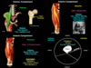
Obturator artery
- Branch of? Except in 20% of population?
- Passes through? Enters?
- Supplies?
Femoral artery
- Primary source?
- Continuation of? Changes names at?
- Directly supplies?
- Femoral pulse can be felt?
- Passes through what to become the popliteal artery?
Deep artery of the thigh
- Largest branch of?
- Perforating branches supply?
Obturator artery
- Typically a branch of the internal iliac artery, however in 20% of the population it arises from the inferior epigastric artery (aberrant obturator artery)
- Passes through obturator canal to enter medial compartment of the thigh
- Supplies medial and posterior compartments of the thigh
Femoral artery
- Primary source of blood to the lower extremtiy
- Continuation of the external iliac artery (changes names at the inguinal ligament)
- Directly supplies anterior and medial compartments of the thigh (supplies other regions via its branched names)
- Femoral pulse can be felt just inferior to the midpoint of the inguinal ligament, with patient in supine position - compression at this location will stop blood flow through the femoral artery
- Distally, the femoral artery passes through the adductor hiatus (gap in adductor magnus) to reach the popliteal fossa on the posterior aspect of the knee, where it becomes the popliteal artery
- Femoral artery –> adductor hiatus –> popliteal artery
Deep artery of the thigh
- Largest branch of the femoral artery
- Perforating branches supply the posterior compartment of the thigh, medial compartment of the thigh, and the lateral aspect of the anterior compartment of the thigh

Lateral and medial circumflex femoral arteries
- Typically are branches of? But can also be branches of?
- What do each supply?
Anterior tibial artery
- Branch of?
- Passes through what and descends along what?
- Supplies?
- Becomes what at the ankle?
Dorsalis pedis artery
- Major source of blood to?
- Anastomoses with?
- What are the major branches?
- Where can the dorsalis pedis pulse be felt?
Lateral and medial circumflex femoral arteries
- Typically are branches of the deep artery of the thigh
- Can also be branches of the femoral artery though
- Lateral circumflex femoral artery supplies the lateral aspect of the thigh
- Medial circumflex femoral artery supplies most of the blood to the femoral head and neck
Anterior tibial artery
- Branch of the popliteal artery
- Passes through gap in superior border of the interosseous membrane and descends along the membrane with the deep fibular nerve
- Supplies the anterior lateral leg
- Terminates at the ankle, where it becomes the dorsalis pedis artery
- Anterior tibial artery –> dorsalis pedis artery
Dorsalis pedis artery
- The major source of blood to the toes
- Anastomoses with the deep plantar arch
- Major branches:
- Arcuate artery
- Dorsal metatarsal arteries
- Dorsal digital arteries
- The dorsalis pedis pulse can be palpated ont he dorsal aspect of the foot, just lateral to the tendon of extensor hallucis longus

Veins
- Dorsal venous arch
- Drains what?
- Anastomoses with?
- Drains into what medially and laterally?
- Perforating veins: connect?
- Deep veins: accompany?
- Femoral vein
- Receives blood from?
- Drains into?
Veins
-
Dorsal venous arch
- Superficial drainage of the dorsal foot
- Anastomoses with the plantar venous arch in the sole of the foot
- Drains into the great saphenous vein medially and the small saphenous vein laterally
- Perforating veins: connect the superficial to the deep veins
- Deep veins: accompany the arteries and share their names (however the is NO dorsalis pedis vein)
-
Femoral vein
-
Receives blood from:
- Popliteal vein
- Deep vein of the thigh
- Great saphenous vein
- Drains into the external iliac vein
- Popliteal vein –> femoral vein
-
Receives blood from:

Femoral triangle
- What are the borders?
- What are the contents?
- Three compartments of the femoral sheath?
- Femoral canal contents?
- Function if femoral canal?
Femoral triangle
- Borders:
- Inguinal ligament (superior)
- Adductor longus (medial)
- Sartorius (lateral)
- Contents:
- NAVEL: nerve, artery, vein, empty space, lymph
- Femoral nerve and its branches
-
Femoral sheath
- Fascial tube enclosing the femoral vessels and femoral canal
- Does NOT enclose the femoral nerve
- Divided into three compartments:
- Lateral contains femoral artery
- Intermediate contains femoral vein
- Medial contains femoral canal
- Femoral canal contains loose connective tissue, fat, lymphatic vessels, and deep inguinal lymph nodes
- Primary function of the femoral canal is to allow expansion of femoral vein when venous return from lower limb is increased (e.g. during strenuous activity)

Femoral hernia
- Location and significance of femoral ring?
- What is the ring normally closed by?
- How is a femoral hernia formed?
- Femoral hernia lies where? Medial to? Inferior to?
- May emerge where?
- More frequent in males or females?
- How do they differ from direct/indirect hernias?
Femoral hernia
- The proximal opening of the femoral canal is called the femoral ring
- The femoral ring is an area of weakness where femoral hernias can occur
- Normally, the ring is closed by extraperitoneal fatty tissue and this fatty tissue is overlain by parietal peritoneum
- Abdominal viscera can protrude through the femoral ring into the femoral canal, forming a femoral hernia
- A femoral hernia lies in the femoral triangle, medial to the femoral vein, and inferior to the inguinal ligament
- A femoral hernia may emerge through the saphenous opening into the subcutaneous tissue of the thigh
- Femoral hernias occur more frequently in females
- NOT to be confused with direct/indirect hernias, which emerge superior to the inguinal ligament

