Posterior Abdominal Wall Flashcards
(24 cards)
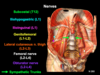
Kidneys
- What type of organ?
- Vertebrae level?
- Which one is higher?
- What do you have to remove to see kidneys?
- Quadrant?
- List layers of fat/fascia from superficial to deep
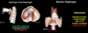
Kidneys
- Primarily retroperitoneal
- Located at T12 - L3 vertebral levels
- RUQ and LUQ
- Left kidney higher due to liver on right side
- Must remove parietal peritoneum to see kidneys
- Surrounded by layers of fat and fascia - from SF to deep:
- Paranephric fat
- Renal fascia
- Prolonged inferiorly along ureters
- Helps prevent spread of infection locally, but acts as a conduit for the spread of these fluids into the pelvis
- Perinephric fat

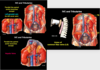
What are the features of the kidneys?
What is anterior, middle, and posterior in renal hilum?
Features of the kidneys
- Superior pole - adjacent to renal hilum
- Inferior pole
- Lateral border - convex
- Medial border - concave, contains renal hilum
- Renal capsule - very thin outer layer
- Renal cortex - deep to the capsule and extends internally to renal columns
- Renal medulla - renal pyramids
Renal hilum
- Arteries enter
- Veins and renal pelvis exit
- Anterior - renal vein
- Middle - renal pelvis
- Posterior - renal pelvis

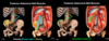
What is the pathway of urine to the ureter?
Pathway of urine to the ureter
- Renal papillae - apices of renal pyramids
- Minor calyx - collects urine from renal papillae
- Major calyx - collects uring from 2-3 minor calices
- Renal pelvis - receives urine from 2-3 major calices and delivers it to the ureter

What are the two anomalous kidneys?
Anomalous kidneys
-
Pelvic kidney
- failure of kidney to ascend during development
-
Horsehoe kidney
- right and left kidneys are united at inferior poles
- ascent stopped by IMA

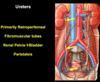
Ureters
- What kind of organ?
- What kind of tubes and where do they extend to?
- How do they transport urine?
Ureters
- Primarily retroperitoneal
- Fibromuscular tubes extending from renal pelvis to bladder
- Transport urine via peristaltic contractions
Adrenal (suprarenal) glands
- What kind of organ?
- Location?
- Quadrant?
- Enclosed by?
Adrenal (suprarenal) glands
- Primarily retroperitoneal
- Located on superomedial aspect of kidneys
- RUQ or LUQ (where kidneys are)
- Enclosed by renal fasica

Lymphatic drainage of the kidneys and adrenal glands
Lymphatic drainage of the kidneys and adrenal glands
- lumbar lymph nodes (along aorta)
- chyle cistern
- thoracic duct

Parasympathetic innervation of kidneys
Parasympathetic innervation of kidneys
- Preganglionic cell bodies: brain
- Preganglionic fibers: vagus nerve (CN X)
- Postganglionic cell bodies: wall of kidney
- Postganglionic fibers: wall of kidney

Sympathetic innervation of kidneys
Sympathetic innervation of kidneys
- Preganglionic cell bodies: lateral horn of the thoracolumbar spinal cord
- Preganglionic fibers: ventral root, spinal nerve, ventral ramus, white ramus communicans, sympathetic trunk, thoracic splanchnic nerve (usually least thoracic splanchnic nerve)
- Postganglionic cell bodies: renal (aorticorenal) ganglia
- Postganglionic fibers: follow arterial branches to target organ

Visceral pain from kidneys and ureters
- Where do you enter spinal cord?
- Where does pain refer to?
Visceral pain from kidneys and ureters
- follow the sympathetic pathway back to the spinal cord but enter dorsal root
- refer to back, flank, groin, and genitals

Sympathetic innervation of adrenal glands
Sympathetic innervation of adrenal glands
- Preganglionic cell bodies: lateral horn of the thoracolumbar spinal cord
- Preganglionic fibers: ventral root, spinal nerve, ventral ramus, white ramus communicans, sympathetic trunk, thoracic splanchnic nerve
- Postganglionic cell bodies: secretory cells of adrenal medulla

Abdominal aorta
- Where does it begin?
- Located left or right of midline?
- Where does it bifurcate and into what?
- What are the unpaired branches?
Abdominal aorta
- begins at aortic hiatus of diaphragm at T12
- located left of the midline
- bifurcates at L4 into common iliac arteries
- unpaired branches:
- celiac trunk
- SMA
- IMA

What are the paired branches of the abdominal aorta? Include branches if applicable
What do they supply?
Paired branches of abdominal aorta
Paired branches supply primarily retroperitoneal organs, gonads, and body wall
- inferior phrenic arteries
- supply inferior surface of diaphragm and adrenal glands via the superior suprarenal arteries
- middle suprarenal arteries
- renal arteries
- inferior suprarenal arteries
- gonadal (testicular or ovarian) arteries
- subcostal arteries
- lumbar arteries

Paired branches of abdominal aorta
Renal arteries
- Which side is longer?
- What are the (possible) branches?
Paired branches of abdominal aorta
Renal arteries
- Right renal artery is longer because it courses posterior to IVC and aorta is left of midline
- Can have accessory renal arteries, which are from a failure of embryonic arteries to degenerate
- Inferior suprarenal branch off

What are the arteries to the adrenal gland and where are they from?
Arteries to adrenal gland
- Superior suprarenal artery
- via inferior phrenic artery
- Middle suprarenal artery
- via aorta
- Inferior suprarenal artery
- via renal artery

Abdominal aortic aneurysm
Abdominal aortic aneurysm
- can rupture
- high mortality rates (90%) if not diagnosed

Inferior vena cava
- Formed by union of? Vertebral level?
- Exits abdomen through what? Vertebral level?
- Located right or left of midline?
- Tributaries of IVC parallel what?
- What are the exceptions?
Inferior vena cava
- formed by a union of common iliac veins at L5 vertebral level
- exits abdomen through caval opening of diaphragm at T8 vertebral level
- located right of the midline
- tributaries of IVC parallel the paired branches of the aorta
- notable exceptions:
- Left gonadal veins - drain to left renal vein
- Hepatic veins - have no arterial complement
Posterior abdominal wall muscles
- What are they?
- Origin for each?
- Insertion for each?
Posterior abdominal wall muscles
-
Iliopsoas
-
Psoas major
- Origin - lumbar vertebrae
-
Iliacus
- Origin - iliac fossa
- Psoas major and iliacus fuse to form iliopsoas, which inserts on the lesser trochanter of the femur
-
Psoas major
-
Quadratus lumborum
- Origin - 12th rib and lumbar vertebrae
- Insertion - iliac crest
What are the nerves of the posterior abdominal wall
Nerves of the posterior abdominal wall
- subcostal nerves
- lumbar plexus
- greater, lesser, and least splanchnic nerves
- sympathetic trunks

Nerves of the posterior abdominal wall
Subcostal nerves
- Ventral rami of what?
- Travel laterally across what?
- Pierce what to enter abdominal wall?
- What innervation to what dermatome?
- Motor innervation to where?
Nerves of the posterior abdominal wall
Subcostal nerves: ventral rami of T12
- travel laterally across quadratus lumborum
- pierce transversus abdominis to enter abdominal wall
- sensory innervation and sympathetic innervation to T12 dermatome
- motor innervation to external obliques, internal obliques, and transverse abdominis
Nerves of the posterior abdominal wall
Lumbar plexus
- Ventral rami of what lumbar spinal nerves?
- List the nerves and vertebral level
Nerves of the posterior abdominal wall
Lumbar plexus: ventral rami of L1 - L5
- iliohypogastric nerve L1
- ilioinguinal nerves L1
- genitofemoral nerve L1 - L2
- lateral cutaneous nerve of the thigh L2 - L3
- femoral nerve L2 - L4
- obturator nerve L2 - L4

Nerves of the posterior abdominal wall
Greater, lesser, and least splanchnic nerves
What do they pass through?
Nerves of the posterior abdominal wall
Greater, lesser, and least splanchnic nerves
Pass through the diaphragm

Nerves of the posterior abdominal wall
Sympathetic trunks
- Continue from where?
- Located?
Nerves of the posterior abdominal wall
Sympathetic trunks
- continue from thorax
- located lateral to the lumbar vertebral bodies along the anterior surface of the sacrum

Diaphragm
- What are the attachments?
- What are the openings?
Diaphragm
Attachments (all peripheral):
- xiphoid process
- costal margin
- ribs 11-12
- lumbar vertebrae (via right and left crura)
Openings:
- caval opening at T8 for IVC
- esophageal hiatus at T10 for esophagus and vagus nerve
- aortic hiatus at T12 for aorta, thoracic duct, and azygos vein