Pelvis II Flashcards
(19 cards)
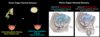
What are the pouches in males and females?
The peritoneal cavity extends inferiorly into the pelvis. The peritoneum drapes the pelvis organs and creates spaces where peritoneal fluid can accumulate
- Males: rectovesicle pouch
- Females: vesicouterine pouch and rectouterine pouch

Ureters
- Descend into pelvis where?
- Course where for females/males?
- Surgical risk?
- How to prevent backflow?
Ureters
- Descend into the pelvis near the bifurcation of the common iliac vessels into external and internal iliac vessels
- “Water under the bridge”
- Course inferior to the uterine artery in the female
- Course inferior to the ductus deferens in the male
- Due to its close proximity to the uterine artery, the ureter is vulnerable to injury during a hysterectomy when the uterine artery is ligated
- Ureters pass obliquely through the posterosuperior bladder wall
- Its oblique passage forms a one way valve, which prevents the backflow of urine into the ureters

Urinary bladder
- Function?
- What are the walls made of?
- Sympathetic and parasympathetic innervation - what do they stimulate and from what nerves?
- When is the rugae present and absent?
- Sphincter function?
- Triangle portion
- Name?
- Defined by?
- Function?
Urinary bladder
- The bladder receives urine from the ureters, stores it, then expels it through the urethra
-
Detrusor muscle - highly stretchable smooth muscle walls
- Parasympathetic innervation (pelvic splanchnic nerve) stimulates contraction of the detrusor muscle, promoting micturation
-
Rugae
- Mucosal folds are prominent when the bladder is empty
- Folds disappear as the bladder distends
-
Internal urethral sphincter
- Smooth muscle sphincter at the neck of the bladder that surrounds the opening of the urethra in males
- Sympathetic innervation (lumbar and sacral splanchnic nerves) stimulates contraction of the sphincter, preventing micturation and preventing sperm from entering the bladder during ejaculation
-
Trigone
- Triangular portion of the posterior wall defined by the two ureteric orifices superiorly and the urethral orifice inferiorly
- Smooth (no rugae)
- Very sensitive to stretch (visceral sensory innervation stimulates urge to void)

Important relationships to the bladder
- What is inferior to the bladder in males?
- What is between the pubis and bladder?
- Location of bladder in babies, children, and adults with distended bladders?
- Surgical significance?
- Risk of this?
Important relationships to the bladder:
- In males, the bladder lies superior to the prostate gland
- The retropubic space lies posterior to the pubis and anterior to the bladder
- In babies and children (until puberty), and adults with a distended bladder, the bladder extends superior to the pubis
- A suprapubic incision can be made to access the bladder without transversing the peritoneum and entering the peritoneal cavity
- Bladder is susceptible to injury in this position (MVA, fall from high place, heavy object falling on lower abdomen) because it can rupture

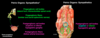
Female pelvic organs
Broad ligament
- What is it?
- Location?
- Function?
- What two structures are on its border?
- What does it enclose?
- What is its extension and what does that enclose?
Female pelvic organs
Broad ligament
- Double layer of peritoneum (mesentery) that extends from the sides of the uterus to the lateral walls and floor of the pelvis
- Holds the uterus in position and conveys vasculature and nerves to/from the uterus
- The uterine fallopian tubes lie in the superior edge of the broad ligament
- The ovaries are attached to the posterolateral border of the broad ligament
- An extension of the broad ligament, the suspensory ligament of the ovary, encloses the ovarian arteries, veins, lymphatic vessels, and nerves
- Broad ligament also encloses the bilateral remnants of the gubernaculum:
- Ovarian ligament - between ovary and uterus
- Round ligament of the uterus - between the uterus and labia majora

Female pelvic organs
Ovaries
- Intra or retro peritoneal?
- Originate where? Attached to what?
- Attach to what after descent?
- Vessels, nerves, lymphatics enclosed in what?
- What happens during ovulation?
Female pelvic organs
Ovaries
- Intraperitoneal
- Originate in the posterior abdominal wall, attached to the gubernaculum
- Descend into the pelvis and become attached to the posterior aspect of the broad ligament
- Ovarian vessels, nerves, and lymphatics that accompanied the descent of the ovary are enclosed within the suspensory ligament of the ovary
- During ovulation, an oocyte is expelled from the ovary into the peritoneal cavity, towards the abdominal orifices of the uterine tubes
Female pelvic organs
Uterine (fallopian) tubes
- Intra or retro peritoneal?
- Where does it open to peritoneal cavity and uterus?
- Why is PID more common in females?
- Most common site of what?
Female pelvic organs
Uterine (fallopian) tubes
- Intraperitoneal (enclosed in the superior edge of the broad ligament)
- Open into the peritoneal cavity at their abdominal orifices, which are surrounded by finger-like fimbriae
- Enter the superolateral walls of the uterus at their uterine orifices
- The peritoneal cavity is open in females and closed in males
- Pelvic inflammatory disease is more common in females, as the vagina, uterus, and uterine tubes provide a pathway for infections into the peritoneal cavity
- Uterine tubes are the most common site of an ectopic pregnancy

Female pelvic organs
Uterus
- Intra or retro peritoneal?
- Location?
- Myometrium
- What is it?
- What happens during pregnancy?
- Endometrium
- What is it?
- Function?
- Body
- What part of the uterus is it?
- What is the superior part?
- Cervix
- Intra or retro peritoneal?
- What part of the uterus is it?
- What are the os’s?
Female pelvic organs
Uterus
- Most of the uterus (the body, specifically) is intraperitoneal (suspended by the broad ligament)
- Located between the bladder and rectum
-
Myometrium
- Smooth muscle of the uterus
- Distends during pregnancy, superior to the pubic symphysis
-
Endometrium
- Internal lining
- Glandular mucosa
- Site of implantation
- Shed each month
-
Body
- Superior 2/3 of the uterus
- Fundus - rounded superior portion
-
Cervix
- Primarily retroperitoneal
- Cylindrical inferior 1/3 of the uterus
- Central canal opens superiorly into the body of the uterus at the internal os
- Central canal opens inferiorly into the vagina at the external os (site of Pap smear)

Female pelvic organs
Vagina
- Intra or retro peritoneal?
- Location?
- Continuous with cervix where?
- Cervix projects into vagina creating what?
- Location and surgical signifcance of posterior fornix?
Female pelvic organs
Vagina
- Primarily retroperitoneal
- Located between the bladder and rectum
- Anterior and posterior walls contact each other
- Continuous with the cervix at the external ox
- The cervix projects into the vagina at its superior end, creating recesses anteriorly, posteriorly, and laterally called the vaginal fornices
- The posterior fornix is the deepest recess and is related to the rectourterine pouch and the rectum posteriorly
- An incision can be made through the posterior fornix to examin the peritoneal cavity endoscopically

Male pelvic organs
Testes
- Originate where?
- What path do they take to descend?
- What does the parietal peritoneum that descends form?
- What is a hydrocele?
- Testis is covered by?
- What is inside testis?
- Path of the spermatozoa?
Male pelvic organs
Testes
- Originate in th posterior abdominal wall, attached to the gubernaculum
- Descend into the inguinal region, pass through the deep ring and inguinal canal, and exit the superficial ring to enter the scrotum
- As the testes descend, some of the parietal peritoneum is drawn into the inguinal canal and scrotum, and forms the tunica vaginalis
- Hydrocele - abnormal accumulation of fluid within the cavity of the tunica vaginalis
- Each testis is covered by a tough fibrous coat called the tunica albuginea
- The testes contain seminiferous tubules inside, which is where the spermatozoa mature
- Spermatozoa are transported from seminiferous tubules to the rete testis, to the efferent ductules, and finally to the epididymis

Male pelvic organs
Epididymis
- Location?
- Connects what to what?
Ductus (vas) deferens
- Pathway of its location?
- In relation to the ureter?
- Where does it form an ampulla?
- What is a vasectomy?
Male pelvic organs
Epididymis
- Highly coiled tube located on the posterior surface of the testis
- Connects the testis to the ductus deferens
Ductus (vas) deferens
- Muscular tube ascends within the spermatic cord, through the superficial ring and inguinal canal, and into the abdomen via the deep inguinal ring (lateral to the inferior epigastric vessels)
- Courses superior to the ureter - “water under the bridge”
- When it reaches the posterior aspect of the bladder, the vas deferens enlarges to form an ampulla
- A vasectomy is performed by making an incision in the superior aspect of the scrotum to expose and ligate the vas deferens

Male pelvic organs
Seminal vesicles
- What are they? Location?
- How is ejaculatory duct formed?
Ejaculatory ducts
- What does it open into?
What is the path to the prostatic urethra?
Male pelvic organs
Seminal vesicles
- Thin walled tubes that are coiled to form a mass that lies between the posterior wall of the bladder and the rectum
- Secrete fluid that mixes with the spermatozoa
- Ducts of the seminal vesicles join the ampullae of the ductus deferens to form the ejaculatory ducts
Ejaculatory ducts
- Open into the prostatic urethra
- Ampulla of ductus deferens + duct of seminal vesicles
- Ejaculatory duct
- Prostatic urethra

Male pelvis organs
Prostate
- Intra or retro peritoneal?
- What does it surround?
- Secretes fluid where and how?
- Where are the base, apex, and lobes?
- Median lobe
- Causes what?
- What happens when this is englarged?
- What is the most common site for prostatic carcinoma?
Male pelvis organs
Prostate
- Primarily retroperitoneal
- Gland surrounds the prostatic urethra
- Secretes fluid into the prostatic urethra via numerous ducts
- Base - superior, surrounds neck of the bladder and the internal urethral sphincter
- Apex - inferior
- Lobes:
-
Anterior lobe
- Anterior to prostatic urethra
-
Lateral lobes (two)
- On either side of prostatic urethra
-
Median (middle) lobe
- Posterior to the prostatic urethra and superior to the ejaculatory ducts
- This lobe is involved in benign hypertrophy of the prostate
- When median lobe is enlarged:
- It stretches the internal urethral sphincter (causing urine leakage)
- Puts pressure on the trigone (creating intense desire to void, especially at night)
- Can obstruct / narrow the prostatic urethra (making it difficult to void)
- Posterior lobe
- Posterior to the prostatic urethra and inferior to the ejaculatory ducts
- The posterior lobe is the most common site of prostatic carcinoma
-
Anterior lobe

Rectal exam - What is the anterior wall of the rectum related to for males and females?
Rectal exam
Males
- The anterior wall of the rectum is related to the prostate (posterior lobe) and seminal vesicles
Females
- The anterior wall of the rectum is related to the vagina

Differences of sex development (DSD) / Intersex
- What is it?
- How many babies are born with DSD?
- What is CAIS?
Differences of sex development (DSD) / Intersex
- Chromosomal, developmental, hormonal, gonadal, or phenotypic variations that influence reproductive and urogenital development
- DSD is also referred to as intersex
- Presence or development of characteristics that reflect non-binary sex genotype and phenotype
- 1 in 1500 babies are born with DSD / intersex characteristics
- Prevalence varies for specific types of DSD
- Those born with Complete Androgen Insensitivity (CAIS) 46, XY karyotypes but cell receptors do not respond to androgens (direct masculinzed sex development).
- Those with CAIS are phenotypically female with incompletely descended testes and no uterus, cervix, or fallopian tube development

What is the parasympathetic innervation of pelvic organs?
Parasympathetic innervation of pelvic organs
- Preganglionic cell bodies: S2-S4 spinal cord (intermediate gray matter)
- Preganglionic fibers: ventral root, spinal nerve, ventral ramus, pelvic splanchnic nerves
- Post ganglionic cell bodies: wall of organ
- Post ganglionic fibers: wall of organ

What is the sympathetic innervation of the pelvic organs?
Sympathetic innervation of the pelvic organs:
* Ovaries / testes are exceptions to this *
- Preganglionic cell bodies: lateral horn of the thoracolumbar spinal cord
- Preganglionic fibers: ventral root, spinal nerve, ventral ramus, white ramus communicans, sympathetic trunk, lumbar or sacral splanchnic nerves
- Post ganglionic cell bodies: superior or inferior hypogastric ganglia (usually the inferior)
- Post ganglionic fibers: follow arterial branches to target organ
What is the sympathetic innervation to the ovaries / testes?
Sympathetic innervation to the ovaries / testes
- Preganglionic cell bodies: lateral horn of the thoracolumbar spinal cord
- Preganglionic fibers: ventral root, spinal nerve, ventral ramus, white ramus communicans, sympathetic trunk, thoracic splanchnic nerves
- Post ganglionic cell bodies: superior mesenteric ganglia
- Post ganglionic fibers: follow gonadal vessels to target organ

Visceral sensory
- Where do they enter spinal cord?
- What are the routes taken for structures superior and inferior to the pelvic pain line?
- Where is the pain line located at the hindgut?
Visceral sensory
- Fibers follow the sympathetic or parasympathetic pathways back to the CNS, but enter the dorsal root
- Remember: visceral sensory bodies are located in the dorsal root ganglia
- The route taken by the sensory fibers differs relative to the pelvic pain line
-
Structures superior to the pelvic pain line:
- Visceral sensory fibers follow the sympathetic fibers back to the inferior thoracic and superior lumbar spinal cord levels
- Structures inferior to the pelvic pain line:
- Visceral sensory fibers follows the parasympathetic fibers (i.e. pelvic splanchnic nerves) back to the S2-S4 spinal cord
-
Structures superior to the pelvic pain line:
- The pain line in the hindgut is located at the midpoint of the sigmoid colon, so the visceral sensory fibers from the distal sigmoid colon and rectum follow the parasympathetic fibers back to the CNS
- Some organs lie superior and inferior to the pelvic pain line (i.e. bladder and uterus), so both visceral sensory pathways will be used
- E.g. sensory fibers from the superior aspect of the bladder will follow the sympathetic fibers, while sensory fibers from the inferior aspect of the bladder will follow the parasympathetic fibers