Pelvis I Flashcards
(28 cards)
What forms the pelvic girdle?
Pelvic girdle
- 2 os coxae (hip bones)
- sacrum
- coccyx
Os coxae (hip bones)
- Formed by?
- What are the two features of the lateral side?
Os coxae (hip bones)
- Formed by the fusion of:
- ilium
- pubis
- ischium
-
Acetabulum
- lateral aspect
- formed by the ilium, pubis, and ishium
- head of femur articulates here, forming the hip joint
-
Obturator foramen
- formed by the ischium and pubis
- big hole

Ilium
- What does the iliac crest connect?
- What is the concave medial surface?
Ilium
- The iliac crest connects the anterior superior iliac spine (ASIS) and posterior iliac spine (PSIS)
- The concave medial surface is the iliac fossa

Pubis
- What are the features of the pubis?
- Four rami
- Two rami form what
- Arch and angle
Pubis
- Pubic tubercles
- Pubic bones articulate at the midline to form the pubic symphysis
- Superior pubic ramus - unites with the ilium and ischium superiorly
- Inferior pubic ramus - unites with the ischial ramus inferiorly, which forms the ischiopubic ramus
- Pubic arch - formed by the two ischiopubic rami
- Subpubic angle - formed by the inferior borders of the two ischiopubic rami

Ischium
- What joins to form the ischiopubic ramus?
- What are the two notches?
- What is for muscle attachment?
Ischium
- Ischiopubic ramus - formed by the ischial ramus and inferior pubic ramus
- Ischial spine - parrot peak projection
- Greater sciatic notch - large concavity superior to the ischial spine
- Lesser sciatic notch - small concavity inferior to the ischial spine (and superior to ischial tuberosity)
- Ischial tuberosity - for muscle attachment

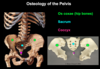
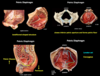
Sacrum
- Base, apex
- SI joint
- Anterior projection
- Foramina
Sacrum
- 5 fused vertebrae
- Base - articulates with L5 / S1 intervertebral disk and L5 vertebrae
- Apex - articulates with coccyx
- Sacroiliac (SI) joint - formed by the lateral surfaces articulating with the ilium on each side (very stable, this joint holds a lot of weight)
- Sacral promontory - anterior projection of S1 vertebrae (obstetrical landmark - limits about of space baby has to come out)
-
Sacral foramina - 4 pairs of anterior and posterior sacral foramina
- Anterior foramina - ventral rami of sacral spinal nerves
- Posterior foramina - dorsal rami of sacral spinal nerves

Coccyx
- How many vertebrae?
- Articulates with sacrum where?
Coccyx
- 4 fused vertebrae
- Articulates with sacrum at the sacrococcygeal joint

Pelvic inlet
- Divides the pelvis into what?
- How is it tilted?
Pelvic inlet
- The pelvic inlet divides the pelvis into:
-
Greater (false) pelvis
- Superior to pelvic inlet
- Occupied by abdominal viscera (hence “false”)
-
Lesser (true) pelvis
- Inferior to pelvic inlet
- Occupied by pelvic viscera
-
Greater (false) pelvis
- The pelvic inlet is tilted 55 degrees from the horizontal plane
- ASIS and anterior border of the pubic symphysis are in the same coronal plane

Pelvic outlet
- Boundaries?
- What is in the pelvic outlet?
- How does it tilt?
Pelvic outlet
- Boundaries:
- Coccyx and sacrum
- Sacrotuberous ligaments
- Ischial tuberosities
- Ischiopubic rami / pubic arch
- Pubic symphysis
- The pelvic outlet is closed by the pelvic diaphragm
- The pelvic outlet is tilted 15 degrees from the horizontal plane
- ASIS and anterior border of the pubic symphysis are in the same coronal plane

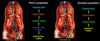
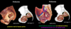
What are the sex differences in the pelvis?
(There are three)
Features different in the female and male pelvis shape (so babies can pass through)
Females have:
- Larger, more round / oval shaped pelvic inlet
- Ischial tuberosities and ischial spines are farther apart
- Subpubic angle is wider
Pelvic inlet
- Females: larger, round / oval shaped
- Males: heart shaped
Ischial tuberosities and ischial spines
- Females: farther apart
Subpubic angle:
- Females: wider, greater than 80 degrees
- Males: acute, less than 70 degrees

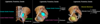
What are the three sacroiliac ligaments?
Sacroiliac ligaments
- Anterior sacroiliac ligaments
- Posterior sacroiliac ligaments
- Interosseous sacroiliac ligaments (deep)
Sacrotuberous and sacrospinous ligaments
- Location of each?
- What do these ligaments do?
Sacrotuberous ligaments
- Extend from the sacrum / coccyx to the ischial tuberosity
Sacrospinous ligaments
- Extend from the sacrum / coccyx to the ischial spine
- Located anterior to the sacrotuberous ligament
Together, these resist the influence of body weight that rotates the sacrum / coccyx posteriorly and superiorly

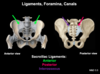
Foramen and canals of the pelvis
- The sacrotuberous and sacrospinous ligaments form what foramen?
- Obturator membrane? Canal? ?
- What do all foramen / canal form passageways to
Foramen and canals of the pelvis
- The sacrotuberous and sacrospinous ligaments form the:
-
Greater sciatic foramen
- Superior to ischial spine
- Passageway to the gluteal region
-
Lesser sciatic foramen
- Inferior to ischial spine
- Passageway to perineum
-
Greater sciatic foramen
-
Obturator membrane
- Fills the obturator foramen, except for a small opening, the obturator canal
- Obturator canal forms passageway to the medial compartment of the thigh
What are the three major muscles of the lesser pelvis?
What part of pelvis does each form?
Muscles of the lesser pelvis:
- Piriformis (forms wall)
- Obturator internus (forms wall)
- Pelvic diaphragm (forms floor)
- Coccygeus
- Levator ani

Piriformis
- Forms what wall of lesser pelvis?
- Originates from? Courses through? Inserts on?
- What nerves pass through this muscle?
- Innervation?
Piriformis
- Helps form the posterior wall of the lesser pelvis
- Originates from the anterior surface of the sacrum
- Courses through the greater sciatic foramen
- Inserts on the femur
- Ventral rami of the sacral spinal nerves emerge from the anterior sacral foramina and pass through piriformis, and meet with the lumbosacral trunk to form the sacral plexus on its internal surface
- Innervation: sacral plexus
Obturator internus
- Forms what wall of lesser pelvis?
- Originates from? Courses through? Inserts on?
- What covers the medial (internal) surface? Thickens to form?
- Innervation?
Obturator internus
- Helps form the lateral walls of the lesser pelvis
- Originates from the obturator membrane and the margin of the obturator foramen
- Courses throught the lesser sciatic foramen
- Inserts on the femur
-
Obturator fascia covers medial (internal) surface
- Obturator fascia thickens centrally as a tendinous arch for attachment of levator ani (muscle of pelvic diaphragm)
- Innervation: sacral plexus

Pelvic diaphragm
- Composed of what two muscles?
- Shaped like?
- Fuse at center to form?
- Fills the? Forms what of the pelvis?
- What is superior and inferior?
- What structures pass through it?
- Weakness of pelvic diaphragm can cause what?
- Innervation?
Pelvic diaphragm
- Shaped like a bowl / hammock
- Composed of:
- Coccygeus
- Levator ani
- Right and left muscles fuse at midline, forming a median raphe
- Fills the inferior pelvic aperture and forms the pelvic floor
- Supports the pelvic viscera on superior surface (peritoneum is inferior to pelvic diaphragm)
- Pelvic structures that pass through to reach perineum:
- Urethra
- Vagina
- Rectum
- Weakness of the pelvis diaphragm (coccygeus and levator ani) can result in:
- Urinary stress incontinence
- Bowel incontinence
- Prolapse of pelvic viscera (more common in females due to childbirth)
- Innervation: branches of the sacral and coccygeal plexuses
Coccygeus muscle of the pelvic diaphragm
Coccygeus muscle of the pelvic diaphragm
- Extends from the ischial spine to the coccyx and inferior sacrum
- Lies on the internal surface of the sacrospinous ligament

Levator ani muscle of the pelvic diaphragm
- Extends from what to what?
- What are the two openings?
- What are the two portions?
- Portion subdivided into?
- How does the puborectalis work?
Levator ani muscle of the pelvic diaphragm
- Extends from the pubis, the tendinous arch (thickening in the oburator fascia), and the ischial spine TO the coccyx and levator ani of the opposite side, along the median raphe
- Two openings:
- Posterior anal aperture
- Anterior urogenital hiatus (passageway for urethra and in females, vagina)
- Portions of the levator ani:
- Iliococcygeus
- Pubococcygeus
- Pubovaginalis (females)
- Puboprostaticus (males)
- Puborectalis
- Puborectalis
- Fibers surrounding the rectum
- Forms a sling around the anorectal junction, maintaining the anorectal flexure and preventing defecation
- This relaxes during defecation, while the rest of the levator ani contracts to prevent pelvic viscer from herniating inferiorly during the increase in intra abdominal pressure

Pelvic arteries
- Gonadal arteries branch off?
- Where do the testicular and ovarian arteries travel?
- How does the abdominal aorta divde? At what vertebral level?
- What does the external iliac artery supply?
Pelvic arteries
-
Gonadal (ovarian or testicular) arteries are branches of the abdominal aorta
- Testicular arteries travel within the spermatic cords inferiorly
- Ovarian arteries travel within the suspensory ligaments of the ovaries
- In addition to supplying the gonads, the ovarian arteries also supply the uterine tubes and the uterus
- Abdominal aorta divides into the common iliac arteries at the L4 vertebral level
- In the pelvis, the common iliac arteries divide into the external and internal iliac arteries
- The external iliac artery supplies the anterolateral abdominal wall and the lower extremity

Internal iliac artery
- What does it supply?
- What are the branches?
Internal iliac artery
- Primary blood supply to the pelvis, perineum, and gluteal region
- Branching pattern varies greatly!
- Major branches (paired)
- Superior gluteal artery
- Inferior gluteal artery
- Internal pudendal artery
- Umbilical artery
- Obturator artery
- Inferior vesicle artery
- Middle rectal artery
- Uterine artery (females)
- Vaginal artery (females)

What is the location of the superior and inferior gluteal arteries of the internal iliac artery?
Superior gluteal artery
- Exits greater sciatic foramen superior to piriformis to supply the gluteal region
Inferior gluteal artery
- Exites greater sciatic foramen inferior to piriformis to supply the gluteal region

Internal pudendal artery of the internal iliac artery
- Location?
- Travels to?
- Gives rise to?
Umbilical artery of the internal iliac artery
- Gives rise to? Which supplies?
- Obliterated portion continues on where as the what?
Internal pudendal artery
- Exits the greater sciatic foramen inferior to the piriformis to enter the gluteal region
- Then travels to the perineum via the lesser sciatic foramen
- Eventually gives rise to the inferior rectal artery
Umbilical artery
- Gives rise to the superior vesical arteries supplying the superior aspect of the bladder (and ductus deferens in males)
- Obliterated portion continues on the internal surface of the anterior abdominal wall as the medial umbilical ligaments

Obturator artery of the internal iliac artery
- Where does it course?
- Where does it exit and go to?
Aberrant obturator artery
- Where does it rise from and course over?
- Clinical significance?
Obturator artery
- Courses along the lateral wall of the pelvis with the obturator vein and nerve
- Exits the pelvis via the obturator canal to enter the medial compartment of the thigh
- In 20% of the population, there is an aberrant obturator artery arising from the inferior epigastric artery (instead of the internal iliac artery)
- The aberrant obrturator artery courses over the superior pubic ramus to exit via the obturator canal
- This artery can be accidently damaged during inguinal hernia repairs