Lower Extremity Overview Flashcards
(14 cards)
What are the five divisions of the lower extremity?
Divisions of the lower extremity
- Gluteal region
- Thigh (hip to knee)
- Popliteal fossa (posterior aspect of knee)
- Leg (knee to ankle)
- Foot (dorsal and plantar aspects)

What are all of the bones of the lower extremity?
Osteology of the lower extremity
- Hip bones
- Ilium
- Ischium
- Pubis
- Femur
- Patella (knee cap)
- Tibia (medial)
- Fibula (lateral)
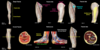
- 7 tarsals
- Calcaneus
- Talus
- Navicular (looks like a boot)
- Cuboid (looks like a cube)
- Medial, intermediate, and lateral cuneiforms
- 5 metatarsals (within body of foot, do not stick out with the toes)
- 14 phalanges
- Proximal
- Middle
- Distal
- (Hallux has proximal and distal phalanx only)

What are the parts of the femur, tibia, and fibula?
Osteology of lower extremity
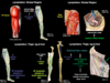
Femur
- Head
- Neck
- Shaft (body) with linea aspera (posterior)
- Medial and lateral femoral condyles (distal)
Tibia
- Medial
- Medial and lateral tibial condyles (proximal)
- Shaft (body)
- Medial malleolus (distal protuberance on medial side of the ankle)
Fibula
- Lateral
- Head
- Shaft (body)
- Lateral malleolus (distal protuberance on lateral side of the ankle)
(Pictures showing right side)

What are the structures and movements of the hip joint and knee joint?
Hip joint
- Acetabulum: articulates with femoral head, ball and socket synovial joint
- Flexion: reduces angle, kick forward
- Extension: increases angle, kick backwards
- Abduction: thigh away from midline
- Adduction: thigh towards midline
- Medial rotation: femur rotates medially (internal rotation)
- Lateral rotation: femur rotates laterally (external rotation)
Knee joint
- Femoral condyles: articulate with tibial condyles, hinge at synovial joint
- Flexion: leg closer to posterior thigh
- Extension: leg away from posterior thigh

What are the structures and movements of the ankle (talocrural) joint and subtalar joint?
Ankle (talocrural) joint
- Talus: articulates with distal tibia and lateral malleolus of the fibula, hinge synovial joint
- Dorsiflexion: brings foot closer to anterior thigh, toes superior
- Plantarflexion: brings foot away from anterior thigh, toes inferior
Subtalar joint
- Synovial joint between the talus and calcaneus
- Eversion: sole of foot faces laterally (uses lateral muscles)
- Inversion: sole of foot faces medially (uses medial muscles)

What are the structures and movements of the metatarsophalangeal (MIP) joints and interphalangeal (IP) joints?
Metatarsophalangeal (MTP) joints
- Synovial joints between the metatarsals and the proximal phalanges
- Flexion: toes towards the ground
- Extension: toes away from the ground
- Abduction: movement away from second digit axis
- Adduction: movement towards second digit axis
Interphalangeal (IP) joints
- Hinge synovial joints between phalanges
- Flexion: toes towards the ground
- Extension: toes away from the ground
- Proximal interphalangeal (PIP) joints: located between the proximal and middle phalanges
- Distal interphalangeal (DIP) joints: located between the middle and distal phalanges
- Hallux: great toe, single IP joint between proximal and distal phalanges

Fascia of the lower extremity
- What are the two layers of fascia?
- What does the first layer contain?
- Second layer
- What kind of tissue?
- How does it cover the muscles?
- Prevents muscles from what?
- Efficient mechanism for what?
- What is it called in the thigh and leg?
Fascia of the lower extremity
-
Superficial fascia
- Contains:
- Fat
- Cutaneous nerves
- Superficial veins (e.g. great saphenous vein)
- Lymphatic vessels
- Lymph nodes
- Contains:
-
Deep fascia
- Dense connective tissue
- Invests the lower extremity muscles like an elastic stocking
- Prevents muscles from bulging during contraction
- Efficient mechanism to pump blood back to the heart
- Thigh: fascia lata
- Leg: crural fascia

Fascia lata
- Location?
- Tightly adhered to?
- Oval shaped window? Located? What passes through?
- Thickening of fascia lata?
- Septa - how many, attach to what? Divide the thigh how?
Crural fascia
- Location?
- Continuous with?
- Septa - how many? Divide leg how?
- Another septum does what?
- Crural fascia form what near the ankle joint? Function?
Fascia lata
- Deep fascia of the thigh
- Tightly adhered to the pelvis (pubis, inguinal ligament, iliac crest, sacrum, coccyx, and ischial tuberosity)
- Saphenous opening: oval shaped window in the fascia, located inferior to the inguinal ligament
- Great saphenous vein and associated lymphatic vessels pass through the saphenous opening
- Iliotibial tract: lateral thicking of fascia lata
- 3 intermuscular septa arise from the fascia lata and attach to the linea aspera of the femur
- These septa divide the thigh into 3 compartments: anterior, posterior, and medial
Crural fascia
- Deep fascia of the leg
- Continuous with the fascia lata
- 2 intermuscular septa arise from the crural fascia and attach to the fibula
- These septa (along with the interosseous membrane between the tibia and fibula) divide the leg into 3 compartments: anterior, posterior, and lateral
- Another septum separates the posterior compartment into superficial and deep layers
- Near the ankle joint, the deep fascia forms the extensor, flexor, and fibular retinacula, which keep the tendons in place around the ankle joint
Veins of the lower extremity
- More valves in superficial or deep veins?
- What are the two superficial veins?
Great saphenous vein
- Arises from?
- Location / path?
- Drains into?
- Anastomoses with?
- How can it be accessed?
- What are three reasons why this vein can be used as a graft in coronary bypass surgery?
Small saphenous vein
- Arises from?
- Location / path?
Veins of lower extremity
- All of these veins have valves, but there are more valves in the deep veins vs superficial veins
Superficial veins
- Great saphenous
- Small saphenous
Great saphenous vein
- Arises from dorsal aspect of foot
- Passes anterior to medial malleolus
- Ascends along the medial aspect of the leg and thigh
- Enters the saphenous opening in the fascia lata
- Drains into the femoral vein
- Receives numerous tributaries as it ascends and anastomoses with the small saphenous vein
- The great saphenous vein can be accessed anterior to the medial malleolus to adminster meds, fluids, etc. This is effective even in infants, obese patients, and dehydrated patients (with collapsed veins)
- Great saphenous vein grafts can be used in coronary bypass surgery because:
- The vein is superficial and easily accessible
- The wall of the vein contains a high percentage of muscular and elastic fibers
- The vein is long with long distances between tributaries, easy to harvest usable lengths
Small saphenous vein
- Arises from lateral side of foot
- Passes posterior to the lateral malleolus of the ankle
- Enters popliteal foassa and drains into the popliteal vein

Veins of the lower extremity:
Deep veins
- Accompany what? Naming?
Perforating veins
- Connect what?
- Function of valves?
Musculovenous pump
- What is it?
- Function of valves in deep veins?
- Function of valves in perforating veins?
- What are varicose veins?
Veins of the lower extremity:
Deep veins
- Accompany major arteries and share the same names
Perforating veins
- Connect superficial and deep veins
- Contain valves that allow blood to only flow from superficial to deep
Musculovenous pump
- When muscles contract, blood in the deep veins is propelled to the femoral and then external iliac veins - this is the musculovenous pump
- Valves in the deep veins prevent reflux of blood inferiorly
- Valves in the perforating veins prevent reflux of blood from the deep to superficial veins
- Varicose veins occur when valves in the perforating veins become incompetent, allowing large amounts of blood to reflux from the deep to superficial veins, which then become distended

Lymphatics of the lower extremity:
Gluteal region
- Superficial tissues?
- Deep tissues?
Thigh, leg, and foot
- Lymphatic vessels that accompany great saphenous vein?
- Lymphatic vessels that accompany small saphenous vein?
- Lymphatic vessels that accompany deep veins?
- Superficial and deep inguinal lymph nodes drain into?
Lymphatics of the lower extremity:
Gluteal region
- Lymph from superficial tissues of gluteal region:
- Superficial inguinal lymph nodes
- External iliac lymph nodes
- Lymph from deep tissues of the gluteal region:
- Superior and inferior gluteal lymph nodes
- Internal iliac lymph nodes
Thigh, leg, and foot
- Superficial lymphatic vessels accompanying the great saphenous vein drain into the superficial inguinal lymph nodes
- Superficial lymphatic vessels accompanying the small saphenous vein drain into the popliteal lymph nodes to the deep inguinal lymph nodes
- Deep lymphatic vessesl accompany the deep veins (e.g. popliteal, femoral) and drain into the deep inguinal lymph nodes
- Both superficial and deep inguinal lymph nodes drain into the external iliac nodes
Dermatomes of the lower extremity
- What are dermatomes?
- Know the dermatome map
Dermatomes of the lower extremity
- Dermatomes are areas of the skin innervated by a single spinal nerve
- Know the following map
- However there is a great deal of overlap between adjacent spinal nerve territories

Lumbar plexus
- Which ventral rami?
- What six nerves compose plexus?
- What vertebral level?
- Sensory / motor innervation to where?
- Location of last three nerves?
Lumbar plexus (ventral rami L1-L4)
-
Iliohypogastric nerve (L1)
- Sensory: skin of inguinal region and superior lateral gluteal region
- Motor: abdominal muscles
-
Ilioinguinal nerve (L1)
- Sensory: skin of inguinal region, scrotum, labia majora, and superior medial thigh
- Motor: abdominal muslces
-
Genitofemoral nerve (L1-L2)
- Femoral branch sensory: skin of superior medial thigh
- Genital branch sensory: skin of scrotum or labia majora
- Genital branch motor: cremaster muscle
-
Lateral cutaneous nerve of the thigh (L2-L3)
- Courses over iliacus muscle
- Sensory: skin of lateral thigh
-
Obturator nerve (L2-L4)
- Emerges medial to psoas major
- Courses along lateral wall of pelvis and exits via the obturator canal
- Sensory: skin of medial thigh
- Motor: medial compartment of the thigh
-
Femoral nerve (L2-L4)
- Emerges lateral to psoas major
- Passes deep to inguinal ligament
- Sensory: skin of anterior medial thigh, leg, and foot
- Motor: anterior compartment of the thigh

Sacral plexus
- What ventral rami?
- What two join to form the sacral plexus?
- What are the four nerves?
- Location / path?
- Sensory / motor innervation?
Sacral plexus (ventral rami L4-S4)
- Lumbosacral trunk (ventral rami L4-L5) descends medial to psoas major and joins the ventral rami of S1-S4 as they emerge from the anterior sacral foramina and through piriformis
-
Superior gluteal nerve
- Exits pelvis via greater sciatic foramen superior to piriformis
- Motor: gluteus medius, gluteus minimus, tensor fascia latae
-
Inferior gluteal nerve
- Exits pelvis via greater sciatic foramen inferior to piriformis
- Motor: gluteaus maximus
-
Sciatic nerve (L4-S3)
- Largest nerve in the body
- Sciatic nerve = common fibular (peroneal) nerve + tibial nerve
- Enters gluteal region but does NOT innervate gluteal muscles
- Sensory: skin of foot and most of leg
- Motor: posterior compartment of the thigh and all compartments of the leg and foot
-
Pudenal nerve (S2-S4)
- Exits pelvis via greater sciatic foramen inferior to piriformis
- Courses around the ischial spine
- Enters the perineum through the lesser sciatic foramen
- Enters the pudendal (Alcock) canal on the lateral wall of the ischioanal fossa
- Sensory: skin of the UG and anal triangles
- Motor: muscles of the UG and anal triangles

