Chest Radiology - slides 36-43 Flashcards
(14 cards)
Echocardiogram Technique
An echocardiogram is a noninvasive procedure used to assess the heart’s function and structures.
- A transducer is placed on the chest at certain locations and angles
- Ultrasonic sound waves move through the skin and other body tissues to the heart tissues, where the waves bounce or “echo” off of the heart structures.
- These sound waves are sent to a computer that can create moving images of the heart walls and valves.
Looking at this echocardiogram, which structures were closest to the probe (transducer) ?

The ventricles are on top of the image because they are closest to the probe.
Is this a normal or pathological echocardiogram of the heart?

NORMAL
Is this a normal or pathological echocardiogram of the heart?

PATHOLOGICAL.
- this patient has Amyloid Deposits in the heart

What is Amyloidosis (Amyloid Deposits) ?
- deposits of an abnormal protein (amyloid) in the heart tissue
- over time, these proteins replace normal heart muscle
- make it hard for the heart to work properly
- may affect the way electrical signals move through the heart (conduction system).
- can lead to abnormal heart beats (arrhythmias) and faulty heart signals (heart block)
- (more common in men than women)*
Identify the Left/Right ventricles and Left/Right atria in this ecocardiogram.


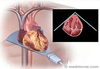
Transesophageal Echocardiogram (TEE)

Transesophageal Echocardiogram (TEE)
- a flexible tube (probe) with a transducer at its tip guided down the throat and into the esophagus
- shows the size and shape of the heart and how well the heart chambers and valves are working
- can pinpoint areas of heart muscle that aren’t contracting well because of poor blood flow or injury from a previous heart attack
- can detect possible blood clots inside the heart, fluid buildup in the pericardium (the sac around the heart), and problems with the aorta

What structure is closest to the probe in a TEE (transesophageal echocardiogram)?
the atria.

The probe is at the upper apex of this image. What are the structures that are identified?

Left Atrium
Left Ventricle
Right Ventricle
Aorta

What is a pulmonary embolism?
- one or more pulmonary arteries in your lungs become blocked
- most commonly caused by blood clots that travel to the lungs from the legs (or rarely other parts of the body)
called deep vein thrombosis, or DVT
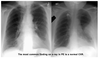
what is a “Hampton’s hump” ?
“a dome-shaped, pleurally-based opacification in the lung due to pulmonary embolism and lung infarction”
(part of the lung is devoid of air from a clot and collapses)
- pregnant women post-delivery can be prone to these clots that migrate to the lungs
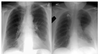
What is the most common finding on a chest x-ray of a pulmonary embolism?
A NORMAL X-RAY
CT Angiography (CTA)
- combines a CT scan with an injection of a special dye called contrast material (injected through an IV line started in your arm or hand)
- produce cross-sectional images of your body, blood vessels and tissues
- used to find: pulmonary embolism, an aneurysm, blood vessels narrowed by atherosclerosis, a blood clot, observes tumors

If you suspect a pulmonary embolism which scans should your alway remember to do?
CT Angiography (CTA)
*(because chest x-rays usually appear normal) *
