CNS 1 (Sept 26) Flashcards

1
Q

What does the peripheral NS consist of?

A

-mixed spinal nerves carrying: a) sensory information from the body, such as muscles and skin (afferent) and b) motor information from CNS to the body to get it to move (efferent)

2
Q

What does central NS consist of?

A

-brain -spinal cord

3
Q

What are glial cells?

A

-they support the neurons -4x more of these than neurons themselves -brain consists both of neurons and these glial cells

4
Q

What are ependymal cells?

A

-inside of lining of ventricular system containing the CSF -form a barrier between CSF and the brain itself -CSF brain barrier -also produce fluid contained in ventricular system (CSF) -found in CNS grey matter

5
Q

What are astrocytes?

A

-make contact with blood vessels and form blood brain barrier -performs barrier to prevent things from poisoning the neurons -can trick astrocytes- dissolve things in membrane then it gets across blood brain barrier (eg. alcohol) -found in CNS grey matter

6
Q

What are microglia cells?

A

-blood brain barrier prevents immune system from getting into brain -provide intrinsic immune capacity of the brain -white matter of CNS

7
Q

What are oligodendrocytes?

A

-need fast conduction of signal -myelin is derived from oligodendrocyte -one oligodendrocyte sends off many arms which wrap around multiple axons to provide myelination -white matter of CNS

8
Q

Which axons are unmyelinated?

A

-neurons carrying pain and temperature

9
Q

What are Schwann cells?

A

-one Schwann cell ensheaths only one axon -myelination in the PNS

10
Q

How is immunity addressed in the PNS?

A

-no blood brain barrier in PNS so you have opportunity for blood to interact with PNS -blood contains T cells, macrophages, etc. -able to be mobilized to fight infection and injury in PNS

11
Q

Why is CNS regeneration limited?

A

-neurons are postmitotic (rare to get new ones growing) -glia inhibit axon growth (injury of CNS, loss for causing any regeneration/making new synapses to make new function) -brain is formed carefully during development so you don’t want to have new axons being formed everywhere -CNS purposefully limits regeneration

12
Q

Describe regeneration in the PNS

A

-1mm/day -neurons can sprout collaterals and regenerate (if you injure axon, axon can regenerate or you can get collaterals growing from adjacent uninjured neurons) -nerve growth factor promotos attraction of new neurons from collaterals or regeneration -glia produce growth factors -macrophages remove debris (from blood- get rid of old dead cells that would inhibit regrowth)

13
Q

What body cavity is CNS located in?

A

-dorsal body cavity -protected by structures that make up the dorsal body cavity which for the head is the skull and for the spinal cord is the vertebral column

14
Q

Label from top to bottom and give description of function of each

A
  • cerebral cortex: outside of the brain, involved in: thinking, memory, voluntary motor movements, sensory perception
  • diencephalon: deep within brain grey matter, sensory/motor relay centre to and from cortex, autonomic functions
  • brainstem: midbrain, pons, and medulla- autonomic functions, cranial nerve nuclei contained in here which are responsible for generating function in head/neck
  • cerebellum: coordination of movement and balance
  • spinal cord: motor output, sensory input, reflexes and interface with PNS
15
Q

What is meninges?

A
  • three layers
  • inside skull
  • protection
16
Q

What is dura mater?

A
  • firmly attached to the skull
  • has no instrinsic pain or ability to feel anything (but meninges does)
17
Q

What is located between the dura and the skull?

A
  • meningeal veins, arteries, and nerves (neurovascular bundle)
  • supplying dura
18
Q

What lines the dura?

A
  • arachnoid mater
  • small spider like extensions that separate the arachoid from brain
19
Q

What membrane covers the brain?

A
  • pia mater
  • covers cerebral vessels
  • thin translucent membrane
20
Q

What is the falx cerebri?

A

-extension of the dura that divides the left and right cerebral hemispheres

21
Q

What is the tentorium cerebelli?

A

-extention of dura that separates cerebellum and cerebrum

22
Q

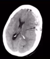
What happens when we get a space occupying lesion in the brain such as a tumour or hematoma?

A
  • herniation
  • causes part of the brain to move into different compartment
  • damages CNS
23
Q

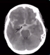
What occurs when you lose pressure within the skull?

A
  1. Brain can drop down and cause tentorial hernation
  2. Coning- brainstem falls down and hits bones (through foramen magnum)
24
Q

What are cerebral vessels and where do they originate from?

A
  • blood vessels on surface of brain
  • derived from internal carotid artery
25
Q

What occurs with pulsations of the meningeal vessels?

A
  • no place to go, stuck up against bone
  • that signals pain
  • irritating nerves of meninges (headache)
26
Q

What is brain freeze?

A
  • put something cold on palette
  • same nerve (trigeminal) carrying back info to brain as from your meninges
  • get confused about this; think that your brain is having a headache but it is actually pain associated with palette
27
Q

What happens with misaligment of temporomandibular joint?

A
  • pain in the joint
  • “wake up with huge headache”
  • getting referred pain to meninges from the jaw
  • same originating nerve carrying and supplying pain information from those 2 areas
28
Q

Where is CSF contained?

A
  • subarachnoid space
  • space between arachnoid mater and the pia mater
29
Q

What happens when meningeal vessels rupture?

A
  • bleeding that results in a clot
  • can diagnose with CT/MRI imaging (remember that these are looking caudal-cranial)
30
Q

What kind of hematoma is this?

A
  • epidural hematoma (*always happens at top)
  • lenticular appearance (looks like a lens)
  • usually happens when you rupture meningeal vessels which separate the dura from the surface of the brain (blunt force to skull)
31
Q

What kind of hematoma is this?

A
  • subdural (*always happens at top)
  • more diffuse
  • blood clot has spread all around surface of brain
  • rupture of bridging veins
  • most of movement when skull goes back and forth is occuring at top of the brain where bridging veins are located (eg. whiplash, shaking baby syndrome, brain shrinks in elderly which puts traction on bridging veins and they pop and bleed)
  • bleed between dura and arachnoid but often times arachnoid is so thin that you can blood in CSF as well
32
Q

What kind of hematoma is this?

A
  • subarachnoid
  • base of brain bleed
  • arteries come in at base and there are often congenitally weakenings in walls of blood vessels (aneurisms)
  • aneurysms pop and allow bleeding in base of brain under the arachnoid in CSF which leads to subarachnoid hematoma
  • usually due to rupturing of cerebral artery
33
Q

What is the difference between the meninges in the brain versus spinal cord?

A
  • epidural fat space
  • spinal cord needs to have bending ability for vetebrae to move
  • fat can be compressed then springs back
  • between the dura and periosteum of bone in vertebrae is location of epidrual fat space
34
Q

How is brain formed during development?

A
  • formed as a tube
  • top surface of embryo is called ectoderm
  • ectoderm gets sucked inwards then two tops of grooves that formed fuse and create a tube which becomes brain with a core filled with fluid
  • fluid filled core– ventricle system
35
Q
A
36
Q

Where are lateral ventricles located?

A
  • one in each hemisphere
  • 1 and 2
37
Q

What does interventricular foramina connect?

A

-connects lateral ventricles to third ventricle which is in midline of diencephalon

38
Q

What does cerebral aqueduct connect?

A

-leads from third ventricle to dilatation of the ventricular system around cerebellum which is fourth ventricle

39
Q

What tube leads from the fourth ventricle?

A
  • central canal
  • goes down spinal cord
40
Q

What are ventricles filled with?

A

-CSF

41
Q

What is the choroid plexus?

A

-system that produces CSF

  • remove cells of blood and only allow for plasma to create CSF
  • begins as tufts of arteries in the ventricles (increase in SA) which are lined by ependymal cells
42
Q

What is the general production/flow of CSF in the ventricle system?

A

-choroid plexus in lateral ventricles which leaks thorugh intervertebral foramen to get into 3rd ventricle

  • 3rd ventricle has more choroid plexus which adds even more CSF
  • goes through cerebral aqueduct into 4th ventricle where there is more choroid plexus which adds more CSF

-goes down central canal of spinal cord

43
Q

How does CSF leak from inside of the brain to the outside?

A
  • apertures in the roof and floor of 4th ventricle where attachment of cerebellum is located
  • allow CSF to leak from inside to outside
  • brain is filled with CSF and is floating in CSF (like unset Jello- if you didn’t have pressure of CSF to contain brain it would ooze everywhere)
44
Q

Where is the stenosis?

A
  • can observe in hydrocephalus dilated lateral ventricles and dilated 3rd ventricle
  • cerebral aqueduct is where stenosis is
45
Q

How many arteries are involved in giving the blood supply to brain?

A

4

46
Q

What artery do the vertebral arteries coalesce to form/

A

-basilar artery

47
Q

Label the diagram and describe the blood supply from each vessel

A
  1. Anterior communicating: connects two internal carotids
  2. Internal carotid: gives off middle cerebral and anterior cerebral artery which supply medial and lateral areas of brain
  3. Posterior communicating: connects posterior cerebral artery to internal carotid
  4. Basilar artery: base of brain, gives off posterior cerebral artery
  5. Anterior cerebral: supplies medial surface of brain
  6. Middle cerebral: supplies lateral surface of brain
  7. Posterior cerebral: supplies underside of brain
  8. Vertebral: come through foramen magnum and coalesce to form basilar artery
48
Q

Describe the normal blood flow through the Circle of Willis

A
  • incoming blood can come from vertebral/basilar and the internal carotid
  • blood in posterior cerebral mainly coming from vertebral artery
  • middle and anterior cerebral artery mainly coming from interal carotid
49
Q

What does the circle of willis do?

A
  • if you have a narrowing of one of the 4 arteries supplying the brain, it doesn’t matter very much
  • blood can flow in the opposite direction within circle to supply entire brain
  • anastomosis
50
Q

How are deep grey matter structures supplied with blood?

A
  • deep cerebral arteries (anterior, posterior, and middle)
  • as Circle of Willis extends out and starts to wrap around brain to supply the cortex, it also gives off deep branches to supply deeper grey structures and white matter tracks
51
Q

Where does blood end up after it’s gone through brain, bridging veins, and into sinuses?

A

-gathers in jugular vein found adjacent to carotid

52
Q

The tentorium cerebelli has in it the _____ in the same way that the falx cerebri has in it the _______

A
  • transverse sinus (brings blood to sides)
  • superior sagittal sinus
53
Q

What does the external jugular do?

A

-drains the face

54
Q

What is a hemorrhagic stroke?

A
  • blood vessel breaks
  • creates hemorrhage
  • hemorrhage allows for blood to leak into brain tissue
  • lead to intracerebral or subpial hematoma
55
Q

What is ischemic stroke?

A
  • blood clots flowing through body
  • once clot goes through and gets into smaller blood vessles/capillaries it will get lodged there
  • prevents brain from receiving its oxygen/glucose
  • stops blood supply to that area of brain
  • death of the cortex/grey matter as well as the white matter tracks connecting the cortex to deeper brain structures or spinal cord
56
Q
A
57
Q

Where does dura mater stop in spinal cord area?

A
  • dura mater surrounds spinal canal and supports lateral roots that lead to the peripheral nerves
  • ends at the exit of these nerves
58
Q

What is the lymphatic system?

A

-lymphatic system drains CSF through lymphatic vessels through neck lymph nodes and back into body where it is drained away by lymphatic system

59
Q

What happens in brain when heart pumps?

A

-pumping of heart pushes CSF around through ventricular system