Sensory Vision Flashcards

1
Q

What muscles do CN 3,4,6 control?

A

Extraocular muscles

2
Q

What does the oculomotor nerve provide a pathway for?

A

-parasympathetic nerves to control the ciliary muscle of the lens and the constrictor of the pupil

3
Q

What does the facial nerve provide a pathway for?

A

-parasympathetic nerves to control secretion of tears from the lacrimal gland

4
Q

Which cranial nerves supply sensory information from the eye?

A

-optic and trigeminal

5
Q

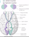
What does the optic nerve do?

A

-relays visual information from the retina to the lateral geniculate nucleus of the thalamus and the superior colliculus

6
Q

What does the opthalmic division of the trigeminal nerve do?

A

-carries sensory information from the cornea (density of sensory receptors in cornea is 500x greater than skin which is why it is so sensitive to touch and pain)

7
Q

What are the two elements of vision?

A
  1. Refractive media: refract the light to focus the image of the eye on the retina 2. Neural detection and transmission to the brain: detect the light and transmit the visual information to the brain
8
Q

What is the vitreous humor?

A

-occupies the posterior segment of the eye behind the lens -consists of 99% water 1% protein -has high refractive index -slowly replaced over time with turnover of about 7 months -attached to retina at back of eye -shrinkage of eye during aging/change of composition can pull on retina and lead to a retinal detachment

9
Q

Where does light first enter the eye? What is the difference in refractive index like here?

A

-light first enters eye through tear film at surface of cornea -this is where difference in refractive index of the air and tissues of the eye is the greatest -this means that most of bending of light occurs at this location

10
Q

Where does light pass through after the cornea?

A

-through the anterior chamber of the eye, the lens, then vitreous humour before being focused on the retina at the back of the eye

11
Q

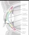
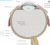
What makes up the outer fibrous coat of the eye? What does this provide?

A

-cornea and sclera -structural support

12
Q

What does the vascular coat consist of?

A

-choroid which is posterior -ciliary body near front -iris

13
Q

What is the choroid?

A

-highly vascularized and pigmented layer -supplies nutrients and oxygen to outer layers of retina and makes eye opaque to light coming from the side

14
Q

What is the ciliary body?

A

-contains a muscle at the front of the eye that changes the shape of the lens -contains blood vessels that produce the aqueous humour immediately behind the cornea -continuous with the iris at the front of the eye

15
Q

What is the iris?

A

-contains variable amount of pigment that blocks light from entering the eye except through the pupil -size of pupil opening in the iris is regulated by contraction of pupilary sphincter muscles at tip of iris and contraction of dilator muscles which open size of pupil that run radially to its base

16
Q

What is the innermost coat of the eye? What does it do?

A

-neural coat -aka retina -contains rod and cone photoreceptors that transduce photons into a neural signal -also contains two layers of inhibitory neurons that process the image; allow image to be processed in both dim and bright light, sharpens image, and extracts features of texture, colour, and edges to be sent to the brain through the optic nerve

17
Q

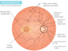
What is the fovea centralis?

A
  • area of retina with highest visual acuity
  • only small cones are located here
  • cones synapse with retinal neurons located in parafoveal retina
  • since blue light is scattered more than other wavelengths, retina around the fovea has deposits of yellow pigment that absorb blue light (macula lutea)
  • blue light also has potential to damage photoreceptors so the macula lutea protects foveal photoreceptors
18
Q

What is the function of the ganglion cells of retina?

A

-have axons that transmit visual info from retina to lateral geniculate nucleus of thalamus and superior colliculus of midbrain

19
Q

What is the lateral geniculate pathway for from the retina? Superior colliculus pathway?

A

-conscious vision -unconscious vision

20
Q

What are included in the connections of the superior colliculus?

A

-the nuclei that regulate pupil size and cerebellum that coordinates muscle movement

21
Q
A
22
Q

Where does bending of light occur in the eye?

A
  • where there is greatest difference in density or refractive index
  • refractory power: this is at the air/water cornea interface (80% occurs here)
  • accomodation: lens is denser than the aqueous and vitreous humour and contributes 20% of refraction occurs here
23
Q

How are the cornea and the lens different in terms of refraction?

A
  • the lens is able to change its shape
  • refraction that occurs when the lens is used to view close objects is higher than when the lens is used to view far objects
  • accomodation: ability to increase refractive power of the lens when viewing close objects
24
Q

What determines where the sharp point of focus of light from the lens and cornea will land?

A

-shape of fibrous coat

25
Q

What is the shape of the eyeball largely determined by?

A

-genetic factors

26
Q

What is hyperopia?

A
  • eyeball is flattened so that the focal point lands behind the eye
  • objects that are far away are sharp but objects that are close are out of focus
  • 60% of the population
  • can be corrected with convex (positive diopter lens) placed in front of the cornea
27
Q

How does positive diopter lens work?

A
  • lens is the same shape as the lens of the eye
  • even though incidence is high, most people at a young age can get away with using their own lens but it can become a problem as you get older when the lens becomes less pliable
28
Q

What is myopia?

A
  • 25% of population
  • eyeball is elongated
  • focal point falls in front of retina
  • objects that are near are sharp, objects that are far are not
  • can be corrected with concave (negative diopter lens) in front of cornea
  • younger people wearing glasses generally have this because the lens doesn’t have the intrinsic ability to change the shape in a negative way
29
Q

What is astigmatism?

A
  • eyeball can be mishapen along meridian plane
  • causes blur in axis of error
  • 30-60% are affected depending on ethnicity
  • corrected by adding appropriate positive or negative diopters in the axis where the error occurs
30
Q

Describe the cornea?

A
  • makes up the anterior transparent part of the fibrous coat
  • it’s composed of layers of collagen secreted by fibroblasts
  • cornea has an epithelial layer (unlike sclera) which is bathed by tear film
  • endothelium is bathed by the aqueous humour
  • epithelium gets its nutrients from surrounding fluids because blood vessels would block vision
31
Q

What are the reasons for the transparency of the cornea?

A
  • dehydration of the tissues because water would disrupt the layers of collagen
  • epithelial cells have ion pumps that expel sodium and chloride ions into the tears
  • water then follows that concentration gradient
32
Q

What is a keratopathy?

A
  • corneal abrasion that penetrates to deepest layers of the epithelium would destroy those pumps and the underlying cornea stroma will swell
  • can also get it from wearing non breathable contacts for too long, not cleaning breathable contacts, chemical toxins, lack of oxygen
33
Q

What does dehydration of corneal stroma do?

A
  • allows for collagen to be closer together and have regular spacing (has property of being crystal)
  • collagen fibres in sclera are further apart and irregularly spaced making it more translucent
34
Q

What maintains the health of the corneal epithelium?

A
  • tears
  • cornea is washed by tears secreted from lacrimal gland in upper lateral quadrant of orbit
  • blinking encourages continuous flow of tears
  • tears wash away microbes, chemicals, and particulates
  • tears contain high concentrations of salt and IgA which inhibits microbial growth
35
Q

What does mucus layer of tear film do?

A

-allows tears to adhere to the corneal epithelium

36
Q

How is evaporation of tears inhibited when eyes are open (eg. other than blinking)

A

-oil layer that is secreted on surface of tear film by meibomian glands located in eyelids

37
Q

Explain steps of LASIK surgery

A
  • razor is used to remove corneal epithelium from stroma
  • laser destroys stromal collagen from the selected areas
  • corneal epithelium is reapplied to corneal stroma and readheres within hours
  • refraction of light at edges of remodelled areas can cause halos of light
  • neuropathic pain can appear weeks/months later from damaged sensory fibres in trigeminal nerve
38
Q

Describe cornea transplant

A
  • avascular nature of cornea allows it to be transplanted with minimal rejection
  • damaged cornea removed and donor cornea is sutured in place
39
Q

What is the conjunctiva?

A
  • at the corneal scleral junction the epithelium transitions to a non-keratinzed columnar epithelium that lines the anterior surface of the sclera
  • contains numerous goblet cells which secrete mucous that contributes to tear film
  • very rich blood supply
40
Q

What is the function of the fornix?

A
  • reservoir for extra tears
  • allows eye to move in eye socket
41
Q

What does conjunctiva on inner eyelid transition to?

A

-keratinized stratified squamous epithelium at edge of eyelids

42
Q

What leads to conjunctivitis?

A
  • allergens, chemicals, microbial infection
  • dilated BVs of inflammed mucosa lead to red eye
43
Q

What can happen to Meibomian glands?

A
  • they secrete the oily top surface of the tear film and have very long ducts that extend most of the length of the eyelids
  • can be seen as yellow streaks when the eyelid is inverted to visualize the mucosal surface of the lid
  • often can be blocked or infected
44
Q

How does the lacrimal gland inhibit inflammation/infection of the conjunctiva?

A
  • secretions wash over eye from lateral to medial
  • tears are drained by openings of two lacrimal canaliculi located above and below the lacrimal caruncle
  • both lead to the lacrimal sac which drains into nasal cavity through lacrimal duct
  • CN VII innervates lacrimal gland
45
Q

What separates the anterior segment containing aqueous humour from the posterior containing vitreous humour?

A

-lens

46
Q

How is the lens produced? What are lens fibres?

A
  • from cells at its equator
  • cells produce crystalline
  • as the cells become layered, the innermost cells lose their organelles so they are then termed lens fibres
  • lens fibres have regular shape with hexagonal cross section making them more transparent
  • have microscopic projections that allow them to adhere to one another, metabolism through glycolysis, share metabolites from aqueous humour pumped through gap junctions
47
Q

What is presbyopia?

A
  • ball and socket projections between lens firbes allow them to move
  • lens is normally flattened by tension placed on it by the pull of the zonule fibres connecting it to the ciliary body
  • to see close objects, ciliary body muscle contracts and pulls the lens closer to the cornea
  • relaxation of zonule fibres allows the lens fibres to move on one another and become rounder; increased diameter of lens allows eye to focus on close objects
  • lens fibre movement is impaired with age (crystalline is non renewable) so reading or keeping close objects in focus requires a positive diopter correcting lens
48
Q

What is cataract?

A
  • crystalline protein can form clumps as it ages
  • reduces transparency
  • at first it might cloud small region of lens but can become bigger to form cataract
  • blurs image reaching retina
  • can be solved with intraocular lens implant
49
Q

What is neural retina? What are its functions?

A
  • innermost layer of the eye
  • only part of CNS that resides outside of the protection of the skull/vertebral column
  • photoreceptors are deeply embedded into pigmented epithelium (shields photoreceptors from scattered light, renews photosensitive pigment in photoreceptors, transfers oxygen and nutrients from the choroid)
  • this association is loose so it is possible for it to detach; may cause death of retinal cells or blindness
  • rod and cone photoreceptors make synapses with 2 layers of neurons that regulate the sensitivity of the retina to varying light levels, detect moving objects, and extract features of the visual scene
  • photoreceptors transduce photons into neural signal that travels to retinal ganglion cells which carry visual scene features in separate axons that project through optic nerve to brain
50
Q

What is the fundus?

A

-optic nerve, macula, and retinal vessels are located here and can be seen with opthalmoscope

51
Q

What can be seen through the pupil?

A
  • macula, optic nerve head, retinal arteries, veins
  • fovea can not be visualized but estimate its location at the centre of the macula
  • optic nerve is covered in layers of the meninges
  • raised intracranial pressure in CNS can therefore be detected as a raised rim around the optic nerve head where subarachnoid space meets sclera
52
Q

What does loss of retinal ganglion cell axons look like?

A

-excavation of optic nerve head and visualized as abnormal cupping of the optic nerve head

53
Q

What is AV nicking?

A
  • retinal arteries always cross over the veins
  • chronic high BP can dilate the arteries resulting in them compressing the underlying veins

-

54
Q

Do retinal arteries and veins cross the fovea?

A

-no, presence of BVs would obscure vision over highest area of visual acuity

55
Q

What disease can result in the death of foveal neurons?

A
  • diabetes
  • damage to small retinal vessels
  • loss of central vision
56
Q

What is AMD?

A
  • BVs deteriorate with age because the fovea is reliant on those BVs to deliver oxygen and nutrients to the central retina
  • one response to reduced oxygen tension in the fovea is to stimulate new BV growth in the parafoveal capillaries
  • new BV growth over the fovea can obscure vision
  • can be inhibited with VEGF inhibitors
57
Q

What is the optic disc?

A
  • blind spot
  • optic nerve head is the exit point of retinal ganglion cell axons so it lacks photoreceptors and the layers of the neuronal retina
  • not in a region of retina that is responsible for high visual activity
58
Q

What happens at optic chiasm to get visual info to the brain?

A
  • retinal ganglion cells that sample the same visual field in each eye are paired
  • axons from one eye are uncrossed and matched with the crossed axons from the other eye resulting in 50% crossing in the chiasm
  • axons from each eye from the same part of the visual field are paired at the optic chiasm and relayed to thalamus and visual cortex
  • paired axons travel to geniculate nucleus of the thalamus through the optic tract
  • modified visual info is transmitted to primary visual cortex in occipital lobe by the optic radiation
  • part of this tract carrying info from inferior visual field travels through the parietal lobe while remaining tract carries information from the superior visual field and travels through the temporal lobe
59
Q

Where is the left visual field detected?

A

-retinal cells in the nasal retina of the left eye and the temporal retina of the right eye

60
Q

Where is the right visual field detected?

A

-retinal cells in the temporal retina of the left eye and the nasal retina of the right eye

61
Q

What is macular sparing?

A
  • seeing entire visual scene requires eye movements to allow the fovea to sample the scene
  • lateral geniculate neuron axons representing the fovea synpase in a larger surface area in the cortex
  • foveal overrepresentation in the cortex represents a cortical magnification of the area of highest acuity
  • injury to the fovea of the eye would result in blindness whereas injury to the cortex would represent a partial visual loss
62
Q

Describe layers of geniculate nucleus and stereopsis

A
  • axons from each eye synapse in alternate layers
  • each layer then projects to adjacent cortical columns in the occipital cortex
  • cortex determines the relationship of objects represented in the cortical columns
  • far objects are represented in adjacent cortical columns while near objects will be represented in cortical columns that are far apart
63
Q

What will damage to the retina or ganglion cell axons before the optic chiasm result in?

A

-partial or complete blindness in one eye

64
Q

What will damage to the retina or ganglion cell axons after the optic chiasm result in?

A

-partial blindness in the same area of both eyes

65
Q

What causes monocular blindness?

A

-injury to the retina or optic nerve of the left eye will cause blindness in the left visual field

66
Q

What will happen with hypertrophy of the pituitary gland?

A
  • may compress the crossing axons of retinal ganglion cells from the nasal retina of each eye
  • causes bitemporal hemianopia
67
Q

What results from an injury to the optic tract between the chiasm and the lateral geniculate nucleus?

A
  • would affect axons from the temporal retina of one eye and the axons from the nasal retina of the other eye
  • lesion of the left optic tract would result in a right homonymous hemianopia
68
Q

What would cause a right homonymous superior quadrantanopia? Right homonymous inferior quadrantanopia?

A
  • right homonymous superior quadrantanopia results from injury to Meyer’s loop on left side of brain
  • right homonymous inferior quadrantanopia results from injury to the parietal lobe on the left side
69
Q

A lesion in the right visual cortex may lead to…

A

-left homonymous hemianopia with macular sparing due to the cortical maginification of the foveal representation or selective blindness of only the foveal representation

70
Q

What is the suprachiasmatic nucleus of the hypothalamus?

A
  • retinal ganglion cells project here
  • sets the daily clock and controls circadian rhythms
  • explains why blue light, especially from electronic devices in the evening, can disrupt sleep rhythms
71
Q

Describe the role of the superior colliculus

A
  • many retinal ganglion cells have axon collaterals to the superior colliculus
  • superior colliculus does not filter visual information for importance and has a uniform representation of the visual field rather than an emphasis on the fovea
  • provides input to cerebellum to allow the cerebellum to make coordinated visually guided movements
  • projection to nucleus of Edinger-Westphal provides information on light intensity; nucleus provides parasympathetic input to CN III to control pupillary constriction
  • projection to pontine reticular formation provides visual input to regulate alertness in response to activity in the visual field
  • projection to inferior olive of the medulla provides input to the system that coordinates head position and helps maintain horizontal visual axis (explains why people who are cortically blind can respond with head movements to approaching objects)
  • projection to the hypothalamus that regulates sympathetic NS can initiate an autonomic sympathetic response to a visual stimulus through the hypothalamic control of the lateral horn of the thoracic spinal cord where preganglionic sympathetic neurons are located
72
Q

What is the anterior segment of the eye filled with? What are its functions?

A
  • aqueous humour
  • provides nutrients to the lens and maintains pressure within the eye
  • internal pressure adheres the retina to the choroid and sclera in the same way that intracranial pressure supports the brain
  • anterior segment is divided by the iris into a posterior and anterior chamber
73
Q

How is aqueous humour produced?

A
  • by dialyzing blood in the ciliary body
  • flows over the anterior surface of the lens and into the anterior chamber through the pupil
  • it is drained into venous circulation or lymphatics at the base of the pupil through a connective tissue sieve, the trabecular meshwork
  • canal of Schlemm runs circumferentially around the corneo-scleral junction and is believed to be the primary vessel into which aqueous humour drains from the trabecular meshwork
  • melanin pigment granules from choroid or iris can become dislodged and block the drainage holes in the trabecular meshwork
  • when pupil dilates at night, tissues of the iris can fold over the drainage openings, and close the outflow angle
74
Q

What can increase in intraocular pressure result in?

A
  • can compress retinal ganglion cell axons
  • retinal ganglion cell survival is dependent on target derived growth factors (BDNF) which flow to ganglion cell somas through the axon
  • decreased axoplamic flow leads to apoptosis
  • loss of retinal cells leads to irreverisble blindness: glaucoma
75
Q

How can loss of retinal ganglion cells in glaucoma be visualized?

A
  • optic nerve cupping is indicative of axon loss
  • accompanying visual field loss starts in the periphery that leads to increasing tunnel vision
76
Q

How is intraocular pressure measured?

A
  • tonometry in routine eye exam (measures the deflection of the anesthetized cornea from a puff of air)
  • can vary with BP, posture, time of day so the measurement is only an indication of chronic raised intraocular pressure
  • in an eye with raised intraocular pressure, cornea will be stiffer
77
Q

How are patients with raised intraocular pressure treated?

A
  • topical application of adrenergic drugs can constrict BVs in the ciliary body and reduce the production of aqueous humour while having a minimal effect on systemic BP
  • if the cause is due to the closure of drainage angle by dilated iris, topical application of cholinergic drugs will constrict the iris
  • surgical approaches involve using a laser to create drainage holes at the base of the iris to allow aqueous humour flow more easily between posterior and anterior chambers
  • implantation of miniature stent through the sclera at the corneo-scleral junction to allow drainage of aqueous humour from the anterior chamber into the tears