MSK2 Flashcards
(43 cards)
What are the terminal nerves of the brachial plexus and are they innervating flexors or extensors?
Flexors: musculocutaneous, median, ulnar
Extensors: radial, axillary

What are the terminal nerves of the lumbar plexus and are they innervating flexors or extensors?
Flexor: obturator (adductors of hip flexors) Extensor: femoral (extensors of knee)

What are the terminal nerves of the sacral plexus and are they innervating flexors or extensors?
-sciatic nerve then splits at knee Flexor: tibial nerve (plantar flexors) Extensor: common fibular nerve (dorsiflexors and extensors)

What deficits do you expect with an proximal injury to a nerve that forms a plexus?
- partial paralysis (paresis) in the muscle
- 1/3 of the dermatome that is supplied by that spinal level being affected
What deficits do you expect with a distal injury in a plexus?
- nerves have joined together from spinal levels already
- complete paralysis of the muscle and dermatomes all affected
What deficits do you expect with a proximal injury to a peripheral nerve?
- before the division of the muscular and sensory branches
- both sensory and motor loss
What deficits do you expect with a distal lesion to a peripheral nerve after it has given off its deep branches?
-sensory loss only
How are bones developed?
- develop from cartilage
- cartilage models in upper and lower limb develop into the bones that make up both limbs between 5 and 6 weeks in utero
- connective tissue on outside of cartilage- perichondrium
- ossification centres: diaphysis and epiphyses
- diaphysis ossification centre begins in the middle and moves towards the epiphysis while the inside of the epiphysis also forms bone
- eventually these will fuse together to give you one bone

What is the epiphyseal plate?
- where diaphysis and epiphysis meet
- after the age of 20, it disappears
- after that you can see the epiphyseal line which is the remnant of the cartilaginous plate

What cavity is found in the diaphysis? What type of marrow is found here?
- completely hollowed out forming the medullary cavity with cortex of bone on the outside
- filled completely with fat
- important storage site for fat and energy
- yellow marrow

What type of marrow is found in the epiphysis? What is the role of this area of the bone?
- in the epiphysis, it is not completely hollowed out
- pillars of bone with spaces in between filled with blood
- spongy bone
- red marrow
- very good blood supply
- role is to form new blood cells

What is periosteum? What does it originate from?
- perichondrium on the cartilage models becomes periosteum once the bone is formed
- connective tissue around the bone
- 2 layers: outer fibrous layer made of collagen fibres running in many directions which allows for the attachment of ligaments/tendons, inner layer where mesenchymal stem cells are located (important for formation and healing of bones)

What do periosteum mesenchymal cells turn into?
- osteoprogenitor cells turn into osteoblasts
- osteoblast secretes ECM (made up of collagen and hydroxyapatite)
- some osteoblasts differentiate into osteocytes

Describe the arrangement of osteocytes and osteoblasts in the compact and spongy bone
- osteocytes have projections off of them- they sit in wells of bones surrounded by bone
- channels run longitudinally around the bone and osteocytes are located around the channel
- spongy bone- osteoblasts located on the surface of bony pillars that are creating the spongy bone

Describe where osteoclasts originate from
-bone marrow derived macrophages fuse to form osteoclasts
What happens during bone reabsorption?
- osteoclasts use acid and collagenase to dissolve bone and release calcium
- under certain circumstances with lots of osteoclastic activity, lots of spongy bone can be removed
- this can happen under the influence of some hormones

Describe the changes to calcium homeostasis when calcium levels are too high
- thyroid releases calcitonin (comes from thyroid gland)
- increases osteoblastic activity and deposition of calcium in the bone
- decreases calcium uptake in the intestines
- decreases calcium reabsorption from the urine
- mobilizes calcium from blood to deposit into the bone

Describe the changes to calcium homeostasis when calcium levels are too low
- parathyroid gland releases PTH
- stimulates osteoclastic activity
- increases calcium release from the bones, uptake in the intestines, and reabsorption from the urine
- calcitriol- skin and GI

What contributes to skeleton renewal?
- balance between osteoblasts and osteoclasts regulated by cytokines
- 10% per year is renewed

-places where a joint is formed remain as cartilage (articular cartilage)

What happens as a result of the good blood supply to the epiphysis? What happens when this is blocked?
- new blood cells are formed
- many foramina in the epiphysis to nutrient arteries to get in and wash newly formed blood cells from the spongy bone and mobilize them into veins
- bone itself is also dependent on the good blood supply; when it is blocked/compressed you don’t have enough supply and the bone will die called avascular necrosis
Describe the mechanical factors that contribute to bone remodelling
- bone is a crystal
- when you put force on a crystal you can generate electricity
- body weight puts force on the crystal
- spongy bone trabeculae form pillars that are oriented to maximize bone strength
- trabeculae are constantly being remodelled by cellular responses to force (piezoelectric)
What is a comminuted fracture?
- break causes it to splinter
- found in extreme trauma
- happens when older person breaks a bone (less cartilage and collagen so bone is more brittle)

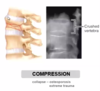
What is a compression fracture?
- osteoporosis and extreme trauma
- vertebrae become smaller and more prone to being crushed
- can happen from jumping off of high things