Muscle Physiology (Sept 19) Flashcards
(47 cards)
What are the three types of muscle?
-skeletal -cardiac -smooth
What type of muscle cell is this? What are some of this muscle type’s functions?

- smooth muscle cell
- storing and moving substances
What type of muscle cell is this? What are some of this muscle type’s functions?

-cardiac muscle
What type of muscle cell is this? What are some of this muscle type’s functions?

- skeletal muscle
- movement, stabilization
- thermoregulation
- contractions account for all body movements
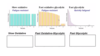
Fill in the chart below


What are the layers of mysium and their functions?
- endomysium: surrounds each individual muscle fibre
- perimysium: surrounds a bundle of muscle fibres
(fascicle: bundle of muscle fibres surrounded by perimysium) - epimysium: surrounds the entire muscle
- all 3 layers contribute to the muscle sheath and become the tendon which connects 2 bones together
- tendon becomes continuous with the periosteum (wrapping around bone)
Myocyte/myofibre structure
- muscle fibre is composed of myofibrils: long tube-like structures, sarcomeres in series (collection of sarcomeres which allow muscles to contract)
- sarcomeres: basic contractile unit of muscle
- made up of a thick filament (myosin) and thin filament (actin)
Label the diagram

- Tendon
- Muscle
- Epimysium
- Perimysium
- Blood vessel
- Endomysium
- Muscle fibre
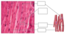
Label the diagram

- Myofibril
- Muscle fibre
- I band
- A band
- Sarcomere
- Z line
- thick filament (myosin)
- Thin filament (actin)

What happens when stressor is put on muscles?
- hypertrophy!
- muscles enlarge with use
- exercise stimulates production of actin and myosin filaments
- increased number of myofilaments expands the fibre causing muscle enlargement and definition
- want more sarcomeres to handle that stress
What happens if you remove stress from muscle?
- atrophy
- have lots of metabolically active tissue but it’s not being used so it takes proteins and sarcomeres away to use for something else
- aging, spinal cord injury, space flight all cause atrophy
What is the sliding filament theory?
- contraction of sarcomeres happens through sliding of filaments
- thin filament slides over thick filament (thick doesn’t move)
- bring Z lines closer together
- when sarcomere shortens, fill H zone and space between 1/2 I bands is being removed (distance between thick filament, A band, and Z line)
Label the diagram

- A band
- 1/2 I band
- H zone
- Relaxed
- Contraction
What is outlined in yellow? What are represented by dark black dots?

- muscle fibre
- thick filament (composed of myosin)
Label the diagram. What function does this structure serve?

- it is a thick filament and the projections are cross bridges
- the cross bridges are what makes the thick filament interact in 3D
- each thick filament can interact with 6 thin filaments
- each thin filament (actin) can interact with 3 thick filaments (myosin)

What is thick filament composed of?
- several myosin molecules with their head sticking up
- tail is lying down in parallel
- wants to grab on to thin filament

What is thin filament composed of? What is the role of each component? Where does Ca2+ come in to play?
- three proteins: actin, tropomyosin, troponin
- actin: form 2 coiled chains (which is the “thin filament”)
- tropomyosin: run along actin blocking cross bridge binding site (tropo: to turn or react)
- troponin: holds tropomyosin in place
- Ca2+ binds troponin and changes its conformation which pulls tropomyosin away from cross bridge binding site which allows for myosin-actin interaction to occur
- when Ca2+ is removed, tropomyosin moves back to block cross bridge binding site (no actin-myosin interaction)

What is this diagram depicting?

- cross bridge cycling
- Ca2+ must be present
- binds and releases
Complete the diagram

-if no ATP is present, can’t release cross bridge so there is tension in the muscle (rigor mortis)

What is excitation?
- motor neuron attaches to every muscle fibre
- action potential comes, ACh comes across NMJ, hits muscle fibre
- muscle fibre propogates action potential
- action potential spreads over muscle fibre’s membrane
- muscle fibres are large and have a large diameter
What are T tubules? Terminal cisternae?
- invaginations of the membrane
- tunnels that go deep into muscle fibre and allow for synchronous release of calcium along the muscle fibre
- T tubule wraps around myofilaments as it goes deep into muscle fibre
- sarcoplasmic reticulum contains calcium in terminal cisternae
- terminal cisternae is up against t tubule
- triad consists of one t tubule with a terminal cisternae on either side

What is the function of the DHP receptor?
- located on t tubule membrane
- reaches out and grabs “plug” on ryanodine receptor and pulls it open with arrival of action potential
- electrically sensitive and undergoes confirmational change
- ryanodine receptor located on terminal cisternae
- calcium rushes out and follows concentration gradient (lots inside terminal cisternae so it rushes out)
What are calcium ATPases?
- bring calcium against its concentration gradient to pull it back into terminal cisternae
- so it’s ready for next action potential and try to stop contraction
- release is fast but this uptake is slower which allows for the contraction to last a certain amount of time
Label the diagram