EX3 Reproductive System Images Flashcards
(61 cards)
1
Q

A
A; mesonephric (Wolffian) duct
B; paramesonephric (Müllerian) duct
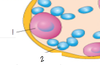
2
Q

A
1; gonadal ridge
2; PCGs
3; dorsal mesentary
4; primitive gonadal cords
3
Q

A
- seminiferous tubules
- PGCs
- Leydig cells
4
Q

A
- seminiferous tubules
- rete testes
- efferent ductules
- duct of epididymis
5
Q
A
- spermatogonium
- Sertoli cell
6
Q

A
- allantosis
- prostatic uticle
- efferent ducts
- epididymis
- vas deferens
- rete testes
- tunica albugenia
- pardidymis
- epididymis
7
Q

A
- vas deferns
- ureter
- seminal vesicle
- prostate
- bulbourethral glands
8
Q

A
- semineferous tubules
- vas deferns
- head of epidiymis
- efferent ductules
- rete testes
9
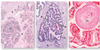
Q
What is this an image of

A
Müllerian duct degenerating
10
Q

A
- gubernaculum
- processus vaginalis
- labiosacral swelling
11
Q
What is the defect

A
congenital inguinal hernia
12
Q
what is the defect

A
hydrocoele
13
Q

A
- cortical cords
- PGCs
14
Q

A
- nucleus of primary oocyte
- flat epitheial (follicular) cell
15
Q

A
- Müllerian duct
- mesonephric duct degenerating
16
Q
What are these remnants of

A
mesonephric duct
- epoöphroron
- paraöphoron
- gartner duct cysts
17
Q
What is the “arrow” pointing to

A
garter’s cyst
18
Q
What is the circled region

A
uterovaginal primordium
19
Q

A
- paraurethral glands
- greater vestibular glands
20
Q
What derives these ligaments

A
gubernaculum
21
Q

A
- genital tubercule
- cloacal fold
- cloacal membrane
- labioscrotal swelling
- anal fold
- uretheral fold
- urogenital membrane
22
Q

A
- genital tubercule
- uretheral folds
- labioscrotal swellings
- penile shaft
- glans penis
- spongy urethera
- scrotum
23
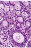
Q

A
- interstitial cells (leydig cell)
- seminiferous tubule
- tubule lumen
- sustentacular cells
- sperm cells
- spermatids
- spermatogonia
24
Q

A
- head of epididymis
- tunica albuginea
- tunica vaginalis
- rete testis
- body of epididymis
25

A; straight tubules
B; rete testes
C; septum
D; mediastinum testes
26

1. tunica albuginea
2. blood vessels
3. septum
4. seminiferous tubules
27

1. leydig cells
2. seminiferous tubule
28
What is happening at the arrow

transition from straight tubule to rete testes
A; vein
B; rete testes
C; seminiferous tubule
29
Why does the edge look "scalloped"

ciliated vs non-ciliated
1; rete testis
2; straight tubule
3; sartoli cells
30
what is this an image of

excretory genital ducts
31
What is this an image of

testes - intratesticular ducts
32
what is this an image of

excretory genital ducts
33
what is this an image of

seminal vesicle
34
what is this an imge of
prostate gland
35
what is this an image of

bulbourethral gland
36
what is this an image of

penis
A; corpus cavernosum
B; tunica albuginea
C; superficial fascia
D; urethra
E; corpus sponguosum
F; venous space
37
What is this an image of

penis
38

A; sartoli cell
B; myogenic cell
C; spermatogonia
D; primary spermatocyte
E; seminiferous tubule
F; leydig cell
39

1. ovarian follicles
2. cortical region
3. medullary region
40
What is this an image of

primordial follicle
41
What is this an image of

early or primary follicle
42
what is this an image of
late or multilayered primary follicle
43
primordial follicle and unilaminar primary follicle
44

multilayered primiary follicle
45
what is this an image of

secondary (antral) follicles
46

1. antrum
2. oocyte
3. zona pellucida
4. granulosa cells
5. theca interna
6. theca externa
7. corona radiata
8. cumulus oophorus
47
what is this an image of

mature or Graafian folicle
1. stratum granulosum
2. cumulus oophorus
3. antrum
4. theca interna
48
what is this an image of

corpus luteum
1. granulosa lutein
2. theca lutein
49

1. theca leutin
2. graulosa lutein
50
what is this an image of
corpus albicans
51
what type of follicle is this

primordial follicles
52
what kind of follicle is this

primary follicle
53
what type of follicle is this

secondary follicle
54
what type of follicle is this

vesicular follicle
55
what kind of follicle is this

corpus luteum
56
what kind of follicle is this

corpus albicans
57
what is this an image of

uterine tubes
58
what is this an image of

uterine tubes
59
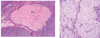
what is this an image of

uterus
1. endometrium
2. myometrium
3. perimetrium
60
what is this an image of

uterus
61
what is this an image of

vagina
