Introduction to the CVS Flashcards
(19 cards)
Why do we need the CVS?
- To pump blood through the lungs and carry oxygen.
- To transport nutrients to the muscles and organs.
- To circulate hormones and immune mediators.
- As a connection to the lymphatic system.
- For human reproduction.
- For temperature regulation.
What is passive diffusion, and what is its relative equation?
- Passive diffusion is the random, undirected thermal movement of molecules.
- The time needed to diffuse a given distance is proportional to the square of the distance: t ∝ d².
- Diffusion is fast in μm but very time consuming over distances > 1 mm and inappropriate for transport throughout the body.

What is the main method of molecule movement within the CVS?
- The CVS uses convection (bulk flow), which is the movement by a pressure gradient (so a pressure gradient is needed for bulk flow but not necessary for diffusion).
- It provides fast and directional transport; however, diffusion is still crucial for transport over short distances.
How is the pressure difference created in the blood vessels? What is the significance of having pressure differences?
- The output of blood at high pressure creates a pressure difference with distant blood vessels.
- Aorta > 100mmHg
- Large veins 5 - 10mmHg
- This pressure difference drives blood flow.
mmHg meaning - millimeter of mercury.
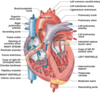
What is systole?
Contraction
Systole is the part of the cardiac cycle during which some chambers (the ventricles) of the heart muscle contract after refilling with blood.

What is diastole?
Relaxation
Diastole is the part of the cardiac cycle during which the heart refills with blood after the emptying done during systole (contraction).

What is the cardiac output?
- The cardiac output is the volume of blood ejected from the heart per minute.
- It is typically around 5 litres/min.
- Cardiac output = heart rate x stroke volume. (CO = HR X SV).
What is stroke volume?
- Stroke volume (SV) is the volume of blood pumped from the left ventricle per beat.
- The term stroke volume can apply to each of the two ventricles of the heart, although it usually refers to the left ventricle.
- The stroke volumes for each ventricle are generally equal, both being approximately 70 mL in a healthy 70-kg man.
- Its value is obtained by subtracting end-systolic volume (ESV) from end-diastolic volume (EDV) for a given ventricle.
- SV = EDV - ESV.
Where does cardiac output go? What two areas are of significance in relation to cardiac output distribution?
- The blood is distributed to various parts of the body, such as the kidneys, liver and GI, etc.
- Two places to take note of are the brain and myocardium (muscular heart tissue). These places are relatively underperfused.
- They have special mechanisms to overcome this problem; however, whenever there is a problem with blood supply, these two places are at risk (stroke, heart attack, etc.).
Perfusion meaning - It is the passage of fluid through the circulatory system or lymphatic system to an organ or a tissue, usually referring to the delivery of blood to a capillary bed in tissue.

What controls cardiac output?
- Filling pressure (Starling’s Law)
- Sympathetic and parasympathetic autonomic nerves
- Chemical factors and hormones (eg. adrenaline)
The starling’s law - It states that the stroke volume of the heart increases in response to an increase in the volume of blood in the ventricles, before contraction (the end diastolic volume), when all other factors remain constant.
Stroke volume meaning - The amount of blood pumped out of one ventricle of the heart as the result of a single contraction.
Why are blood flow and blood pressure important?
- Blood flow and blood pressure are critical for correct bodily functioning as they are linked in the proper distribution of blood (for eg. if there is poor perfusion of the kidney, it could cause renal failure and death).
What is the equation for calculating blood flow?
- Blood flow = (Pa - Pv) / resistance
- Pa is the pressure at the artery, while Pv is the pressure at the vein.
- Unit of blood flow is cm3/s.

What are the two properties of blood flow?
Blood flow is :-
- Proportional to pressure across blood vessel.
- Inversely proportional to resistance of blood flow.
Why does blood slow down in the capillaries?
The velocity of the blood is much slower in the capillaries to allow for gaseous/nutrient exchange to occur.

What is the equation for calculating blood velocity?
Blood velocity (cm/s) = blood flow (cm³/s) / cross-sectional area (cm²)
- To calculate cross-sectional area, we do the number of vessels times the πr² per vessel.

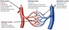
Describe how dual circulation can be in parallel or in series.
- IN SERIES (PORTAL) -
- Same blood supply between organs - lower perfusion pressures.
- Medically significant if the first organ is being underperfused.
- IN PARALLEL: -
- Cardiac output is split up.
- Safeguards O₂ supply in organs.
- Most organs are supplied this way.

List differences between the structures of arteries and veins.
-
ARTERIES: -
- Thicker elastic wall to maintain blood pressure.
- Have high pressure that ensures blood flows in one direction.
-
VEINS: -
- Thinner elastic walls.
- Have valves to ensure no blood backflow.

What are the four main functional groups of blood vessels?
-
ELASTIC VESSELS: ARTERIES
- Large arteries accomodate stroke volume and convert intermittent ejection into continuous flow.
-
RESISTANCE VESSELS: ARTERIOLES
- Control arterial blood pressure and regulate local blood flow.
-
EXCHANGE VESSELS: CAPILLARIES
- Nutrient delivery to cells and tissues for water and lymph formation, and removal of metabolic waste.
-
CAPACITANCE VESSELS: VENULES AND VEINS
- Control the filling pressure and provide a reservoir of blood, so the veins can constrict and send blood to the heart if needed.
Where is most of the blood volume distributed?
- It is mostly found in large and small veins and venules.
- Systemic veins and venules serve as a reservoir, holding about 65% of the volume.

