Joints: Upper & Lower Limb Flashcards
(100 cards)
the pelvic girdle is the union of the sacrum and 2 hip bones at the (three joints)
the pelvis can be divided into the ____ and _____
sacroilliac joints and pubic symphasis
The pelvis is divided into Greater (false) and Lesser (True) pelvises by terminal line.
the false pelvis is the superior half, the true would be the inferior half due to the contents of the pelvic organs
10

iliofemoral ligament
strongest ligaments in the human body, part of the extracapsular ligaments
13

pubofemoral ligament
part of the extracapsular ligaments
11
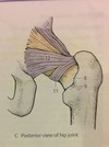
zona orbiculuaris
12

ischiofemoral ligament
part of the extracapsular ligaments
the sacroilliac joint is composed of two parts:
Composed of 2 parts:
- Anterior Synovial joint (Amphiarthrosis) between the auricular surfaces of sacrum and ilium, the joint capsule is very taut and encloses the almost immobile joint and reinforced by ligaments.
- Posterior Syndesmosis between the tuberosities of sacrum and ilium

1 and 2

superior and inferior pubic ligaments
Pubic Symphysis is what kind of joint?
Pubic Symphysis:
non-synovial joint
- Fibrous cartilaginous joint consists of a fibrocartilaginous interpubic disc.
- The disc is generally wider in women, containing a small Non-Synovial cavity
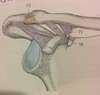
the hip joint is what kind of joint? describe the capsule
- Ball & Socket (Spheroidal) Multiaxial free moving type of joint.
Joint capsule, strong yet loose, has an external fibrous layer and an internal Synovial
membrane. Proximally it attaches around the Acetabular rim and transverse Acetabular
ligament, and distally it attaches to the femoral neck only at the Intertrochanteric line
anteriorly, but posteriorly it crosses the neck to attach to the Intertrochanteric crest.
There are 4 extracapsular ligaments and 1 intercapsular ligament (the femoral ligament)

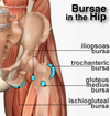
name the 4 hip joint Bursa

- Iliopectineal Bursa: large bursa between the Iliopsoas muscle and the hip joint anteriorly
- Obturator Externus Bursa: a Synovial protrusion beyond the margin of joint capsule into the posterior aspect of the femoral neck. For the tendon of Obturator Externus
- Obturator Internus Bursa
- Gluteus Maximus, Medius and Minimus Bursae: Also called Trochanteric Bursae, found between tendons and greater and lesser trochanters
What are the movements of the hip joint?
- Movements: Free moving, Multiaxial joint, 3 axes:
Anteversion (Flexion) / Retroversion (Extension), at a transverse axis through the head of femur
Abduction / Adduction, at an Anterior-Posterior (Sagittal) axis through the head of femur
Medial / Lateral Rotation, at a vertical axis through the head of femur and the medial femoral condyle
Combined movements of the above produce Circumduction
** Note: the degree of flexion/extension possible at the hip joint depends on the position of the knee. If the knee is flexed (hamstring muscles are relaxed), the
thigh can be actively flexed until it reaches the anterior wall of abdomen. Extension is very limited due to the strong Iliofemoral ligament. Lateral rotation is more powerful than medial rotation.
The hip joint uses what muscles to aid in Flexion? What is the maximum degree of movement?
flexion/Anteversion
Iliopsoas
Rectus Femoris
Pectineus
Sartorius
(With flexed knee Max. 130-140°)
The hip joint uses what muscles to aid in extension? What is the maximum degree of movement?
extension/Retroversion
Gluteus Maximus
Semimembranosus
Semitendinosus
Biceps Femoris- Long Head
(15°, with abduction 45°)
The hip joint uses what muscles to aid in abduction? What is the maximum degree of movement?
Abduction
Gluteus Medius
Gluteus Minimus
Tensor Fascia L
(50°, in flexion 80°)
The hip joint uses what muscles to aid in adduction? What is the maximum degree of movement?
Adduction
Adductor Magnus
Adductor Longus
Adductor Brevis
Pectineus
Gracilis
(10°, in flexion 30°)
The hip joint uses what muscles to aid in lateral rotation? What is the maximum degree of movement?
Lateral Rot.
Gluteus Maximus
Gluteus Medius- Dorsal part
Gluteus Minimus- Dorsal part
Obturator internus
Piriformis
Quadratus Femoris
(15°, in flexion 60°)
The hip joint uses what muscles to aid in medial rotation? What is the maximum degree of movement?
Medial Rot.
Gluteus Medius
Ventral part
Gluteus Minimus
Ventral part
Adductor Longus
Iliopsoas
(30°, in flexion 40°)
The knee joint is what kind of joint and which bones/ surfaces are involved?
mainly a hinge joint combined with gliding
and rolling and with rotation (Torchoid) about a vertical axis, 2 axes joint.
The articular surfaces are large sized, complicated & incongruent shapes. We have to distinguish 3 articulations in the knee joint:
2 Femortibial articulations (lateral & medial) between the lateral and the medial femoral and Tibial condyles
1 Intermediate Femoropatellar articulation between patella and the femur
The fibula is not involved in the joint
2

Patellar ligament:
distal part of quadriceps muscle, thick fibrous band passing from apex of patella to the Tibial tuberosity. On both sides it receives the lateral and medial patellar retinacula (expansions of Vastus muscles and overlying
fascia) which make up the joint capsule on both sides of the patella. Maintain the patella in appropriate articular position with the femur.
5

lateral patellar retinaculum
6

medial patellar retinaculum
9

tibial collateral ligament
strong, flat capsular band
that extends from medial epicondyle of femur to the medial condyle of tibia (&medial superior surface). Its deep fibers are strongly attached and fused to the
medial meniscus and joint capsule. It is weaker than the fibular collateral ligament and more often damaged.
15

Fibular collateral ligament (lateral collateral ligament):
cord-like strong extracapsular ligament from lateral epicondyle of femur to the lateral surface of
head of fibula. It is separated from the lateral meniscus and capsule by the tendon of popliteus and it splits the tendon of Biceps Femoris into 2.
22

suprapatellar bursa