Lab Quiz 1: Labs 1, 2, 3 Flashcards Preview

BIO 151 A&P II > Lab Quiz 1: Labs 1, 2, 3 > Flashcards

Flashcards in Lab Quiz 1: Labs 1, 2, 3 Deck (85)
Loading flashcards...
1
Q

3 factors that determine the quality of a microscopic image

A
  1. Contrast: distinguish objects, such as cells, from the background
  2. Magnification: increase in image size
  3. Resolution: distinguish fine detail
2
Q

4 objective lens sizes

A
  • 4X, 10X, 40X, and 100X
  • Note
    • When focusing on a slide, start on the scanning, or low power, objective lens
    • The lower the power objective, the greater the field of view. The field of view is the amount of the slide you are able to see through the eyepiece.
3
Q

ABO system of blood typing

  • Describe
  • How is type determined?
A
  • Determined by the presence or absence of specific surface antigens on RBCs
    • A: when a blood cell has the A antigen on its surface
    • B: when a blood cell has the B antigen
    • O: neither A or B antigens are present
    • AB: when both A and B antigens are present
4
Q

Agglutination

A
  • When the cells clump together due to the antibody / antigen interaction
5
Q

Antibodies

A
  • Immune system proteins that binds to antigens to aid in creating immune responses
6
Q

Antigen

A
  • Any substance that is capable of causing an immune reaction
  • Usually proteins, glycoproteins, or glycolipids
7
Q

Impact of Aperture

A
  • As you move the aperture to decrease the light to the slide, the contrast increases
  • As you move the aperture to increase the light to the slide, the contrast decreases

Note: The sliding bar on the condenser lens is the aperture

8
Q

Basophils

  • Indication of
  • Appearance (nucleus & color)
  • Frequency in WBCs
A
  • WBC granulocyte
    • Fight fungal or bacterial infections and viruses
    • Nucleus is usually bilobed
    • Black or dark purple granules
  • 0-2% of WBCs
    • The least abundant in all mammals
9
Q

Blood

  • What is it composed of?
A
  • Liquid connective tissue
  • Matrix is composed of plasma surrounding the formed elements
10
Q

Blood doping

  • Describe
  • Normal ranges
A
  • Methods used to increase the blood oxygen-carrying capacity
  • Ranges
    • Male: 14–18 grams Hb/100 mL
    • Female: 12–16 grams Hb/100 mL
11
Q

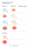
Blood type identification

A
12
Q

How do you convert cm to m?

A
  • cm * 0.01; OR
  • cm / 100
13
Q

Eosinophils

  • Indication of
  • Appearance (nucleus & color)
  • Frequency in WBCs
A
  • WBC granulocyte that promotes inflammation
    • Parasitic infection, allergic reaction, cancer
    • Nucleus is usually bilobed
    • Bright red granules
  • 0-5% of WBCs
14
Q

Fields of study where a microscope is a necessary tool

A
  • Cytology: the study of cells
  • Histology: the study of tissues
  • Pathology: the study of disease
15
Q

Formed elements

A
  • Platelets (thrombocytes)
  • RBCs (erythrocytes)
  • WBCs (leukocytes)
    • Granular
      1. Basophils
      2. Eosinophils
      3. Neutrophils
    • Agranular
      1. Lymphocytes
      2. Monocytes
16
Q

Hematocrit (HCT)

  • Description
  • Indication of
  • Generally accepted ranges
A
  • Measures packed RBC cell volume
  • Determined by separating the formed elements from the plasma
  • Indication of high altitude, elevated testosterone levels, blood doping
  • Ranges
    • Adult males: 42% – 52%
    • Adult females: 37% – 47%
17
Q

Hematocrit formula

A

(RBC x MCV) / 10 = HCT

Note: MCV is the average volume (size) of the patient’s RBCs

18
Q

Hematology

A

The study of the blood and the organs that produce it

19
Q

Hemoglobin

  • Describe
  • How is it measured?
A
  • One measure of the oxygen-carrying capacity of the blood
    • RBCs are about ⅓ hemoglobin
    • Measured using a hemoglobinometer
20
Q

How do the coarse and fine focus knobs work on a Brightfield microscope?

A

They move the stage up and down, allowing the user to move the slide into focus

21
Q

How do you adjust field of view?

A
  • Top stage knob moves the stage forward and back
  • Bottom stage knob moves it side-to-side
22
Q

How do you read a graduated cylinder?

A
  • Water adheres to the glass walls of a graduated cylinder, creating a curved appearance termed a meniscus
  • Accurate volume measurements are taken at the bottom of the meniscus with your eye level parallel to the meniscus
23
Q

How is blood type determined?

A
  • By the antigens that are present on the surface of RBCs
    • ABO blood type is determined by the presence of A, B, both (AB), or neither (O) of the A or B antigens
    • Rh blood type is determined by the presence of (+) or lack of (-) the D antigen
24
Q

How to focus on a specimen

A
  1. Place the slide on the stage
  2. Make sure that the scanning (4x) objective is in place
  3. Using the coarse adjustment knob, adjust the stage to its highest position (closest to the specimen)
  4. Using the mechanical stage place the specimen in the path of light
  5. Looking through the ocular, first use the coarse adjustment knob to bring the specimen in view. The ocular is usually 10-15x
  6. Use the fine adjustment knob to focus more clearly on the specimen
  7. Only now can you move up to the next objective (low power 10x)
  8. Repeat steps 7 and 8 to move to the 40x (high dry) objective

*Remember to use the fine adjustment knob only for both the 10x and 40x objectives.

25
Q

How to handle the microscope

A
  • Always carry it by the ARM with one hand on the BASE, always using both hands
26
Q

How to measure circles

A
  • Place the zero of the ruler at the left edge of the circle
  • The ruler should bisect the midpoint of the circle to measure the maximum length (diameter) of the circle
27
Q

How to put away a microscope

A
  1. Remove the slide and put it back in the slide tray
  2. Center the stage arm
  3. Place the voltage regulator to the lowest level
  4. Lower the stage fully
  5. Turn off the microscope
  6. Put the scanning objective into place
  7. Secure the power cord
  8. Put the cover on the microscope and put it away in its proper location
28
Q

Lab clean up procedures

A
  1. The laboratory is to be left the same or cleaner than when you entered it
  2. You must disinfect your bench BEFORE you put any of your own materials on it
  3. All materials used by yourself and / or your group are to be put away properly before leaving
  4. Your equipment and bench should be cleaned and disinfected before leaving the laboratory
  5. You must take extra care not to clog the sinks
  6. You should disinfect your laboratory aprons before AND after using them.
  7. You must disinfect your bench AFTER you are finished
  8. When you are completely cleaned up – the last thing you MUST do before you leave is wash your hands
29
Q

Impact of light intensity on magnification

A

Decreases with increased magnification

30
Q

Lymphocytes

  • Description
  • Indication of
  • Types
  • Presence rates
A
  • WBC agranulocyte
    • Made in the bone marrow
    • Found in blood and lymph tissue
    • Small cells with a round nucleus that takes up most of the cell volume
  • Indication of viral infection, Mononucleosis, AIDS
  • Types
    • B lymphocytes
    • T lymphocytes
      • AIDS infects T-lymphocytes (T-cells) and reduces their numbers and inhibit immune system function
  • Rates
    • Normal: 15-40%
    • Atypical: 0-1%
31
Q
A
32
Q
A
33
Q

Microscopy

  • Goal of microscopy
  • How do Brightfield microscopes accomplish this
A
  • Create a magnified image of objects too small to be seen with the eye alone
  • Use a combination of glass lenses and light to view the specimen
34
Q

mm to cm Formula Conversion

A

Multiply the millimeter value by 0.1 or divide by 10

35
Q

mm to m Conversion Formula

A

Multiply the millimeter value by 0.001 or divide by 1000

36
Q

Monocytes

  • Description
  • Indication of
  • Frequency in WBCs
A
  • WBC agranulocyte and phagocyte
    • Large cells
    • Bean-shaped nucleus that takes up half to nearly all the volume of the cell
  • Indication of chronic or sub-acute infections
    • Linked with some types of cancer, especially leukemia
    • High counts occur when you are recovering from an acute infection
  • 0-10% of WBCs
37
Q

Name the test

A

Hematocrit

38
Q

Neutrophils

  • Indication of
  • Appearance (nucleus & color)
  • Frequency in WBCs
A
  • WBC granulocyte
    • Type of elevated cell found in bacterial infections
    • Contain light pink and/or purple granules
    • The nucleus is multilobed
  • Types
    • Segmented: 50-60%
    • Banded: 0-5%
39
Q

Positive & negative results

A
  • Does NOT mean “good.” In testing, positive means what you are testing for is present
  • Does NOT mean “bad.” In testing, negative means what you are testing for is absent
40
Q

Preparation of blood smear

A
  1. Place microscope slide on staining rack
  2. Add drop of blood to slide using pipette. Dispose of pipette
  3. Take another slide from box and use it to smear blood drop by sliding it from right to left. Let air dry for 1 minute. Dispose of slide
  4. Add Wright stain to smear. Wait 2 minutes
  5. Add distilled water to smear. Wait 4 minutes
  6. Wash slide with filtered water. Wait 1 minute
  7. Place slide on microscope stage
41
Q

RBC function

A

Carry oxygen bound to hemoglobin and deliver it to the tissues

42
Q

Reasons to wear gloves

A
  • Protect yourself and others from harmful chemicals (do not contaminate your work area)
  • Prevent contamination of the chemicals
43
Q

Rh blood group

A
  • Determined by the presence of (+) or lack of (-) the D antigen
    • Antigen named D, or more commonly Rh antigen, that can be present (Rh+) or absent (Rh-) from the surface of the RBCs
    • Normally a person does not create antibodies towards the Rh antigen unless they are exposed to it directly
44
Q

Role of condenser lens

A

Focuses the light on the slide

45
Q

Safety Resources

A
  • Nearest telephone
  • Emergency Telephone Numbers
  • Emergency exits
  • Fire extinguisher
  • Emergency Shower
  • Safety kit (not a first aid kit)
  • Eyewash station
  • Safety Data Sheets
  • Fire alarm location
46
Q

Serum/Sera

A

Fluid containing antibodies.

47
Q

Basic units of the metric system

  • 4 units and what they measure
  • Why is metric system used
A
  • Units
    • Grams (g) for weight
    • Meters (m) for length
    • Liters (L) for volume
    • Degrees Celsius (°C) for temperature
  • Why
    • Gives all scientists the same standards of measurement when they conduct experiments.
48
Q

Total magnification formula

A

ocular magnification x objective magnification = total magnification

49
Q
A
50
Q

Transfusion reaction

A
  • When a recipient receives blood from an incompatible donor the antibodies in their plasma cause agglutination and hemolysis of the donated RBCs
51
Q

Transporting slides

A
  • Do not place slides on the bench or pile them up together
  • When transporting more than one or two slides, they should always be carried in a slide box
52
Q

Types of personal protective equipment

A
  • Lab coat or apron
  • Gloves
  • Goggles or lab safety glasses
  • Closed toe and covered heel shoes
  • Long hair should be tied up to meet proper laboratory safety protocols
53
Q

Using the following image and a calculator, calculate the hematocrit

A
  • (10.5mm / 32mm) x 100
  • 0.328 x 100 = 32.8%
54
Q

What do atypical lymphocytes suggest?

A

Mononucleosis

55
Q

What do high banded neutrophils suggest?

A

Acute bacterial infection

56
Q

What do high eosinophil levels suggest?

A

Parasitic worms or allergy

57
Q

What do high lymphocytes suggest?

A

Viral infection

58
Q

What do high neutrophil levels suggest?

A

Bacterial infection

59
Q

What do low lymphocytes suggest?

A

AIDS

60
Q

What do you label on a blood smear slide

A
  • Blood sample number
  • Serum type
61
Q

What is a normal range of hematorcrit for females?

A

37-47%

62
Q

What is a normal range of hematorcrit for males?

A

42-52%

63
Q

What is the advantage of starting with the 4X objective before moving to the 10X and then the 40X objective?

A

The lower power objective has a larger field of view and is easier to focus.

64
Q

What is the correct order of steps to view a specimen at 40X magnification?

A
  1. Click power switch to turn on light
  2. Adjust light
  3. Focus at 4X using coarse focus knob
  4. Adjust ocular lenses to view one image
  5. Focus using fine focus knob
  6. Adjust the stage
  7. Put 10X objective in place
  8. Focus using fine focus knob
  9. Put 40X objective in place
  10. Adjust using fine focus knob
65
Q

What is the purpose of the hemolysis applicator?

A

It breaks down the cell membranes of the RBCs releasing the hemoglobin into solution

66
Q

What is the total magnification of a specimen using the 40X objective?

A

400X

67
Q

What is this and what is it used for?

A
  • Microcentrifuge
  • Spins the capillary tube to pack the RBCs
68
Q

What is this and what is it used for?

A
  • Critoseal
  • Clay to plug one open end of the capillary tube during centrifugation
69
Q

What is this and what is it used for?

A
  • Heparinized capillary tubes
  • Holds the blood sample
70
Q

Identify the WBC and its characteristics

A
  • Lymphocyte
  • Small cell with a round nucleus taking up most of the volume of the cell
71
Q

Identify the WBC and its characteristics

A
  • Neutrophil
  • Light pink and/or purple granules
  • Nucleus is multilobed
72
Q

Identify the WBC and its characteristics

A
  • Monocyte
  • Large cell with a bean-shaped nucleus taking up half to most of the volume of the cell
73
Q

Identify the WBC and its characteristics

A
  • Eosinophil
  • Bright red granules
  • Bilobed nucleus
74
Q

Identify the WBC and its characteristics

A
  • Basophils
  • Dark purple or black granules
  • Bilobed nucleus
75
Q

What is used to measure your blood pressure?

A

Sphygmomanometer

76
Q

What tool is used to measure hemoglobin concentration?

A
  • Hemoglobinometer
  • Measures the amount of hemoglobin in grams per deciliter (g/dL)
77
Q

What WBC type do you expect to find the most?

A
  • Neutrophil
  • Depending on the many factors, the normal range for neutrophils is 30–75% of all WBCs
78
Q

Which of the 3 factors affecting image quality is altered by the light source?

A

Contrast

79
Q

Which patient is likely to have an allergy or parasitic infection?

A

Patient A- high eosinophils

80
Q

Which patient is likely to have mononucleosis?

A

Patient C: atypical lymphocytes

81
Q

Which patient is likely to have HIV/AIDS?

A

Patient B: low lymphocytes

82
Q

Why are accurate measurements necessary in scientific research?

A
  • Allows for good hypothesis testing
  • Ability to replicate findings is helped by taking accurate measurements
83
Q

Why are metric units used in scientific research?

A

Ensure consistency of collected data.

84
Q

Why is volume displacement used to calculate the volume of some objects?

A
  • Because it can be difficult to accurately measure oddly shaped objects
  • Measuring the length of a cube or the diameter of a sphere could give a volume, but odd shapes make the mathematics much more difficult
85
Q

Why should the fine focus knob typically be used with the 10x and 40x objective lenses?

A

Using the coarse focus knob would result in a large change in focus, when only a small change is needed