Liver Histopath Flashcards
(46 cards)
Major causes of cirrhosis (3) & less common causes (3)
- Alcoholic fatty liver disease
- Non alcoholic fatty liver disease
- Chronic hepatitis infection
Autoimmune hepatitis
Drugs e.g. Methotrexate
Biliary causes: PBC & PSC
Genetic causes of cirrhosis (5)
1) haemochromatosis - HFE gene chr 6
2) Wilson’s disease - ATP7B gene chr 13
3) alpha 1 anti-trypsin deficiency (pulmonary & hepatic dysfunction)
4) galactosaemia
5) Glycogen storage disease
Difference between micronodular & macronodular cirrhosis & 2 examples of each
Micronodular indicates regenerating nodules (of regenerating hepatocytes) < 3mm e.g. alcoholic & biliary tract disease
Macro > 3mm e.g. Viral, Wilson’s
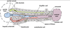
Briefly describe pathophysiology of cirrhosis (Refer to picture)
1) chronic inflammation activates the normally quiescent stellate cells
2) convert to myofibroblasts and deposit collagen in space of disse
3) when myofibroblasts contract they cause constriction of the sinusoids - increasing vascular resistance
4) undamaged hepatocytes regen in nodules between fibrous septa

5 criteria of the child’s Pugh score - indicates prognosis of cirrhosis
Ascites (none, mild, severe)
Encephalopathy (none, mild, severe)
Bilirubin (<34, 34-50, >50)
Albumin (>35, 28-35, 6)
PTT (seconds more than normal) (<4, 4-6, >6)
Score 1,2 & 3 respectively
Score boundaries for Child Pugh A, B & C (Indicates cirrhosis prognosis)
- < 7 (A) = 45% 5 yr survival
- 7-9 (B) = 20% 5 yr survival
- 10+ (C) = <20% 5 yr survival
Portal htn (>10-12mmHg) venous system dilates, where do collateral vessels form? (6)
1) gastro-oesophageal
2) Rectal
3) Umbilical
4) retroperotneal
5) diaphragm
6) left renal vein
Causes of portal HTN (pre hepatic, hepatic & post hepatic)
Pre-hepatic: portal vein thrombosis (e.g, Factor V Leiden)
Hepatic:
- Pre sinusoidal: schistomasis, PBC, Sarcoidosis
- Sinusoidal: Cirrhosis
- Post-sinusoidal: veno-occlusive disease Post-hepatic: Budd-Chiari syndrome
Give some causes of Budd-Chiari & treatment
Occlusion of hepatic vein!
Causes: 30% idiopathic, thrombophilia, OCP, leukaemias, compression by renal tumours etc.
Treatment: Thrombolytic, treat underlying cause, TIPS (transjugular intrahepatic portosystemic shunt)
Triad of Budd Chiari
Pain + ascites + hepatomegaly
3-4 features of cirrhosis
1) hepatocyte necrosis
2) nodules of regenerating hepatocytes
3) fibrosis
4) disrupted liver architecture> increased resistance to blood flow through liver > portal hypertension
Microscopic characteristics of hepatic steatosis (fatty liver)
Steatosis - fatty droplets in hepatocytes. Fibrosis in late stage if chronic exposure reversible if alcohol avoided
microscopic characteristics of alcoholic cirrhosis
MICRONODULAR cirrhosis - small nodules + bands of fibrous tissue
Microscopic characteristics of alcoholic hepatitis (2)
Hepatocyte BALLOONING & necrosis due to accumulation of fat, water & proteins
MALLORY BODIES (damaged intermidiate filaments within hepatocytes)
fibrosis
seen acutely after night of heavy drinking
What antibodies are present in Type 1 autoimmune hepatitis? (4)
ANA (anti nuclear ab), anti-SMA (anti smooth muscle ab), anti-actin ab, anti-soluble liver antigen ab
What antibody is present in Type 2 autoimmune hep?
Anti-LKM Ig (anti-liver-kidney-microsomal Ig)
How is autoimmune hepatitis managed?
Immune suppression until transplant, (but disease returns in up to 40%)
Associations of autoimmune hepatitis: Who gets it? M or F? HLA association?
People with autoimmune conditions females (78%) HLA-DR3
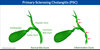
What is main tissue destroyed by autoimmune inflammation in PBC?
medium sized INTRAHEPATIC bile ducts (this leads to cholestasis which very SLOWLY leads to development of cirrhosis over many years)
Ratio of F:M in PBC
10:1
What will the investigations/ histology show in PBC? (4)
High ALP, high cholesterol, high IgM, ANTI-MITOCHONDRIAL Abs in 90%
US - NO bile duct dilatation
histology - bile duct loss with GRANULOMA formation
Presentation of PBC?
Fatigue, pruritis, abdo discomfort
Secondary symptoms of PBC (give 3)
xanathelasma, skin pigmentation, steatorrhoea, inflammatory arthropathy
Treatment of PBC
Ursodeoxychilic acid (slows down absorption of cholesterol from intestine) in early phase (25% remission)