Morphological Plan of the Lower Limb Flashcards
Posterior compartment of thigh
- The muscles in the posterior compartment of the thigh are collectively known as the hamstrings. They consist of the:
- Laterally - biceps femoris (long head from ischial tuberosity and short head from femur - form tendon inserting into fibula)
- Medially - semi… and semi… (origin - ischial tuberosity, insert into tibia)
- Supplied by sciatic nerve
- Extend thigh, flex leg, medially rotate and laterally rotate
- The muscles in the posterior compartment of the thigh are collectively known as the hamstrings. They consist of the:
- Laterally - biceps femoris (long head from ischial tuberosity and short head from femur - form tendon inserting into fibula)
- Medially - semitendinosus and semimembranosus (origin - ischial tuberosity, insert into tibia)
- Supplied by sciatic nerve
- Extend thigh, flex leg, medially rotate and laterally rotate

Femoral triangle
- Femoral artery enters anterior compartment of thigh under inguinal ligament - into femoral triangle region
- Borders - inguinal ligament, medial border of adductor longus, sartorius
- Floor - pectineus, iliopsoas, and adductor longus muscles.
- Passing through the triangle - lateral to medial - Femoral nerve - femoral artery - femoral vein - lymphatics (within the femoral canal)
- Artery, vein and femoral canal are surrounded by … …
- Femoral nerve (lateral to artery) not surrounded by … …

- Femoral artery enters anterior compartment of thigh under inguinal ligament - into femoral triangle region
- Borders - inguinal ligament, medial border of adductor longus, sartorius
- Floor - pectineus, iliopsoas, and adductor longus muscles.
- Passing through the triangle - lateral to medial - Femoral nerve - femoral artery - femoral vein - lymphatics (within the femoral canal)
- Artery, vein and femoral canal are surrounded by femoral sheath
- Femoral nerve (lateral to artery) not surrounded by femoral sheath

Blood supply to the Lower Limb


How many metatarsals?
5
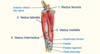
Quadriceps
- Four individual muscles: Rectus …, vastus …, vastus …, vastus …
- Rectus … - extension of leg and flexion of thigh - Origin - anterior inferior iliac spine, insert into tibial tuberosity
- 3 Vastus muscles - extension of leg only - Origin - femur, insert into tibial tuberosity
- All innervated by femoral nerve
- Four individual muscles: Rectus femoris, vastus lateralis, vastus medialis, vastus intermedius
- Rectus femoris - extension of leg and flexion of thigh - Origin - anterior inferior iliac spine, insert into tibial tuberosity
- 3 Vastus muscles - extension of leg only - Origin - femur, insert into tibial tuberosity
- All innervated by femoral nerve

Gluteus Maximus
- The gluteus maximus is the largest of the gluteal muscles. It is also the most superficial, producing the shape of the buttocks.
- Attachments: Originates from the ilium. It slopes across the buttock at a 45 degree angle, then inserts into the iliotibial tract and the gluteal tuberosity of the femur.
- Actions: It is the main extensor of the thigh, and assists with lateral rotation.
- Innervation: … … nerve.
- The gluteus maximus is the largest of the gluteal muscles. It is also the most superficial, producing the shape of the buttocks.
- Attachments: Originates from the ilium. It slopes across the buttock at a 45 degree angle, then inserts into the iliotibial tract and the gluteal tuberosity of the femur.
- Actions: It is the main extensor of the thigh, and assists with lateral rotation.
- Innervation: Inferior gluteal nerve.

Movements of the thigh - Abduction
- Performed by:
- Gluteus …
- Gluteus …
- Performed by:
- Gluteus medius
- Gluteus minimus

Venous drainage - Lower Limb
- Deep veins follow arteries
- Two major superficial veins:
- Great or long saphenous vein
- Small or short saphenous vein
- These Drain dorsal venous arch of foot
- Clinically - varicose veins (damage to valves in perforating veins leads to pooling of blood in superficial veins - varicosities (perforating veins … … to … veins)
- These Drain dorsal venous arch of foot
- Deep veins follow arteries
- Two major superficial veins:
- Great or long saphenous vein
- Small or short saphenous vein
- These Drain dorsal venous arch of foot
- Clinically - varicose veins (damage to valves in perforating veins leads to pooling of blood in superficial veins - varicosities (perforating veins connect superficial to deep veins)
- These Drain dorsal venous arch of foot
The greater sciatic foramen is an opening (foramen) in the posterior human pelvis. It is formed by the … and … ligaments. The … nerve passes through here
The greater sciatic foramen is an opening (foramen) in the posterior human pelvis. It is formed by the sacrotuberous and sacrospinous ligaments. The sciatic nerve passes through here

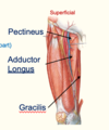
Medial compartment of thigh
- Adductors of thigh
- Muscles are (superficial to deep) Gracilis, Pectineus, Adductor longus, Adductor Brevis, Adductor Magnus (2 parts - … part and … part)
- Adductors of thigh
- Muscles are (superficial to deep) Gracilis, Pectineus, Adductor longus, Adductor Brevis, Adductor Magnus (2 parts - adductor part and hamstring part)
Motor Functions of Sciatic Nerve
- The sciatic nerve innervates the muscles in the … compartment of the … and the hamstring portion of the adductor magnus.
- The sciatic nerve also indirectly innervates several other muscles, via its two terminal branches:
- Tibial nerve – the muscles of the posterior leg (calf muscles), and some of the intrinsic muscles of the foot.
- Common fibular nerve – the muscles of the anterior leg, lateral leg, and the remaining intrinsic foot muscles.
- Overall - innervates muscles of the … …, entire leg and entire foot.
- The sciatic nerve innervates the muscles in the posterior compartment of the thigh and the hamstring portion of the adductor magnus.
- The sciatic nerve also indirectly innervates several other muscles, via its two terminal branches:
- Tibial nerve – the muscles of the posterior leg (calf muscles), and some of the intrinsic muscles of the foot.
- Common fibular nerve – the muscles of the anterior leg, lateral leg, and the remaining intrinsic foot muscles.
- Overall - innervates muscles of the posterior thigh, entire leg and entire foot.

Sciatic nerve
- Fill in the blanks


Quadriceps
- Fill in the blanks


Dermatomes - Lower Limb
main - L1,L2,L3,L4,L5 and S1, S2

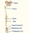
Skeleton (Lower limb)
- Label the diagram

Short saphenous vein
- Drains lateral side of arch
- Pierces deep … fascia
- Drains into … vein
- Drains lateral side of arch
- Pierces deep popliteal fascia
- Drains into popliteal vein

Label the Hip bones


The most superficial and largest gluteal muscle is the … …
gluteus maximus
Sciatic nerve
- Fill in the blanks


Biceps Femoris
- Like the biceps brachii in the arm, the biceps femoris muscle has two heads – a long head and a short head.
- It is the most lateral of the muscles in the posterior thigh
- Attachments: The long head originates from the ischial tuberosity of the pelvis. The short head originates from the femur. Together, the heads form a tendon, which inserts into the head of the …
- Actions: Main action is flexion at the knee. It also extends the thigh at the hip, and laterally rotates at the hip and knee.
- Innervation: Sciatic nerve
- Like the biceps brachii in the arm, the biceps femoris muscle has two heads – a long head and a short head.
- It is the most lateral of the muscles in the posterior thigh
- Attachments: The long head originates from the ischial tuberosity of the pelvis. The short head originates from the femur. Together, the heads form a tendon, which inserts into the head of the fibula.
- Actions: Main action is flexion at the knee. It also extends the thigh at the hip, and laterally rotates at the hip and knee.
- Innervation: Sciatic nerve

Varicose veins- The … … vein is a superficial vein. The deep veins (posterior tibial, anterior tibial, fibular, popliteal, femoral) are separated from the superficial veins by a series of valves. These valves ensure blood flows from the superficial system to the deep system i.e. prevents backflow. The incompetence of these valves results in varicose veins, which are engorged tortuous veins, that can be tender to the touch. Causes include genetic inheritance, pregnancy, chronic heart disease, obesity and prolonged standing.
Varicose veins- The greater saphenous vein is a superficial vein. The deep veins (posterior tibial, anterior tibial, fibular, popliteal, femoral) are separated from the superficial veins by a series of valves. These valves ensure blood flows from the superficial system to the deep system i.e. prevents backflow. The incompetence of these valves results in varicose veins, which are engorged tortuous veins, that can be tender to the touch. Causes include genetic inheritance, pregnancy, chronic heart disease, obesity and prolonged standing.
Motor Functions of Sciatic Nerve
- The sciatic nerve innervates the muscles in the posterior compartment of the thigh and the hamstring portion of the adductor magnus.
- The sciatic nerve also indirectly innervates several other muscles, via its two terminal branches:
- …. nerve – the muscles of the posterior leg (calf muscles), and some of the intrinsic muscles of the foot.
- … … nerve – the muscles of the anterior leg, lateral leg, and the remaining intrinsic foot muscles.
- Overall - innervates muscles of the posterior thigh, entire leg and entire foot.
- The sciatic nerve innervates the muscles in the posterior compartment of the thigh and the hamstring portion of the adductor magnus.
- The sciatic nerve also indirectly innervates several other muscles, via its two terminal branches:
- Tibial nerve – the muscles of the posterior leg (calf muscles), and some of the intrinsic muscles of the foot.
- Common fibular nerve – the muscles of the anterior leg, lateral leg, and the remaining intrinsic foot muscles.
- Overall - innervates muscles of the posterior thigh, entire leg and entire foot.

Movements of the thigh - Flexion at hip joint
- Performed by:
- Rectus femoris
- Sartorius
- … (Iliacus and psoas major) - insert into lesser trochanter
- Performed by:
- Rectus femoris
- Sartorius
- Iliopsoas (Iliacus and psoas major) - insert into lesser trochanter

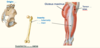
The Femur
- The femur is the only bone in the thigh and the longest bone in the body.
- Proximal part - It consists of a head and neck, and two bony processes – the … and … …
- The shaft of the femur descends in a slight medial direction. On the posterior surface of the femoral shaft, there are roughened ridges of bone, called the linea aspera. Proximally, the medial border of the linea aspera becomes the pectineal line. The lateral border becomes the gluteal tuberosity, where the gluteus maximus attaches.
- Distal - the … tubercle is found distally on the femur and is formed from the termination of the medial supracondylar line.
- Medial and lateral condyles – rounded areas at the end of the femur.
- Medial and lateral epicondyles – bony elevations on the non-articular areas of the condyles.
- The femur is the only bone in the thigh and the longest bone in the body.
- Proximal part - It consists of a head and neck, and two bony processes – the greater and lesser trochanters.
- The shaft of the femur descends in a slight medial direction. On the posterior surface of the femoral shaft, there are roughened ridges of bone, called the linea aspera. Proximally, the medial border of the linea aspera becomes the pectineal line. The lateral border becomes the gluteal tuberosity, where the gluteus maximus attaches.
- Distal - the adductor tubercle is found distally on the femur and is formed from the termination of the medial supracondylar line.
- Medial and lateral condyles – rounded areas at the end of the femur.
- Medial and lateral epicondyles – bony elevations on the non-articular areas of the condyles.

Nerve supply to the Lower Limb
- Nerves innervating lower limb structures originate from either lumbar or sacral plexi
- Lumbar plexus formed from anterior primary rami (L1 to L4)
- Sacral plexus formed from anterior primary rami L4 and L5 (via … trunk) and anterior primary rami … to …
- Femoral Nerve - L2 to L4 (lumbar plexus)
- Obturator Nerve - L2 to L4 (lumbar plexus)
- Sciatic nerve - L4-S3 (sacral plexus)
- Nerves innervating lower limb structures originate from either lumbar or sacral plexi
- Lumbar plexus formed from anterior primary rami (L1 to L4)
- Sacral plexus formed from anterior primary rami L4 and L5 (via lumbosacral trunk) and anterior primary rami S1 to S4
- Femoral Nerve - L2 to L4 (lumbar plexus)
- Obturator Nerve - L2 to L4 (lumbar plexus)
- Sciatic nerve - L4-S3 (sacral plexus)

Femoral triangle
- Femoral artery enters anterior compartment of thigh under inguinal ligament - into femoral triangle region
- Borders - … ligament, medial border of … …, s… muscle
- Floor - pectineus, iliopsoas, and adductor longus muscles.
- Passing through the triangle - lateral to medial - Femoral nerve - femoral artery - femoral vein - lymphatics (within the femoral canal)
- Artery, vein and femoral canal are surrounded by femoral sheath
- Femoral nerve (lateral to artery) not surrounded by femoral sheath

- Femoral artery enters anterior compartment of thigh under inguinal ligament - into femoral triangle region
- Borders - inguinal ligament, medial border of adductor longus, sartorius
- Floor - pectineus, iliopsoas, and adductor longus muscles.
- Passing through the triangle - lateral to medial - Femoral nerve - femoral artery - femoral vein - lymphatics (within the femoral canal)
- Artery, vein and femoral canal are surrounded by femoral sheath
- Femoral nerve (lateral to artery) not surrounded by femoral sheath

Biceps Femoris
- Like the biceps brachii in the arm, the biceps femoris muscle has two heads – a long head and a short head.
- It is the most lateral of the muscles in the posterior thigh
- Attachments: The long head originates from the ischial tuberosity of the pelvis. The short head originates from the femur. Together, the heads form a tendon, which inserts into the head of the fibula.
- Actions: Main action is … at the knee. It also extends the thigh at the hip, and … rotates at the hip and knee.
- Innervation: Sciatic nerve
- Like the biceps brachii in the arm, the biceps femoris muscle has two heads – a long head and a short head.
- It is the most lateral of the muscles in the posterior thigh
- Attachments: The long head originates from the ischial tuberosity of the pelvis. The short head originates from the femur. Together, the heads form a tendon, which inserts into the head of the fibula.
- Actions: Main action is flexion at the knee. It also extends the thigh at the hip, and laterally rotates at the hip and knee.
- Innervation: Sciatic nerve
Motor Functions of Sciatic Nerve
- The sciatic nerve innervates the muscles in the posterior compartment of the thigh and the hamstring portion of the … …
- The sciatic nerve also indirectly innervates several other muscles, via its two terminal branches:
- Tibial nerve – the muscles of the posterior leg (calf muscles), and some of the intrinsic muscles of the foot.
- Common fibular nerve – the muscles of the anterior leg, lateral leg, and the remaining intrinsic foot muscles.
- Overall - innervates muscles of the posterior thigh, entire leg and entire foot.
- The sciatic nerve innervates the muscles in the posterior compartment of the thigh and the hamstring portion of the adductor magnus.
- The sciatic nerve also indirectly innervates several other muscles, via its two terminal branches:
- Tibial nerve – the muscles of the posterior leg (calf muscles), and some of the intrinsic muscles of the foot.
- Common fibular nerve – the muscles of the anterior leg, lateral leg, and the remaining intrinsic foot muscles.
- Overall - innervates muscles of the posterior thigh, entire leg and entire foot.

Gluteus medius and minimus
- The gluteus medius muscle is fan-shaped and lies between to the gluteus maximus and the minimus. It is similar in shape and function to the gluteus minimus. Originates from the gluteal surface of the ilium and inserts into the lateral surface of the … trochanter. Abducts and medially rotates the lower limb. During locomotion, it secures the pelvis, preventing pelvic drop of the opposite limb. Innervation: … … nerve.
- The gluteus minimus is the deepest and smallest of the superficial gluteal muscles. It is similar in shape and function to the gluteus medius. Originates from the ilium and converges to form a tendon, inserting to the anterior side of the … trochanter. Abducts and medially rotates the lower limb. During locomotion, it secures the pelvis, preventing pelvic drop of the opposite limb. Innervation: … … nerve.
- The gluteus medius muscle is fan-shaped and lies between to the gluteus maximus and the minimus. It is similar in shape and function to the gluteus minimus. Originates from the gluteal surface of the ilium and inserts into the lateral surface of the greater trochanter. Abducts and medially rotates the lower limb. During locomotion, it secures the pelvis, preventing pelvic drop of the opposite limb. Innervation: Superior gluteal nerve.
- The gluteus minimus is the deepest and smallest of the superficial gluteal muscles. It is similar in shape and function to the gluteus medius. Originates from the ilium and converges to form a tendon, inserting to the anterior side of the greater trochanter. Abducts and medially rotates the lower limb. During locomotion, it secures the pelvis, preventing pelvic drop of the opposite limb. Innervation: Superior gluteal nerve.
Quadriceps
- Fill in the blanks


Lymphatic drainage of the Lower Limb
- Follows general pattern of … and … veins
- … inguinal nodes
- Drain skin and superficial fascia of lower limb
- … inguinal nodes
- Beside … vein
- Follows general pattern of superficial and deep veins
-
Superficial inguinal nodes
- Drain skin and superficial fascia of lower limb
-
Deep inguinal nodes
- Beside femoral vein

Nerve supply to the Lower Limb
- Nerves innervating lower limb structures originate from either lumbar or sacral plexi
- Lumbar plexus formed from anterior primary rami (L1 to L4)
- Sacral plexus formed from anterior primary rami L4 and L5 (via lumbosacral trunk) and anterior primary rami S1 to S4
- Femoral Nerve - L2 to L4 (lumbar plexus)
- Obturator Nerve - L2 to L4 (lumbar plexus)
- Sciatic nerve - …-… (sacral plexus)
- Nerves innervating lower limb structures originate from either lumbar or sacral plexi
- Lumbar plexus formed from anterior primary rami (L1 to L4)
- Sacral plexus formed from anterior primary rami L4 and L5 (via lumbosacral trunk) and anterior primary rami S1 to S4
- Femoral Nerve - L2 to L4 (lumbar plexus)
- Obturator Nerve - L2 to L4 (lumbar plexus)
- Sciatic nerve - L4-S3 (sacral plexus)

Femoral nerve
- From lumbar plexus (Roots L2-L4)
- Passes under inguinal ligament to enter anterior compartment of thigh
- Supplies quadriceps, supply sartorius and pectineus muscles
- Nerve supplies skin over anterior thigh, knee, medial side of leg and foot
- Produces longest cutaneous nerve in body - … nerve - innervates skin over medial leg and foot
- From lumbar plexus (Roots L2-L4)
- Passes under inguinal ligament to enter anterior compartment of thigh
- Supplies quadriceps, supply sartorius and pectineus muscles
- Nerve supplies skin over anterior thigh, knee, medial side of leg and foot
- Produces longest cutaneous nerve in body - saphenous nerve - innervates skin over medial leg and foot

Lower limb comparison with upper limb
- Lower limbs closer together / under trunk
- Extensors are anterior
- Flexors are posterior
- … rotation during development brings posterior compartment anterior
- Lower limbs closer together / under trunk
- Extensors are anterior
- Flexors are posterior
- Medial rotation during development brings posterior compartment anterior

Gluteus medius and minimus
- The gluteus medius muscle is fan-shaped and lies between to the gluteus maximus and the minimus. It is similar in shape and function to the gluteus minimus. Originates from the gluteal surface of the ilium and inserts into the lateral surface of the greater trochanter. … and … rotates the lower limb. During locomotion, it secures the pelvis, preventing pelvic drop of the opposite limb. Innervation: Superior gluteal nerve.
- The gluteus minimus is the deepest and smallest of the superficial gluteal muscles. It is similar in shape and function to the gluteus medius. Originates from the ilium and converges to form a tendon, inserting to the anterior side of the greater trochanter. … and … rotates the lower limb. During locomotion, it secures the pelvis, preventing pelvic drop of the opposite limb. Innervation: Superior gluteal nerve.
- The gluteus medius muscle is fan-shaped and lies between to the gluteus maximus and the minimus. It is similar in shape and function to the gluteus minimus. Originates from the gluteal surface of the ilium and inserts into the lateral surface of the greater trochanter. Abducts and medially rotates the lower limb. During locomotion, it secures the pelvis, preventing pelvic drop of the opposite limb. Innervation: Superior gluteal nerve.
- The gluteus minimus is the deepest and smallest of the superficial gluteal muscles. It is similar in shape and function to the gluteus medius. Originates from the ilium and converges to form a tendon, inserting to the anterior side of the greater trochanter. Abducts and medially rotates the lower limb. During locomotion, it secures the pelvis, preventing pelvic drop of the opposite limb. Innervation: Superior gluteal nerve.
Great saphenous vein
- … vein in body
- Drains medial side of arch
- Medial side of limb
- Drains into femoral vein in femoral triangle
- Longest vein in body
- Drains medial side of arch
- Medial side of limb
- Drains into femoral vein in femoral triangle

Piriformis
- Origin: internal surface of sacrum
- Insert: greater trochanter
- Supplied by branches from … plexus
- Sciatic nerve under piriformis
- Origin: internal surface of sacrum
- Insert: greater trochanter
- Supplied by branches from sacral plexus
- Sciatic nerve under piriformis

Adductor brevis muscle (medial compartment of thigh)
- Origin: …
- Inserts into linea aspera
- Innervated by obturator nerve
- Hip joint: Thigh flexion, Thigh adduction, Thigh external rotation; Pelvis stabilization
- Origin: Pubis
- Inserts into linea aspera
- Innervated by obturator nerve
- Hip joint: Thigh flexion, Thigh adduction, Thigh external rotation; Pelvis stabilization

Femoral nerve
- From lumbar plexus (Roots L2-L4)
- Passes under … ligament to enter … compartment of thigh
- Supplies quadriceps, supply sartorius and pectineus muscles
- Nerve supplies skin over anterior thigh, knee, medial side of leg and foot
- Produces longest cutaneous nerve in body - saphenous nerve - innervates skin over medial leg and foot
- From lumbar plexus (Roots L2-L4)
- Passes under inguinal ligament to enter anterior compartment of thigh
- Supplies quadriceps, supply sartorius and pectineus muscles
- Nerve supplies skin over anterior thigh, knee, medial side of leg and foot
- Produces longest cutaneous nerve in body - saphenous nerve - innervates skin over medial leg and foot

The Femur
- The femur is the only bone in the thigh and the longest bone in the body.
- Proximal part - It consists of a head and neck, and two bony processes – the greater and lesser trochanters.
- The shaft of the femur descends in a slight medial direction. On the posterior surface of the femoral shaft, there are roughened ridges of bone, called the … … Proximally, the medial border of the … … becomes the pectineal line. The lateral border becomes the gluteal tuberosity, where the gluteus maximus attaches.
- Distal - the adductor tubercle is found distally on the femur and is formed from the termination of the medial supracondylar line.
- Medial and lateral condyles – rounded areas at the end of the femur.
- Medial and lateral epicondyles – bony elevations on the non-articular areas of the condyles.
- The femur is the only bone in the thigh and the longest bone in the body.
- Proximal part - It consists of a head and neck, and two bony processes – the greater and lesser trochanters.
- The shaft of the femur descends in a slight medial direction. On the posterior surface of the femoral shaft, there are roughened ridges of bone, called the linea aspera. Proximally, the medial border of the linea aspera becomes the pectineal line. The lateral border becomes the gluteal tuberosity, where the gluteus maximus attaches.
- Distal - the adductor tubercle is found distally on the femur and is formed from the termination of the medial supracondylar line.
- Medial and lateral condyles – rounded areas at the end of the femur.
- Medial and lateral epicondyles – bony elevations on the non-articular areas of the condyles.

Medial compartment of thigh
- Adductors of thigh
- Muscles are (superficial to deep) Gracilis, Pectineus, Adductor longus, Adductor Brevis, Adductor Magnus (2 parts - adductor part and hamstring part)
- Adductors of thigh
- Muscles are (superficial to deep) Gracilis, Pectineus, Adductor longus, Adductor Brevis, Adductor Magnus (2 parts - adductor part and hamstring part)
All muscles in the medial compartment of the thigh (Adductors) are supplied by … nerve (except pectineus + Hamstring part of AM)
All muscles in the medial compartment of the thigh (Adductors) are supplied by obturator nerve (except pectineus + Hamstring part of AM)
Obturator nerve
- Fill in the blanks


Medial compartment of thigh
- Adductors of thigh
- Muscles are (superficial to deep) …, Pectineus, Adductor longus, Adductor Brevis, Adductor … (2 parts - adductor part and hamstring part)
- Adductors of thigh
- Muscles are (superficial to deep) Gracilis, Pectineus, Adductor longus, Adductor Brevis, Adductor Magnus (2 parts - adductor part and hamstring part)
Nerve supply to the Lower Limb
- Nerves innervating lower limb structures originate from either lumbar or sacral plexi
- Lumbar plexus formed from anterior primary rami (… to …)
- Sacral plexus formed from anterior primary rami … and … (via lumbosacral trunk) and anterior primary rami S1 to S4
- Femoral Nerve - L2 to L4 (lumbar plexus)
- Obturator Nerve - L2 to L4 (lumbar plexus)
- Sciatic nerve - L4-S3 (sacral plexus)
- Nerves innervating lower limb structures originate from either lumbar or sacral plexi
- Lumbar plexus formed from anterior primary rami (L1 to L4)
- Sacral plexus formed from anterior primary rami L4 and L5 (via lumbosacral trunk) and anterior primary rami S1 to S4
- Femoral Nerve - L2 to L4 (lumbar plexus)
- Obturator Nerve - L2 to L4 (lumbar plexus)
- Sciatic nerve - L4-S3 (sacral plexus)

Nerve supply to the Lower Limb
- Nerves innervating lower limb structures originate from either lumbar or sacral plexi
- Lumbar plexus formed from anterior primary rami (L1 to L4)
- Sacral plexus formed from anterior primary rami L4 and L5 (via lumbosacral trunk) and anterior primary rami S1 to S4
- Femoral Nerve - L2 to L4 (lumbar plexus)
- … Nerve - L2 to L4 (lumbar plexus)
- Sciatic nerve - L4-S3 (sacral plexus)
- Nerves innervating lower limb structures originate from either lumbar or sacral plexi
- Lumbar plexus formed from anterior primary rami (L1 to L4)
- Sacral plexus formed from anterior primary rami L4 and L5 (via lumbosacral trunk) and anterior primary rami S1 to S4
- Femoral Nerve - L2 to L4 (lumbar plexus)
- Obturator Nerve - L2 to L4 (lumbar plexus)
- Sciatic nerve - L4-S3 (sacral plexus)

Posterior compartment of thigh
- The muscles in the posterior compartment of the thigh are collectively known as the hamstrings. They consist of the:
- Laterally - … … (long head from ischial tuberosity and short head from femur - form tendon inserting into fibula)
- Medially - semitendinosus and semimembranosus (origin - ischial tuberosity, insert into tibia)
- Supplied by sciatic nerve
- Extend thigh, flex leg, medially rotate and laterally rotate
- The muscles in the posterior compartment of the thigh are collectively known as the hamstrings. They consist of the:
- Laterally - biceps femoris (long head from ischial tuberosity and short head from femur - form tendon inserting into fibula)
- Medially - semitendinosus and semimembranosus (origin - ischial tuberosity, insert into tibia)
- Supplied by sciatic nerve
- Extend thigh, flex leg, medially rotate and laterally rotate

Pectineus muscle (medial compartment of thigh)
- Origin: Pubis
- Inserts into linea aspera
- Supplied by … nerve
- Hip joint: Thigh flexion, Thigh adduction, Thigh external rotation, Thigh internal rotation; Pelvis stabilization
- Origin: Pubis
- Inserts into linea aspera
- Supplied by femoral nerve
- Hip joint: Thigh flexion, Thigh adduction, Thigh external rotation, Thigh internal rotation; Pelvis stabilization

Acronym for the contents of the femoral triangle (lateral to medial) – NAVEL:
Acronym for the contents of the femoral triangle (lateral to medial) – NAVEL: Nerve, Artery, Vein, Empty space (allows the veins and lymph vessels to distend to accommodate different levels of flow), Lymph nodes.

Great saphenous vein
- Longest vein in body
- Drains medial side of arch
- Medial side of limb
- Drains into … vein in … triangle
- Longest vein in body
- Drains medial side of arch
- Medial side of limb
- Drains into femoral vein in femoral

Skeleton (Lower limb)
- Label the diagram


Femoral triangle
- Femoral artery enters anterior compartment of thigh under inguinal ligament - into femoral triangle region
- Borders - inguinal ligament, medial border of adductor longus, sartorius
- Floor - p…, ilio…, and … … muscles.
- Passing through the triangle - lateral to medial - Femoral nerve - femoral artery - femoral vein - lymphatics (within the femoral canal)
- Artery, vein and femoral canal are surrounded by femoral sheath
- Femoral nerve (lateral to artery) not surrounded by femoral sheath

- Femoral artery enters anterior compartment of thigh under inguinal ligament - into femoral triangle region
- Borders - inguinal ligament, medial border of adductor longus, sartorius
- Floor - pectineus, iliopsoas, and adductor longus muscles.
- Passing through the triangle - lateral to medial - Femoral nerve - femoral artery - femoral vein - lymphatics (within the femoral canal)
- Artery, vein and femoral canal are surrounded by femoral sheath
- Femoral nerve (lateral to artery) not surrounded by femoral sheath

Most hip adductors are innervated by the obturator nerve (L2-L4) except for the pectineus (… nerve (L2-L3))
Most hip adductors are innervated by the obturator nerve (L2-L4) except for the pectineus (femoral nerve (L2-L3))
Sciatic nerve
- Fill in the blanks


Short saphenous vein
- Drains … side of arch
- Pierces deep popliteal fascia
- Drains into … vein
- Drains lateral side of arch
- Pierces deep popliteal fascia
- Drains into popliteal vein

Most hip adductors are innervated by the … nerve (L2-L4) except for the pectineus (femoral nerve (L2-L3))
Most hip adductors are innervated by the obturator nerve (L2-L4) except for the pectineus (femoral nerve (L2-L3))

Joints and movements of the Lower Limb
- What joints are there? (4) What movements can be done?
- Hip joint - ball and socket joint - extend,flex, abduct, adduct, circumduct, medially and laterally rotate
- Knee joint - hinge joint - extend, flex, lateral and medial rotate
- Ankle joint - Dorsiflexion (or extension) and plantarflexion (or flexion) of foot at ankle joint
- Joints of the foot - Inversion, eversion, extension, flexion, supination, pronation

Gluteus Maximus
- The gluteus maximus is the largest of the gluteal muscles. It is also the most superficial, producing the shape of the buttocks.
- Attachments: Originates from the ilium. It slopes across the buttock at a 45 degree angle, then inserts into the iliotibial tract and the gluteal tuberosity of the femur.
- Actions: It is the main … of the thigh, and assists with … rotation.
- Innervation: Inferior gluteal nerve.
- The gluteus maximus is the largest of the gluteal muscles. It is also the most superficial, producing the shape of the buttocks.
- Attachments: Originates from the ilium. It slopes across the buttock at a 45 degree angle, then inserts into the iliotibial tract and the gluteal tuberosity of the femur.
- Actions: It is the main extensor of the thigh, and assists with lateral rotation.
- Innervation: Inferior gluteal nerve.

Gluteus maximus is supplied by the … … nerve, whereas gluteus medius and minimus is supplied by the … … nerve
Gluteus maximus is supplied by the inferior gluteal nerve, whereas gluteus medius and minimus is supplied by the superior gluteal nerve
Adductor magnus muscle (medial compartment of thigh)
- Split into two parts: Adductor part and Hamstring part
- Adductor part: Origin - … ramus, inserts into linea aspera, innervation - obturator nerve
- Hamstring part: Origin - … …, inserts into adductor tubercle, innervation - tibial division of sciatic nerve
- Hip joint: Thigh flexion, Thigh adduction, Thigh external rotation (adductor part), Thigh extension, Thigh internal rotation (hamstring part); Pelvis stabilization
- Split into two parts: Adductor part and Hamstring part
- Adductor part: Origin - Ischiopubic ramus, inserts into linea aspera, innervation - obturator nerve
- Hamstring part: Origin - Ischial tuberosity, inserts into adductor tubercle, innervation - tibial division of sciatic nerve
- Hip joint: Thigh flexion, Thigh adduction, Thigh external rotation (adductor part), Thigh extension, Thigh internal rotation (hamstring part); Pelvis stabilization

Blood supply to the Lower Limb


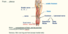
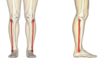
Tibia, fibula and foot
- Label the diagram


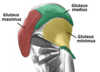
Identify the gluteal muscles


Gluteus Maximus
- Fill in the blanks


Label the Femur


Quadriceps
- Fill in the blanks


Medial compartment of thigh
- The muscles located here are all … of the thigh
- The muscles located here are all adductors of the thigh

Tibia, fibula and foot
- Label the diagram


The Pelvis
- Fill in the blanks


Movements of the thigh - Flexion at hip joint
- Performed by:
- Rectus femoris
- Sartorius
- Iliopsoas (Iliacus and psoas major) - insert into lesser trochanter
- Performed by:
- Rectus femoris
- Sartorius
- Iliopsoas (Iliacus and psoas major) - insert into lesser trochanter

Posterior compartment of thigh
- The muscles in the posterior compartment of the thigh are collectively known as the … They consist of the:
- Laterally - biceps femoris (long head from ischial tuberosity and short head from femur - form tendon inserting into fibula)
- Medially - semitendinosus and semimembranosus (origin - ischial tuberosity, insert into tibia)
- Supplied by … nerve
- Extend thigh, flex leg, medially rotate and laterally rotate
- The muscles in the posterior compartment of the thigh are collectively known as the hamstrings. They consist of the:
- Laterally - biceps femoris (long head from ischial tuberosity and short head from femur - form tendon inserting into fibula)
- Medially - semitendinosus and semimembranosus (origin - ischial tuberosity, insert into tibia)
- Supplied by sciatic nerve
- Extend thigh, flex leg, medially rotate and laterally rotate

Medial compartment of thigh: Adductors - movements
- Adductors
- …. rotation
- … of thigh (hamstring part)
- Adductors
- Medial rotation
- Extension of thigh (hamstring part)
gluteus … also supports the extended knee via … tract
gluteus maximus also supports the extended knee via iliotibial tract
Gluteus maximus is an … and laterally rotates the thigh, wheres gluteus medius and minimus … and medially rotates the thigh
Gluteus maximus is an extenser and laterally rotates the thigh, wheres gluteus medius and minimus abducts and medially rotates the thigh
Biceps Femoris
- Like the biceps brachii in the arm, the biceps femoris muscle has two heads – a long head and a short head.
- It is the most lateral of the muscles in the posterior thigh
- Attachments: The long head originates from the ischial tuberosity of the pelvis. The short head originates from the … Together, the heads form a tendon, which inserts into the head of the fibula.
- Actions: Main action is flexion at the knee. It also extends the thigh at the hip, and laterally rotates at the hip and knee.
- Innervation: Sciatic nerve
- Like the biceps brachii in the arm, the biceps femoris muscle has two heads – a long head and a short head.
- It is the most lateral of the muscles in the posterior thigh
- Attachments: The long head originates from the ischial tuberosity of the pelvis. The short head originates from the femur. Together, the heads form a tendon, which inserts into the head of the fibula.
- Actions: Main action is flexion at the knee. It also extends the thigh at the hip, and laterally rotates at the hip and knee.
- Innervation: Sciatic nerve

Femoral triangle
- Femoral artery enters anterior compartment of thigh under inguinal ligament - into femoral triangle region
- Borders - inguinal ligament, medial border of adductor longus, sartorius
- Floor - pectineus, iliopsoas, and adductor longus muscles.
- Passing through the triangle - lateral to medial - Femoral nerve - femoral artery - femoral vein - lymphatics (within the femoral canal)
- Artery, vein and femoral canal are surrounded by femoral sheath
- Femoral nerve (lateral to artery) not surrounded by femoral sheath

Adductor magnus muscle (medial compartment of thigh)
- Split into two parts: Adductor part and Hamstring part
- Adductor part: Origin - … …, inserts into linea aspera, innervation - obturator nerve
- Hamstring part: Origin - Ischial …, inserts into adductor tubercle, innervation - tibial division of sciatic nerve
- Hip joint: Thigh flexion, Thigh adduction, Thigh external rotation (adductor part), Thigh extension, Thigh internal rotation (hamstring part); Pelvis stabilization
- Split into two parts: Adductor part and Hamstring part
- Adductor part: Origin - Ischiopubic ramus, inserts into linea aspera, innervation - obturator nerve
- Hamstring part: Origin - Ischial tuberosity, inserts into adductor tubercle, innervation - tibial division of sciatic nerve
- Hip joint: Thigh flexion, Thigh adduction, Thigh external rotation (adductor part), Thigh extension, Thigh internal rotation (hamstring part); Pelvis stabilization

Femoral artery
- Femoral artery runs under … before passing through the adductor …
- Femoral artery runs under sartorius before passing through the adductor hiatus

Obturator nerve
- Fill in the blanks


Nerve supply to the Lower Limb
- Nerves innervating lower limb structures originate from either lumbar or sacral plexi
- Lumbar plexus formed from anterior primary rami (L1 to L4)
- Sacral plexus formed from anterior primary rami L4 and L5 (via lumbosacral trunk) and anterior primary rami S1 to S4
- … Nerve - L2 to L4 (lumbar plexus)
- Obturator Nerve - L2 to L4 (lumbar plexus)
- Sciatic nerve - L4-S3 (sacral plexus)
- Nerves innervating lower limb structures originate from either lumbar or sacral plexi
- Lumbar plexus formed from anterior primary rami (L1 to L4)
- Sacral plexus formed from anterior primary rami L4 and L5 (via lumbosacral trunk) and anterior primary rami S1 to S4
- Femoral Nerve - L2 to L4 (lumbar plexus)
- Obturator Nerve - L2 to L4 (lumbar plexus)
- Sciatic nerve - L4-S3 (sacral plexus)

Piriformis
- Origin: internal surface of …
- Insert: greater …
- Supplied by branches from sacral plexus
- Sciatic nerve under piriformis
- Origin: internal surface of sacrum
- Insert: greater trochanter
- Supplied by branches from sacral plexus
- Sciatic nerve under piriformis

Femoral nerve
- From … plexus (Roots …-…)
- Passes under inguinal ligament to enter anterior compartment of thigh
- Supplies quadriceps, supply sartorius and pectineus muscles
- Nerve supplies skin over anterior thigh, knee, medial side of leg and foot
- Produces longest cutaneous nerve in body - saphenous nerve - innervates skin over medial leg and foot
- From lumbar plexus (Roots L2-L4)
- Passes under inguinal ligament to enter anterior compartment of thigh
- Supplies quadriceps, supply sartorius and pectineus muscles
- Nerve supplies skin over anterior thigh, knee, medial side of leg and foot
- Produces longest cutaneous nerve in body - saphenous nerve - innervates skin over medial leg and foot

Sciatic nerve
- Fill in the blanks

Quadriceps
- Four individual muscles: Rectus femoris, vastus lateralis, vastus medialis, vastus intermedius
- Rectus femoris - extension of leg and flexion of thigh - Origin - anterior inferior iliac spine, insert into … tuberosity
- 3 Vastus muscles - extension of leg only - Origin - femur, insert into … tuberosity
- All innervated by … nerve
- Four individual muscles: Rectus femoris, vastus lateralis, vastus medialis, vastus intermedius
- Rectus femoris - extension of leg and flexion of thigh - Origin - anterior inferior iliac spine, insert into tibial tuberosity
- 3 Vastus muscles - extension of leg only - Origin - femur, insert into tibial tuberosity
- All innervated by femoral nerve

Posterior compartment of thigh - Hamstrings
- Fill in the blanks


Identify the gluteal muscles


Blood supply to the Lower Limb


Gracilis muscle (medial compartment of thigh)
- Origin - Pubis
- Inserts into …
- Innervated by obturator nerve
- Adducts thigh, … leg at knee joint
- Origin - Pubis
- Inserts into tibia
- Innervated by obturator nerve
- Adducts thigh, flexes leg at knee joint

Obturator nerve
- Fill in the blanks


The Femur
- The femur is the only bone in the thigh and the longest bone in the body.
- Proximal part - It consists of a head and neck, and two bony processes – the greater and lesser trochanters.
- The shaft of the femur descends in a slight medial direction. On the posterior surface of the femoral shaft, there are roughened ridges of bone, called the linea aspera Proximally, the medial border of the linea aspera becomes the pectineal line. The lateral border becomes the gluteal tuberosity, where the gluteus maximus attaches.
- Distal - the adductor tubercle is found distally on the femur and is formed from the termination of the medial supracondylar line.
- Medial and lateral … – rounded areas at the end of the femur.
- Medial and lateral … – bony elevations on the non-articular areas of the …
- The femur is the only bone in the thigh and the longest bone in the body.
- Proximal part - It consists of a head and neck, and two bony processes – the greater and lesser trochanters.
- The shaft of the femur descends in a slight medial direction. On the posterior surface of the femoral shaft, there are roughened ridges of bone, called the linea aspera Proximally, the medial border of the linea aspera becomes the pectineal line. The lateral border becomes the gluteal tuberosity, where the gluteus maximus attaches.
- Distal - the adductor tubercle is found distally on the femur and is formed from the termination of the medial supracondylar line.
- Medial and lateral condyles – rounded areas at the end of the femur.
- Medial and lateral epicondyles – bony elevations on the non-articular areas of the condyles.

Posterior compartment of thigh
- The muscles in the posterior compartment of the thigh are collectively known as the hamstrings. They consist of the:
- Laterally - biceps femoris (long head from ischial tuberosity and short head from femur - form tendon inserting into fibula)
- Medially - semitendinosus and semimembranosus (origin - ischial tuberosity, insert into tibia)
- Supplied by sciatic nerve
- Extend thigh, flex leg, medially rotate and laterally rotate

Adductor magnus muscle (medial compartment of thigh)
- Split into two parts: … part and … part
- … part: Origin - Ischiopubic ramus, inserts into linea aspera, innervation - obturator nerve
- … part: Origin - Ischial tuberosity, inserts into adductor tubercle, innervation - tibial division of sciatic nerve
- Hip joint: Thigh flexion, Thigh adduction, Thigh external rotation (… part), Thigh extension, Thigh internal rotation (… part); Pelvis stabilization
- Split into two parts: Adductor part and Hamstring part
- Adductor part: Origin - Ischiopubic ramus, inserts into linea aspera, innervation - obturator nerve
- Hamstring part: Origin - Ischial tuberosity, inserts into adductor tubercle, innervation - tibial division of sciatic nerve
- Hip joint: Thigh flexion, Thigh adduction, Thigh external rotation (adductor part), Thigh extension, Thigh internal rotation (hamstring part); Pelvis stabilization

Posterior surface of the distal right femur - label


Great saphenous vein
- Longest vein in body
- Drains … side of arch
- … side of limb
- Drains into femoral vein in femoral triangle
- Longest vein in body
- Drains medial side of arch
- Medial side of limb
- Drains into femoral vein in femoral

Small muscles of the gluteal region
- Equivalent to rotator cuff
- Stabilize …
- … rotation of thigh
- Origin: sacrum (piriformis) and ischium/ischiopubic ramus
- Inserts into: Greater Trochanter
- Equivalent to rotator cuff
- Stabilize hip
- Lateral rotation of thigh
- Origin: sacrum (piriformis) and ischium/ischiopubic ramus
- Inserts into: Greater Trochanter

Obturator nerve
- Fill in the blanks


Gluteus Maximus
- The gluteus maximus is the largest of the gluteal muscles. It is also the most superficial, producing the shape of the buttocks.
- Attachments: Originates from the … It slopes across the buttock at a 45 degree angle, then inserts into the … tract and the gluteal tuberosity of the femur.
- Actions: It is the main extensor of the thigh, and assists with lateral rotation.
- Innervation: Inferior gluteal nerve.
- The gluteus maximus is the largest of the gluteal muscles. It is also the most superficial, producing the shape of the buttocks.
- Attachments: Originates from the ilium. It slopes across the buttock at a 45 degree angle, then inserts into the iliotibial tract and the gluteal tuberosity of the femur.
- Actions: It is the main extensor of the thigh, and assists with lateral rotation.
- Innervation: Inferior gluteal nerve.

Functional compartments of the Lower Limb
- Fill in the blanks

The muscles that form the quadriceps femoris unite proximal to the knee and attach to the patella via the quadriceps tendon. In turn, the patella is attached to the tibia by the patella ligament. The quadriceps femoris is the main … of the knee.
The muscles that form the quadriceps femoris unite proximal to the knee and attach to the patella via the quadriceps tendon. In turn, the patella is attached to the tibia by the patella ligament. The quadriceps femoris is the main extensor of the knee.

Lower limb comparison with upper limb
- Lower limbs closer together / under trunk
- Extensors are …
- Flexors are …
- Medial rotation during development brings posterior compartment anterior
- Lower limbs closer together / under trunk
- Extensors are anterior
- Flexors are posterior
- Medial rotation during development brings posterior compartment anterior

Gluteal muscles
- Three major muscles:
- Gluteus … - Extension + lateral rotation of thigh
- Gluteus … - Abduction + medial rotation of thigh
- Gluteus … - Abduction + medial rotation of thigh
- Gluteus maximus also supports the extended knee
- Via … tract
- Important muscles for locomotion
- Three major muscles:
- Gluteus maximus - Extension + lateral rotation of thigh
- Gluteus medius - Abduction + medial rotation of thigh
- Gluteus minimus - Abduction + medial rotation of thigh
- Gluteus maximus also supports the extended knee
- Via iliotibial tract
- Important muscles for locomotion
Pectineus muscle (medial compartment of thigh)
- Origin: Pubis
- Inserts into … …
- Supplied by femoral nerve
- Hip joint: Thigh flexion, Thigh adduction, Thigh external rotation, Thigh internal rotation; Pelvis stabilization
- Origin: Pubis
- Inserts into linea aspera
- Supplied by femoral nerve
- Hip joint: Thigh flexion, Thigh adduction, Thigh external rotation, Thigh internal rotation; Pelvis stabilization

… muscle (medial compartment of thigh)
- Origin: Pubis
- Inserts into linea aspera
- Supplied by femoral nerve
- Hip joint: Thigh flexion, Thigh adduction, Thigh external rotation, Thigh internal rotation; Pelvis stabilization
Pectineus muscle (medial compartment of thigh)
- Origin: Pubis
- Inserts into linea aspera
- Supplied by femoral nerve
- Hip joint: Thigh flexion, Thigh adduction, Thigh external rotation, Thigh internal rotation; Pelvis stabilization

Femoral nerve
- From lumbar plexus (Roots L2-L4)
- Passes under inguinal ligament to enter anterior compartment of thigh
- Supplies quadriceps, supply sartorius and pectineus muscles
- Nerve supplies skin over anterior thigh, knee, medial side of leg and foot
- Produces longest cutaneous nerve in body - saphenous nerve - innervates skin over …
- From lumbar plexus (Roots L2-L4)
- Passes under inguinal ligament to enter anterior compartment of thigh
- Supplies quadriceps, supply sartorius and pectineus muscles
- Nerve supplies skin over anterior thigh, knee, medial side of leg and foot
- Produces longest cutaneous nerve in body - saphenous nerve - innervates skin over medial leg and foot

Biceps Femoris
- Like the biceps brachii in the arm, the biceps femoris muscle has two heads – a long head and a short head.
- It is the most lateral of the muscles in the posterior thigh
- Attachments: The long head originates from the … … of the pelvis. The short head originates from the femur. Together, the heads form a tendon, which inserts into the head of the fibula.
- Actions: Main action is flexion at the knee. It also extends the thigh at the hip, and laterally rotates at the hip and knee.
- Innervation: … nerve
- Like the biceps brachii in the arm, the biceps femoris muscle has two heads – a long head and a short head.
- It is the most lateral of the muscles in the posterior thigh
- Attachments: The long head originates from the ischial tuberosity of the pelvis. The short head originates from the femur. Together, the heads form a tendon, which inserts into the head of the fibula.
- Actions: Main action is flexion at the knee. It also extends the thigh at the hip, and laterally rotates at the hip and knee.
- Innervation: Sciatic nerve

Blood supply to the Lower Limb


Pectineus muscle (medial compartment of thigh)
- Origin: …
- Inserts into linea aspera
- Supplied by femoral nerve
- Hip joint: Thigh flexion, Thigh adduction, Thigh external rotation, Thigh internal rotation; Pelvis stabilization
- Origin: Pubis
- Inserts into linea aspera
- Supplied by femoral nerve
- Hip joint: Thigh flexion, Thigh adduction, Thigh external rotation, Thigh internal rotation; Pelvis stabilization

The medial malleolus is the prominence on the inner side of the ankle, formed by the lower end of the … The lateral malleolus is the prominence on the outer side of the ankle, formed by the lower end of the …
The medial malleolus is the prominence on the inner side of the ankle, formed by the lower end of the tibia. The lateral malleolus is the prominence on the outer side of the ankle, formed by the lower end of the fibula.

Posterior compartment of thigh - Hamstrings
- Fill in the blanks


Adductor longus muscle (medial compartment of thigh)
- Origin: pubis
- Inserts into linea aspera
- Innervated by … nerve
- Hip joint: Thigh flexion, Thigh adduction, Thigh external rotation; Pelvis stabilization
- Origin: pubis
- Inserts into linea aspera
- Innervated by obturator nerve
- Hip joint: Thigh flexion, Thigh adduction, Thigh external rotation; Pelvis stabilization

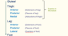
What bone is this?
Fibula
The Femur
- The femur is the only bone in the thigh and the longest bone in the body.
- Proximal part - It consists of a head and neck, and two bony processes – the greater and lesser trochanters.
- The shaft of the femur descends in a slight medial direction. On the posterior surface of the femoral shaft, there are roughened ridges of bone, called the linea aspera. Proximally, the medial border of the linea aspera becomes the pectineal line. The lateral border becomes the … tuberosity, where the … … attaches.
- Distal - the adductor tubercle is found distally on the femur and is formed from the termination of the medial supracondylar line.
- Medial and lateral condyles – rounded areas at the end of the femur.
- Medial and lateral … – bony elevations on the non-articular areas of the condyles.
- The femur is the only bone in the thigh and the longest bone in the body.
- Proximal part - It consists of a head and neck, and two bony processes – the greater and lesser trochanters.
- The shaft of the femur descends in a slight medial direction. On the posterior surface of the femoral shaft, there are roughened ridges of bone, called the linea aspera. Proximally, the medial border of the linea aspera becomes the pectineal line. The lateral border becomes the gluteal tuberosity, where the gluteus maximus attaches.
- Distal - the adductor tubercle is found distally on the femur and is formed from the termination of the medial supracondylar line.
- Medial and lateral condyles – rounded areas at the end of the femur.
- Medial and lateral epicondyles – bony elevations on the non-articular areas of the condyles.

Adductor longus muscle (medial compartment of thigh)
- Origin: pubis
- Inserts into … …
- Innervated by obturator nerve
- Hip joint: Thigh flexion, Thigh adduction, Thigh external rotation; Pelvis stabilization
- Origin: pubis
- Inserts into linea aspera
- Innervated by obturator nerve
- Hip joint: Thigh flexion, Thigh adduction, Thigh external rotation; Pelvis stabilization

Gluteus medius and minimus
- Fill in the blanks


The adductor … gap between the adductor magnus muscle and the femur that allows the passage of the … vessels from the anterior thigh to the posterior thigh and then the popliteal fossa
The adductor hiatus gap between the adductor magnus muscle and the femur that allows the passage of the femoral vessels from the anterior thigh to the posterior thigh and then the popliteal fossa

The muscles that form the quadriceps femoris unite proximal to the knee and attach to the … via the quadriceps tendon. In turn, the … is attached to the tibia by the … ligament. The quadriceps femoris is the main extensor of the knee.
The muscles that form the quadriceps femoris unite proximal to the knee and attach to the patella via the quadriceps tendon. In turn, the patella is attached to the tibia by the patella ligament. The quadriceps femoris is the main extensor of the knee.

Gluteus medius and minimus
- Fill in the blanks


Movements of the thigh - Medial and Lateral Rotation
- Performed by:
- … muscles
- H…
- A…
- Short …
- Performed by:
- Gluteal muscles
- Hamstrings
- Adductors
- Short rotators

Quadriceps
- Four individual muscles: Rectus femoris, vastus lateralis, vastus medialis, vastus intermedius
- Rectus femoris - … of leg and … of thigh - Origin - anterior inferior iliac spine, insert into tibial tuberosity
- 3 Vastus muscles - … of leg only - Origin - femur, insert into tibial tuberosity
- All innervated by femoral nerve
- Four individual muscles: Rectus femoris, vastus lateralis, vastus medialis, vastus intermedius
- Rectus femoris - extension of leg and flexion of thigh - Origin - anterior inferior iliac spine, insert into tibial tuberosity
- 3 Vastus muscles - extension of leg only - Origin - femur, insert into tibial tuberosity
- All innervated by femoral nerve

Pectineus muscle (medial compartment of thigh)
- Origin: Pubis
- Inserts into linea aspera
- Supplied by femoral nerve
- Hip joint: Thigh flexion, Thigh adduction, Thigh external rotation, Thigh internal rotation; Pelvis stabilization
- Origin: Pubis
- Inserts into linea aspera
- Supplied by femoral nerve
- Hip joint: Thigh flexion, Thigh adduction, Thigh external rotation, Thigh internal rotation; Pelvis stabilization

Skeleton (Lower limb)
- Label the diagram


Gluteus Maximus
- Fill in the blanks

Label the Femur


Joints and movements of the Lower Limb
- What joints are there? (4) What movements can be done?
- Hip joint - ball and socket joint - extend,flex, abduct, adduct, circumduct, medially and laterally rotate
- Knee joint - hinge joint - extend, flex, lateral and medial rotate
- Ankle joint - Dorsiflexion (or extension) and plantarflexion (or flexion) of foot at ankle joint
- Joints of the foot - Inversion, eversion, extension, flexion, supination, pronation

Gluteal muscles
- Three major muscles:
- Gluteus … - Extension + lateral rotation of thigh
- Gluteus … - Abduction + medial rotation of thigh
- Gluteus … - Abduction + medial rotation of thigh
- Gluteus maximus also supports the extended knee
- Via iliotibial tract
- Important muscles for locomotion
- Three major muscles:
- Gluteus maximus - Extension + lateral rotation of thigh
- Gluteus medius - Abduction + medial rotation of thigh
- Gluteus minimus - Abduction + medial rotation of thigh
- Gluteus maximus also supports the extended knee
- Via iliotibial tract
- Important muscles for locomotion

Biceps Femoris
- Like the biceps brachii in the arm, the biceps femoris muscle has two heads – a long head and a short head.
- It is the most …. of the muscles in the posterior thigh
- Attachments: The long head originates from the ischial tuberosity of the pelvis. The short head originates from the femur. Together, the heads form a tendon, which inserts into the head of the fibula.
- Actions: Main action is flexion at the knee. It also extends the thigh at the hip, and laterally rotates at the hip and knee.
- Innervation: Sciatic nerve
- Like the biceps brachii in the arm, the biceps femoris muscle has two heads – a long head and a short head.
- It is the most lateral of the muscles in the posterior thigh
- Attachments: The long head originates from the ischial tuberosity of the pelvis. The short head originates from the femur. Together, the heads form a tendon, which inserts into the head of the fibula.
- Actions: Main action is flexion at the knee. It also extends the thigh at the hip, and laterally rotates at the hip and knee.
- Innervation: Sciatic nerve

Adductor … muscle (medial compartment of thigh)
- Split into two parts: Adductor part and Hamstring part
- Adductor part: Origin - Ischiopubic ramus, inserts into linea aspera, innervation - obturator nerve
- Hamstring part: Origin - Ischial tuberosity, inserts into adductor tubercle, innervation - tibial division of sciatic nerve
- Hip joint: Thigh flexion, Thigh adduction, Thigh external rotation (adductor part), Thigh extension, Thigh internal rotation (hamstring part); Pelvis stabilization
Adductor magnus muscle (medial compartment of thigh)
- Split into two parts: Adductor part and Hamstring part
- Adductor part: Origin - Ischiopubic ramus, inserts into linea aspera, innervation - obturator nerve
- Hamstring part: Origin - Ischial tuberosity, inserts into adductor tubercle, innervation - tibial division of sciatic nerve
- Hip joint: Thigh flexion, Thigh adduction, Thigh external rotation (adductor part), Thigh extension, Thigh internal rotation (hamstring part); Pelvis stabilization

Clinically, the femoral triangle region is important. Femoral … (femoral canal), … (insert stent or balloon into artery), femoral nerve …, venepuncture
Clinically, the femoral triangle region is important. Femoral hernias (femoral canal), angioplasty (insert stent or balloon into artery), femoral nerve block, venepuncture

Adductor magnus muscle (medial compartment of thigh)
- Split into two parts: Adductor part and Hamstring part
- Adductor part: Origin - Ischiopubic ramus, inserts into linea aspera, innervation - … nerve
- Hamstring part: Origin - Ischial tuberosity, inserts into adductor tubercle, innervation - tibial division of … nerve
- Hip joint: Thigh flexion, Thigh adduction, Thigh external rotation (adductor part), Thigh extension, Thigh internal rotation (hamstring part); Pelvis stabilization
- Split into two parts: Adductor part and Hamstring part
- Adductor part: Origin - Ischiopubic ramus, inserts into linea aspera, innervation - obturator nerve
- Hamstring part: Origin - Ischial tuberosity, inserts into adductor tubercle, innervation - tibial division of sciatic nerve
- Hip joint: Thigh flexion, Thigh adduction, Thigh external rotation (adductor part), Thigh extension, Thigh internal rotation (hamstring part); Pelvis stabilization

The femoral artery, vein and canal are contained within a fascial compartment – known as the femoral …
The femoral artery, vein and canal are contained within a fascial compartment – known as the femoral sheath.

Piriformis
- Origin: internal surface of sacrum
- Insert: greater trochanter
- Supplied by branches from sacral plexus
- … nerve under piriformis
- Origin: internal surface of sacrum
- Insert: greater trochanter
- Supplied by branches from sacral plexus
- Sciatic nerve under piriformis

Movements of the thigh - Adduction
- Performed by:
- Adductor …
- Adductor …
- Adductor part of adductor …
- P..
- G…
- Performed by:
- Adductor longus
- Adductor brevis
- Adductor part of adductor magnus
- Pectineus
- Gracilis

Functional compartments of the Lower Limb
- Fill in the blanks


Tibia, fibula and foot
- Label the diagram


Quadriceps
- Fill in the blanks


Popliteal vessels
- Femoral … enters the popliteal … as the popliteal artery
- Femoral artery enters the popliteal fossa as the popliteal artery

Quadriceps
- Four individual muscles: Rectus femoris, vastus lateralis, vastus medialis, vastus intermedius
- Rectus femoris - extension of leg and flexion of thigh - Origin - anterior inferior iliac spine, insert into … …
- 3 Vastus muscles - extension of leg only - Origin - femur, insert into … …
- All innervated by femoral nerve
- Four individual muscles: Rectus femoris, vastus lateralis, vastus medialis, vastus intermedius
- Rectus femoris - extension of leg and flexion of thigh - Origin - anterior inferior iliac spine, insert into tibial tuberosity
- 3 Vastus muscles - extension of leg only - Origin - femur, insert into tibial tuberosity
- All innervated by femoral nerve
Blood supply to the Lower Limb


Label the Femur


Movements of the thigh - Extension
- Performed by:
- Gluteus …
- H…
- Hamstring part of adductor …
- Performed by:
- Gluteus maximus
- Hamstrings
- Hamstring part of adductor magnus

Femoral nerve
- Fill in the blanks


Sciatic nerve
- Fill in the blanks


The muscles that form the quadriceps femoris unite proximal to the knee and attach to the patella via the quadriceps … In turn, the patella is attached to the tibia by the patella ligament. The quadriceps femoris is the main extensor of the knee.
The muscles that form the quadriceps femoris unite proximal to the knee and attach to the patella via the quadriceps tendon. In turn, the patella is attached to the tibia by the patella ligament. The quadriceps femoris is the main extensor of the knee.

Blood supply to the Lower Limb


Tibial arteries
- …. artery divides in leg as anterior/posterior tibial arteries
- Popliteal artery divides in leg as anterior/posterior tibial arteries

Movements of the thigh - Flexion at hip joint
- Performed by:
- … femoris
- S…
- Iliopsoas (Iliacus and psoas major) - insert into lesser trochanter
- Performed by:
- Rectus femoris
- Sartorius
- Iliopsoas (Iliacus and psoas major) - insert into lesser trochanter

Femoral triangle
- Fill in the blanks


The Femur
- The femur is the only bone in the … and the … bone in the body.
- Proximal part - It consists of a head and neck, and two bony processes – the greater and lesser trochanters.
- The shaft of the femur descends in a slight medial direction. On the posterior surface of the femoral shaft, there are roughened ridges of bone, called the linea aspera Proximally, the medial border of the linea aspera becomes the pectineal line. The lateral border becomes the gluteal tuberosity, where the gluteus maximus attaches.
- Distal - the adductor tubercle is found distally on the femur and is formed from the termination of the medial supracondylar line.
- Medial and lateral condyles – rounded areas at the end of the femur.
- Medial and lateral epicondyles – bony elevations on the non-articular areas of the condyles.
- The femur is the only bone in the thigh and the longest bone in the body.
- Proximal part - It consists of a head and neck, and two bony processes – the greater and lesser trochanters.
- The shaft of the femur descends in a slight medial direction. On the posterior surface of the femoral shaft, there are roughened ridges of bone, called the linea aspera Proximally, the medial border of the linea aspera becomes the pectineal line. The lateral border becomes the gluteal tuberosity, where the gluteus maximus attaches.
- Distal - the adductor tubercle is found distally on the femur and is formed from the termination of the medial supracondylar line.
- Medial and lateral condyles – rounded areas at the end of the femur.
- Medial and lateral epicondyles – bony elevations on the non-articular areas of the condyles.

Gracilis muscle (medial compartment of thigh)
- Origin - …
- Inserts into tibia
- Innervated by obturator nerve
- Adducts thigh, flexes leg at knee joint
- Origin - Pubis
- Inserts into tibia
- Innervated by obturator nerve
- Adducts thigh, flexes leg at knee joint
Adductor longus muscle (medial compartment of thigh)
- Origin: …
- Inserts into linea aspera
- Innervated by … nerve
- Hip joint: Thigh flexion, Thigh adduction, Thigh external rotation; Pelvis stabilization
- Origin: pubis
- Inserts into linea aspera
- Innervated by obturator nerve
- Hip joint: Thigh flexion, Thigh adduction, Thigh external rotation; Pelvis stabilization

The Pelvis
- Fill in the blanks


Gracilis muscle (medial compartment of thigh)
- Origin - …
- Inserts into tibia
- Innervated by … nerve
- Adducts thigh, flexes leg at knee joint
- Origin - Pubis
- Inserts into tibia
- Innervated by obturator nerve
- Adducts thigh, flexes leg at knee joint

Quadriceps
- Four individual muscles: Rectus …, vastus …, vastus …, vastus …
- Rectus … - extension of leg and flexion of thigh - Origin - anterior inferior iliac spine, insert into tibial tuberosity
- 3 Vastus muscles - extension of leg only - Origin - femur, insert into tibial tuberosity
- All innervated by femoral nerve
- Four individual muscles: Rectus femoris, vastus lateralis, vastus medialis, vastus intermedius
- Rectus femoris - extension of leg and flexion of thigh - Origin - anterior inferior iliac spine, insert into tibial tuberosity
- 3 Vastus muscles - extension of leg only - Origin - femur, insert into tibial tuberosity
- All innervated by femoral nerve

Movements of the thigh - Flexion at hip joint
- Performed by:
- Rectus femoris
- Sartorius
- Iliopsoas (Iliacus and psoas major) - insert into … …
- Performed by:
- Rectus femoris
- Sartorius
- Iliopsoas (Iliacus and psoas major) - insert into lesser trochanter

Quadriceps
- Four individual muscles: Rectus femoris, vastus lateralis, vastus medialis, vastus intermedius
- Rectus femoris - extension of leg and flexion of thigh - Origin - … … … spine, insert into tibial tuberosity
- 3 Vastus muscles - extension of leg only - Origin - …, insert into tibial tuberosity
- All innervated by femoral nerve
- Four individual muscles: Rectus femoris, vastus lateralis, vastus medialis, vastus intermedius
- Rectus femoris - extension of leg and flexion of thigh - Origin - anterior inferior iliac spine, insert into tibial tuberosity
- 3 Vastus muscles - extension of leg only - Origin - femur, insert into tibial tuberosity
- All innervated by femoral nerve

Functional compartments of the Lower Limb
- Fill in the blanks


The … is the longest muscle in the body. It is long and thin, running across the thigh in a inferomedial direction. The … is positioned more superficially than the other muscles in the leg. (Origin - anterior superior iliac spine, insert into tibia)
The sartorius is the longest muscle in the body. It is long and thin, running across the thigh in a inferomedial direction. The sartorius is positioned more superficially than the other muscles in the leg. (Origin - anterior superior iliac spine, insert into tibia)

Femoral triangle
- Fill in the blanks


Clinically, the … … region is important. Femoral hernias (femoral canal), angioplasty (insert stent or balloon into artery), femoral nerve block, venepuncture
Clinically, the femoral triangle region is important. Femoral hernias (femoral canal), angioplasty (insert stent or balloon into artery), femoral nerve block, venepuncture

The sartorius is the … muscle in the body. It is long and thin, running across the thigh in a inferomedial direction. The sartorius is positioned more … than the other muscles in the leg. (Origin - anterior superior iliac spine, insert into tibia)
The sartorius is the longest muscle in the body. It is long and thin, running across the thigh in a inferomedial direction. The sartorius is positioned more superficially than the other muscles in the leg. (Origin - anterior superior iliac spine, insert into tibia)

Nerve supply to the Lower Limb
- Nerves innervating lower limb structures originate from either lumbar or sacral plexi
- Lumbar plexus formed from anterior primary rami (L1 to L4)
- Sacral plexus formed from anterior primary rami L4 and L5 (via lumbosacral trunk) and anterior primary rami S1 to S4
- Femoral Nerve - L2 to L4 (lumbar plexus)
- Obturator Nerve - L2 to L4 (lumbar plexus)
- … nerve - L4-S3 (sacral plexus)
- Nerves innervating lower limb structures originate from either lumbar or sacral plexi
- Lumbar plexus formed from anterior primary rami (L1 to L4)
- Sacral plexus formed from anterior primary rami L4 and L5 (via lumbosacral trunk) and anterior primary rami S1 to S4
- Femoral Nerve - L2 to L4 (lumbar plexus)
- Obturator Nerve - L2 to L4 (lumbar plexus)
- Sciatic nerve - L4-S3 (sacral plexus)

Posterior compartment of thigh
- The muscles in the posterior compartment of the thigh are collectively known as the hamstrings. They consist of the:
- Laterally - biceps femoris (long head from ischial tuberosity and short head from femur - form tendon inserting into fibula)
- Medially - semitendinosus and semimembranosus (origin - ischial tuberosity, insert into tibia)
- Supplied by … nerve
- Extend thigh, flex leg, medially rotate and laterally rotate
- The muscles in the posterior compartment of the thigh are collectively known as the hamstrings. They consist of the:
- Laterally - biceps femoris (long head from ischial tuberosity and short head from femur - form tendon inserting into fibula)
- Medially - semitendinosus and semimembranosus (origin - ischial tuberosity, insert into tibia)
- Supplied by sciatic nerve
- Extend thigh, flex leg, medially rotate and laterally rotate

The Pelvis
- Fill in the blanks


Motor Functions of Sciatic Nerve
- The sciatic nerve innervates the muscles in the posterior compartment of the thigh and the hamstring portion of the adductor magnus.
- The sciatic nerve also indirectly innervates several other muscles, via its two terminal branches:
- … nerve – the muscles of the posterior leg (calf muscles), and some of the intrinsic muscles of the foot.
- … … nerve – the muscles of the anterior leg, lateral leg, and the remaining intrinsic foot muscles.
- Overall - innervates muscles of the posterior thigh, entire leg and entire foot.
- The sciatic nerve innervates the muscles in the posterior compartment of the thigh and the hamstring portion of the adductor magnus.
- The sciatic nerve also indirectly innervates several other muscles, via its two terminal branches:
- Tibial nerve – the muscles of the posterior leg (calf muscles), and some of the intrinsic muscles of the foot.
- Common fibular nerve – the muscles of the anterior leg, lateral leg, and the remaining intrinsic foot muscles.
- Overall - innervates muscles of the posterior thigh, entire leg and entire foot.

Sciatic nerve
- Fill in the blanks


Gluteal muscles
- Three major muscles:
- Gluteus maximus - … + lateral rotation of thigh
- Gluteus medius - … + medial rotation of thigh
- Gluteus minimus - … + medial rotation of thigh
- Gluteus maximus also supports the extended knee
- Via iliotibial tract
- Important muscles for locomotion
- Three major muscles:
- Gluteus maximus - Extension + lateral rotation of thigh
- Gluteus medius - Abduction + medial rotation of thigh
- Gluteus minimus - Abduction + medial rotation of thigh
- Gluteus maximus also supports the extended knee
- Via iliotibial tract
- Important muscles for locomotion

What muscle is this?

Sartorius (origin - anterior superior iliac spine, inserts into tibia, supplied by femoral nerve)
Obturator nerve
- Fill in the blanks


Pectineus muscle (medial compartment of thigh)
- Origin: Pubis
- Inserts into linea aspera
- Supplied by femoral nerve
- Hip joint: Thigh flexion, Thigh adduction, Thigh external rotation, Thigh internal rotation; Pelvis stabilization
- Origin: Pubis
- Inserts into linea aspera
- Supplied by femoral nerve
- Hip joint: Thigh flexion, Thigh adduction, Thigh external rotation, Thigh internal rotation; Pelvis stabilization

Gracilis muscle (medial compartment of thigh)
- Origin - Pubis
- Inserts into …
- Innervated by obturator nerve
- Adducts thigh, flexes leg at knee joint
- Origin - Pubis
- Inserts into tibia
- Innervated by obturator nerve
- Adducts thigh, flexes leg at knee joint

Gluteus medius and minimus
- Fill in the blanks


Short saphenous vein
- Drains … side of arch
- Pierces deep popliteal fascia
- Drains into popliteal vein
- Drains lateral side of arch
- Pierces deep popliteal fascia
- Drains into popliteal vein

Label the Femur


Gluteal muscles
- Three major muscles:
- Gluteus maximus - Extension + … rotation of thigh
- Gluteus medius - Abduction + … rotation of thigh
- Gluteus minimus - Abduction + … rotation of thigh
- Gluteus maximus also supports the extended knee
- Via iliotibial tract
- Important muscles for …
- Three major muscles:
- Gluteus maximus - Extension + lateral rotation of thigh
- Gluteus medius - Abduction + medial rotation of thigh
- Gluteus minimus - Abduction + medial rotation of thigh
- Gluteus maximus also supports the extended knee
- Via iliotibial tract
- Important muscles for locomotion

What bone is shown here?

Tibia
Posterior compartment of thigh
- The muscles in the posterior compartment of the thigh are collectively known as the … They consist of the:
- Laterally - biceps femoris (long head from ischial tuberosity and short head from femur - form tendon inserting into fibula)
- Medially - semitendinosus and semimembranosus (origin - ischial tuberosity, insert into tibia)
- Supplied by sciatic nerve
- Extend thigh, flex leg, medially rotate and laterally rotate
- The muscles in the posterior compartment of the thigh are collectively known as the hamstrings. They consist of the:
- Laterally - biceps femoris (long head from ischial tuberosity and short head from femur - form tendon inserting into fibula)
- Medially - semitendinosus and semimembranosus (origin - ischial tuberosity, insert into tibia)
- Supplied by sciatic nerve
- Extend thigh, flex leg, medially rotate and laterally rotate

Adductor magnus muscle (medial compartment of thigh)
- Split into two parts: Adductor part and Hamstring part
- Adductor part: Origin - Ischiopubic ramus, inserts into … …, innervation - obturator nerve
- Hamstring part: Origin - Ischial tuberosity, inserts into … tubercle, innervation - tibial division of sciatic nerve
- Hip joint: Thigh flexion, Thigh adduction, Thigh external rotation (adductor part), Thigh extension, Thigh internal rotation (hamstring part); Pelvis stabilization
- Split into two parts: Adductor part and Hamstring part
- Adductor part: Origin - Ischiopubic ramus, inserts into linea aspera, innervation - obturator nerve
- Hamstring part: Origin - Ischial tuberosity, inserts into adductor tubercle, innervation - tibial division of sciatic nerve
- Hip joint: Thigh flexion, Thigh adduction, Thigh external rotation (adductor part), Thigh extension, Thigh internal rotation (hamstring part); Pelvis stabilization

Lower limb comparison with upper limb
- Lower limbs closer together / under trunk
- …. are anterior
- …. are posterior
- Medial rotation during development brings posterior compartment anterior
- Lower limbs closer together / under trunk
- Extensors are anterior
- Flexors are posterior
- Medial rotation during development brings posterior compartment anterior

Adductor brevis muscle (medial compartment of thigh)
- Origin: Pubis
- Inserts into linea aspera
- Innervated by … nerve
- Hip joint: Thigh flexion, Thigh adduction, Thigh external rotation; Pelvis stabilization
- Origin: Pubis
- Inserts into linea aspera
- Innervated by obturator nerve
- Hip joint: Thigh flexion, Thigh adduction, Thigh external rotation; Pelvis stabilization

Quadriceps
- Fill in the blanks


Obturator nerve
- Fill in the blanks


Adductor longus muscle (medial compartment of thigh)
- Origin: pubis
- Inserts into linea aspera
- Innervated by obturator nerve
- Hip joint: Thigh flexion, Thigh adduction, Thigh external rotation; Pelvis stabilization
- Origin: pubis
- Inserts into linea aspera
- Innervated by obturator nerve
- Hip joint: Thigh flexion, Thigh adduction, Thigh external rotation; Pelvis stabilization
