Pathology Flashcards
(96 cards)
Elevated CRP levels indicates what?
What does CRP stand for?
Acute inflammation
C reactive protein
What does Increased eosinophils indicate?
allergy and parasitic infections
How is haematocrit calculated?
volume RBC/volume blood
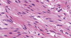
What type of tissue is this?

Smooth muscle
What does haematoxylin bind to?
Acidic or anionic structures
What type of ttissue is this?

Skeletal muscle
What colour does eosin stain?
Pink/orange
What is type III collagen also known as?
Reticulin
What is the function of myoepithelial cells?
surround some exocrine glands to squeeze out contents
What type of tissue is this?

Cardiac muscle
What is the maximum resolving power of a light microscope and of an electron microscope?
- 2 um
- 2 nm
What are simple squamous epithelia specialised for?
Diffusion and protection from abrasion

A = Z disc
B = myosin = thick filament
C = actin = thin filament
D = Sarcomere
What type of tissue is this?

Cardiac muscle
In which 4 locations are simple squamous epithelia found?
Endothelium,
mesothelium,
alveoli,
glomerulus
What type of tissue is this?

Cardiac muscle
What type of tissue is this?

Cardiac muscle
What are simple columnar epithelia specialised for?
secretion and absorption
Pink on an H and E slide indicates what kind of compound?
Cationic and eosinophilic
Between which 2 layers is the basement membrane found?
Epithelium and underlying connective tissue
What type of tissue is this?

Skeletal muscle
What are the 3 types of fibre in connective tissue?
Collagen
Elastin
Reticulin
What is the function of myofibroblasts?
pull together damaged connective tissue to promote wound healing.
Blue on an H and E slide indicates what kind of compound?
Acidic or anionic