Peripheral Blood Morphology Flashcards
(40 cards)
the area of the slide that you would exam on a peripheral smear
from the monolayer to the feathered edge
RBC should be the size of a _______ nucleus
lymphocyte
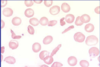
the ares of the central pallor in a RBC should be ___ of the total RBC diameter
1/3
anisocytosis
refers to the red cells which vary widely in size
RDW
red cell distribution width; measures the range of red cell sizes ; measures anisocytosis
microcytosis
refers to RBC that are small
MCV
mean cell volume; measures the individuals volume of red blood cells; measures microcytosis and macrocytosis
differential diagnosis for microcytosis (6)
- Iron deficiency 2. Anemia of chronic disease 3. Thalassemia 4. Hemoglobin C disease 5. Lead Poisoning 6. Sideroblastic anemia
macrocytosis
RBC are too large; use MCV
differential diagnosis of macrocytosis (8)
- B12/FOLATE DEFICIENCY 2. Liver disease 3. Thyroid disease 4. MDS myelodysplastic syndrome 5. Anti-retrovirals 6. Aplastic anemia 7. Chemotherapy 8. Elevated reticulocyte count
hypochromasia
RBC with too little hemoglobin
hypochromasia RBC have a central pallor that is less/more than 1/3 the total red cell diameter
more
hypochromasia is measured by
MCH mean cell hemoglobin
polychromasia
RBC that have more of a blueish tinge; probably reticulocytes
poikilocytosis
RBC that vary in shape
target cells
look like bulls-eyes
differential diagnosis of target cell (4)
- liver disease (most common) 2. thalassemias 3. hemoglobin C 4. After a splenectomy
spherocytes
have loss of central pallor
Spherocytes can be seen in
- autoimmune hemolysis 2. hereditary spherocytosis
schistocytes
red cell fragments with sharp edges
schistocytes are hallmark of
microangiopathic hemolytic anemia (MAHA)
sickle cell
seen in sickle cell anemia
echinocytes
- aka burr cell - small, regular projections - seen in renal disease
acanthocytes
- aka spur cells - large, irregular projections - seen in liver disease