Respiratory system Flashcards

1
Q

What are the functions of the respiratory organs?

A
  • gas exchange
  • protection of resp. apparatus
  • phonation
  • olfactory perception
2
Q

Which organs belong to the upper, which to the lower airways?

A

upper airways: head → larynx

  • nasal cavities
  • paranasal sinuses
  • pharynx

lower airways: larynx → pleural cavity

  • larynx
  • trachea
  • bronchial tree (lung)
3
Q

Which bones and cartilages form the skeletal framework of the external nose?

A

BONES:

  • 2 ossa nasalia
  • 2 procc. frontales maxillae

⇒ apertura piriformis

CARTILAGES:

  • 2 procc. laterales ⇒ dorsum
  • parts of nasal septum
  • cartilago alaris major (on each side)
    • ⇒ crus laterale/crus mediale
      ⇒ apex nasi
  • 3-4 cartilagines alares minores
4
Q

How do you call the hairs inside the nose?

A

vibrissae

5
Q

Which vessels supply, drain the external nose, resp.?

Which nerves are responsible for the sensory and motor innervation?

A

supply:

  • a. facialis → a. angularis
  • a. ophtalmica → a. dorsalis nasi
  • a. maxillaris → a. infraorbitalis

drainage:

  • v. facialis → v. ophtalmica superior

innervation: (cf. supply)

  • sensory: n. opthalmicus, n. maxillaris
  • motor: n. facialis via rr. buccales
6
Q

What may cause a venous sinus thrombosis and how?

A

vv. draining into v. facialis/ophtalmica anastomose btw medial angle of the eye and the root of the nose

in case of inflammation involving lat. part of face + external nose bacteria can reach deep venous sinuses → venous sinus thrombosis

7
Q

1 - 5

A

1) sinus frontalis
2) os nasale
3) proc. lateralis
4) cartilago alaris major, crus laterale
5) limen nasi

8
Q

6 - 10

A

6) vestibulum nasi
7) concha nasalis media
8) cellula ethmoidalis post.
9) concha nasalis sup.
10) sinus sphenoidalis

9
Q

11 - 17

A

11) n. nasopalatinus
12) concha nasalis inferior
13) maxilla
14) os frontale
15) tonsilla pharyngea
16) palatum molle
17) palatum durum

10
Q

Label the access routes to the 3 nasal meatus.

Which structure is located posteriorly behind the superior nasal meatus?

A

olfactory organ

  • dark blue**: sinus sphenoidalis

meatus nasi superior

  • yellow: post. ethmoidal air cells

meatus nasi medius

  • green: sinus frontalis
  • light blue: ant. + med. ethmoidal air cells
  • red: sinus maxillaris

meatus nasi inferior

  • purple: ductus nasolacrimalis
11
Q

Which vessels supply, drain the nasal cavity, resp.?

Which nerves are responsible for the sensory innervation?

A

supply:

  • ant. 1/3: a. ophthalmica → a. ethmoidalis
  • post. 2/3: a. maxillaris → a. sphenopalatina (aa. nasales post. lateral. + rr. septales post.)

drainage:

  • ant: v. facialis, vv. ethmoidales → v. ophthalmica sup.
  • post: vv. ethmoidales → plexus pterygoideus

innervation: (cf. supply)

  • sensory: n. trigeminus
    • ant. 1/3: n. ophthalmicus
    • post. 2/3: n. maxillaris
12
Q

What is the most frequent site for epistaxis (= nose bleeding)?

A

plexus cavernosus conchae (= Kiesselbach’s plexus, locus Kiesselbachi, esp. a. sphenopalatina) on middle and inferior concha

13
Q

Explain how nasal sprays work.

A
  • PNS → swelling of plexus cavernosus conchae
  • SNS → detumescing of plexus cavernosus conchae

⇒ nasal sprays bind to α-adrenoreceptors → activation of SNS → vasoconstriction

14
Q

What are the 4 paranasal sinuses?

What is their function?

A
  • cellulae ethmoidales
  • sinus maxillaris
  • sinus frontalis
  • sinus sphenoidalis

lightweight construction of head + resonance to voice

15
Q

What are the boundaries of sinus frontalis?

  • ant. - post.
  • roof - floor
  • med.

Access route to nasal cavity via .. ?

A
  • ant.: arcus superciliaris
  • med.: septum
  • roof + post.: anterior cranial fossa
  • floor: orbit

⇒ can be accessed via meatus nasi med.

16
Q

What are characteristics unique to ethmoidal air cells?

What are their boundaries?

  • med. - lat.
  • floor - roof

Access route to nasal cavity via .. ?

A

can be grouped into ant./med./post. ethmoidal air cells

⇒ biggest: bulla ethmoidalis

BORDERS

  • medially: upper part of nasal cavity
  • laterally: orbit
  • roof: anterior cranial fossa
  • floor: maxillary sinus

⇒ can be accessed via meatus nasi sup./med.

17
Q

What are the boundaries of sinus maxillaris?

  • roof - floor
  • dorsal - ventral
  • med.

Why are they clinically relevant?

Access route to nasal cavity via .. ?

A

biggest paranasal sinus (12 - 15 ml)

  • roof: orbita
  • ventrally: facial surface of maxilla
  • dorsally: tuber maxillae
  • medially: nasal cavity
  • floor: dental arch of maxilla ⇒ inflammation can cause toothache, ALSO: extraction of teeth can cause infection of sinus max.

⇒ can be accessed via meatus nasi med.

18
Q

Any characteristics unique to sinus sphenoidalis?

What are its boundaries?

  • vent. - caud.
  • lat.

Why are they clinically important?

Access route to nasal cavity via .. ?

A

divided into right and left sinuisoidal sinus by a septum

  • anteriorly: ethmoidal air cells, canalis opticus
  • posteriorly: fossa hypophysialis ⇒ used for access to pituitary gland in case of tumors
  • laterally: sulcus caroticus (a. carotis int., sinus cavernosus)

⇒ can be accessed via rec. sphenoethmoidalis

19
Q

On which vertebral level can we find the larynx?

Where does it open into?

A
  • newborns: C2-4
  • adults: C5-7 (in men lower than in women)

⇒ opens into hypgopharynx AKA laryngopharynx

20
Q

What are the 2 main functions of the larynx?

A

closure of lower airways during swallowing

larynx retracted below corpus adiposum preepiglotticum

phonation

  1. closure + tension of vocal folds
  2. expirational pressure → vibration of vocal folds
  • pitch depends on tension of vocal folds:
    the more tense, the higher the tone
  • loudness depends on volume + velocity of exhaled air
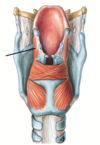
21
Q

1 - 5

A

1) epiglottis
2) hyoid bone
3) cartilago thryoidea
4) trachea
5) arcus cart. cricoideae

22
Q

6 - 10

A

6) lig. thyrohyoideum lat.
7) lig. thyrohyoideum medianum
8) lig. cricotracheale
9) membrana thyrohyoidea
10) cart. triticea

23
Q

11 - 14

What are the 2 parts of #11?

Another name for #12.

A

11) m. cricothyorideus (pars recta + pars obliqua)
12) lig. cricothryoideum medianum (= lig. conicum)
13) n. laryngeus sup. r. int.
14) a. laryngea sup.

24
Q

1 - 5

Another name for #4.

A

1) lig. hyoepiglotticum
2) lig. thyrohyoideum medianum
3) lig. thyroepiglotticum
4) lig. cricothryoideum medianum (= lig. conicum)
5) membrana triangularis

25
Q

6 - 10

A

6) membrana quadrangularis
7) plicae vocales
8) plicae vestibularis
9) plica aryepiglottica
10) cart. cuneiformis

26
Q

11 - 16

No #12.

A

11) cart. corniculata
13) m. arytenoideus transversus
14) m. cricoarytenoideus lat.
15) m. cricoarytenoideus post.
16) hyoid bone

27
Q

17 - 22

A

17) epiglottis
18) cart. thyroidea
19) cart. cricoidea
20) ventriculus larnygis
21) m. thyroarythenoideus
22) m. arytenoideus obliquus

28
Q

What are the parts of cavitas laryngis?

A
  • sup.: vestibulum laryngis → plicae vestibulares
  • med.: ventriculus laryngis → plicae vocales
  • inf.: cavitas infraglottica → trachea
29
Q

Which structures form the glottis AKA rima glottidis?

What are its parts?

A

plicae vocales

  • ant. part: pars intermembranacea
  • post. part: pars intercartilaginea
30
Q

Which muscles cause an elevation or depression of the larynx?

A

ELEVATION

suprahyoid mm.

  • m. digastricus
  • m. stylohyoideus
  • m. mylohyoideus
  • m. geniohyoideus

add:

  • m. thyrohyoideus
  • m. palato-, stylopharyngeus
  • m. constrictor pharyngis inf.

DEPRESSION (infrahyoid mm., exc. thyrohyoideus)

  • m. sternohyoideus
  • m. omohyoideus
  • m. sternothyroideus
31
Q

Which cartilages make up the laryngeal skeleton?

What happens to them at the end of puberty?

A
  • cartilago thyroidea
  • cartilago cricoidea
  • cartilago arytenoidea
  • epiglottis

ossify at the end of puberty (except epiglottis bc elastic, not hyaline cart.!!)

32
Q

1 - 6

Another name for #6.

A

1) incisura thyroidea superior/inferior
2) lamina dextra/sinistra
3) tuberculum thyroideum superius
4) cornu superius/inferius
5) linea obliqua
6) prominentia laryngea (= Adam’s apple)

33
Q

1 - 7

A

1) cartilago corniculata
2) proc. muscularis
3) facies articularis thyroidea
4) lig. cricoarytenoideum posterius
5) lamina cartilaginis cricoideae
6) cartilago arytenoidea
7) arcus cartilaginis cricoideae

34
Q

8 - 13

A

8) collicus
9) proc. vocalis; lig. vocale
10) crista arcuata
11) fovea triangularis
12) proc. muscularis
13) fovea oblonga

35
Q

What are the names of the small cartilages located in the larynx in craniocaudal direction?

A
  • cart. triticea (in lig. thyrohyoideum lat.)
  • cart. cuneiformis (in plica aryepliglottica)
  • cart. corniculata (sitting on apex of cart. arytenoida)
36
Q

1 - 2

A

1) cartilago epiglottica (= epiglottis)
2) petiolus epiglottidis

37
Q

What are the 2 laryngeal joints?

A
  • art. cricothyroidea
  • art. cricoarytenoidea
38
Q

Describe art. cricothyroidea.

A
  • type: bilateral ginglymus
  • articulating surfaces:
    • cornu inferius of thyroid cart.
    • lamina of cricoid cart.

⇒ regulation of length/tension of ligg. vocalia

39
Q

Describe art. cricoarytenoidea

A
  • type: bilateral trochoginglymus with add. sliding
  • articulating surfaces:
    • base of arytenoid cart.
    • lamina of cricoid cart
  • ligaments: lig. cricoarytenoideum

opening and closure of rima glottidis (for forced inhalation, phonation), only pars intercartilaginea open during normal breathing

40
Q

The submucosal conn. tissue of the larynx forms special structures, which?

A

membrana fibroelastica laryngea forms conus elasticus in cavitas infraglottica

  • med. part: lig. cricothryoideum medianum (= lig. conicum)
  • lower lat. part: membrana triangularis
  • ​upper lat. part: membrana quadrangularis
  • plicae vestibulares → ligg. vestibularia
  • *plicae vocales** → ligg. vocalia
41
Q

Explain the process of voice break.

A
  • *growth of vocal ligg. + larynx** → deeper voice
  • (in males more than in females obv)*
42
Q

Why is lig. conicum clinically important?

A

is cut in case of conicotomy in emergency medicine if upper airways are obstructed (= coniotomy, cricothyrotomy)

prominentia laryngea (= Adam’s apple) serves as important landmark

43
Q

What is a laryngeal edema?

What could be possible causes?

A

loose conn. tissue in mucosa of the laryngeal inlet permits build-up of considerable amounts of fluid from the vascular system

e.g. due to inflammation/insect stings → life-threatening mucosal swelling

44
Q

What is to note when using a laryngoscope?

A

image is inverted

45
Q

Which muscles act upon the rima glottidis?

A

open rima glottidis = abduction

  • m. cricoarytenoideus posterior (!!)

close rima glottidis = adduction

  • m. cricoarytenoideus lateralis
  • m. (inter)arytenoideus transversus
  • m. arytenoideus obliquus
  • m. vocalis
46
Q
A

m. circothyroideus (pars recta + pars obliqua)

  • ORIGIN: cricoid cart.
  • INSERTION: cornus inf. of thyroid cart.
  • ACTION: most important tensor of vocal cords → pitch
47
Q
A

m. cricoarytenoideus post. (posticus)

  • ORIGIN: lamina of cricoid cart.
  • INSERTION: proc. muscular of arytenoid cart. on same side
  • ACTION: only m. that opens rima glottidis
48
Q
A

m. cricoarytenoideus lateralis (lateralis)

  • ORIGIN: arch of cricoid cart.
  • INSERTION: proc. muscularis of arytenoid cart. on same side
  • ACTION: closure of pars intermembranacea
49
Q
A

m. (inter)arytenoideus transversus

  • ORIGIN: post. surface of arytenoid cart.
  • INSERTION: same spot on 2nd arytenoid cart.
  • FUNCTION: closure of pars intercartilaginea
50
Q
A

m. arytenoideus obliquus

  • ORIGIN: proc. muscularis of arytenoid cart.
  • INSERTION: apex of 2nd arytenoid cart.
  • ACTION: closure of pars intercartilaginea
51
Q

+ all parts

A

m. thyroarytenoideus

  • ORIGIN: inner side of thyroid cart.
    • INSERTION: arytenoid cart. (int. part)
    • ACTION: closure pars intermembranacea
    • INSERTION: proc. vocalis (m. vocalis)
    • ACTION: closure of vocal cords → fine tuning of tension
    • INSERTION: epiglottis (m. thyroepiglotticus)
    • ACTION: opens aditus laryngis
52
Q

Which nn. are responsible for sensory and motor innervation of the larynx?

A

motor innervation:

  • n. laryngeus sup. r. ext. → m. cricothyroideus
  • n. laryngeus recurrens → n. laryngeus inf. → REST

sensory innervation:

  • sup. part: n. laryngeus sup. r. int.
  • inf. part: n. laryngeus recurrens

⇒ division sup. and inf. to rima glottidis for innervation and supply/drainage

53
Q

Why is the innervation of the larynx clinically important?

A

surgeries on thyroid gland can possibly injure n. laryngeus recurrens → failure of n. cricoarytenoideus post.

  • unilateral damage → hoarseness + paralysis of vocal cord
  • bilateral damage → serious breathing problems
54
Q

Which vessels supply, drain the larynx, resp.?

A

supply:

  • a. carotis ext. → a. thyroidea sup. → a. laryngea sup.
  • a. subclavia → a. thyroidea inf. → a. laryngea inf.

drainage:

  • cf. supply

⇒ division btw sup. and inf. region at rima glottidis for supply/drainage + innervation

55
Q

How long is the trachea?

Where does it start, where does it end?

What are its 2 parts?

A
  • 10 - 13 cm, can expand up to 15 - 18 cm during deep inhalation
  • C7 → T4/5
  • 2 parts:
    • pars cervicalis: behind thyroid gland
    • pars thoracica
56
Q

How do you call the structure btw the cartilagines tracheales?

How do you call the non-cartilaginous part?

A

ligg. annularia

paries membranaceus

57
Q

What is important mention abt the tracheal bifurcation?

Clinical relevance?

A
  • in bifurcation, spine = carina tracheae
  • angle btw main bronchi btw 55 - 65°
    • left angle usually: 35 - 45°
    • right angle usually: 15 - 25°

⇒ aspirated foreign bodies are more likely to enter right main bronchus

58
Q

Which vessels supply, drain the trachea, resp.?

Which nerves are responsible for the innervation?

A

supply:

  • a. thyroidea inf. → rr. tracheales
  • thoracic aorta → rr. bronchiales
  • a. thoracica int. → rr. bronchiales

drainage:

  • plexus thryoideus impar → v. thyroidea inf.
  • vv. esophageales → v. hemi-/azygos

innervation:

  • parasymp.: n. laryngeus recurrens → rr. tracheales, n. vagus (X) → rr. bronchiales
  • symp.: truncus symphaticus
59
Q

What might be the effect of a goiter (swelling of thyroid gland)?

A

= struma

→ can cause compression of trachea → suffocation (= asphyxia)

60
Q

What happens in case of smoking to the cilia in the trachea?

A
  • transition from resp. epithelium to str. squamous epithelium (= squamous metaplasia)
  • adherence + immobility of cilia → removal of pollutants impaired (= impairance of mucociliar clearance)
61
Q

What is the shape of the lung?

Volume?

Surface area?

A
  • half cone shape
  • total volume 2 - 3l, after max. inhalation 5 - 8l
  • *left: 10 - 20 % less**
  • total surface area: 70 - 140 m<span>2</span>
62
Q

1 - 5

A

1) impression for a. subclavia dex.
2) impression for esophageus
3) impression for v. azygos + IVC
4) fissura obliqua
5) fissura horizontalis

63
Q

6 - 10

A

6) impression for a. subclavia sin.
7) impression for aorta / arcus aorticus
8) impressio cardiaca
9) lingula pulmonis sinistri
10) vv. pulmonales

64
Q

11 - 17

A

11) lig. pulmonale
12) right main bronchus in eparterial position
13) left main bronchus in hyparterial position
14) a. pulmonalis dex.
15) a. pulmonalis sin.
16) incisura cardiaca pulmonis sin.
17) hilum pulmonis

65
Q

Which impressions can you find on which lung?

A

right lung:

  • impression for a. subclavia dex.
  • impression for v. azygos
  • impression for IVC
  • impression for esophagus

left lung:

  • impression for a. subclavia sin.
  • impression for arcus aorticus
  • impression for thoracic aorta
66
Q

How do you call the 3 surfaces of the lung?

A
  • facies costalis (= biggest)
  • facies mediastinalis
  • facies diaphragmatica
67
Q

In which position are the bronchi of the right and left lung?

A
  • right main bronchus in eparterial position
    = sup. to a. pulmonalis
  • left main bronchus in hyparterial position
    = inf. to a. pulmonalis
68
Q

How do you call all vessels + nn. in the hilum?

A

radix pulmonis

69
Q

What can possible cause a pneumothorax?

A

injury / central venous catheter in v. subclavia → perforation of cupula pleurae → air evades through perforated lung → lung collapses due to low pressure in pleural cavity

70
Q

Explain the segmentation of the lungs.

A

right lung:​ main bronchi → 3 lobar bronchi:

  • sup. lobar bronchus → 3 segmental bronchi
  • mid. lobar bronchus → 2 segmental bronchi
  • inf. lobar bronchus → 5 segmental bronchi

10 segmental bronchi

left lung: main bronchi → 2 lobar bronchi:

  • sup. bronchus → 5 segmental bronchi
  • inf. bronchus → 4 segmental bronchi

9 segmental bronchi (7th missing due to incisura cardiaca)

REMEMBER HAND RULE: ​ know that right lobe 10 seg./3 bronchi, left 9/2 <br></br>- right hand: 3 fingers (1st) + 2 fingers (2nd) = 5 fingers (3rd)<br></br>- left: 9 (total) - right hand (1st) = 4 (2nd)

71
Q

Explain the structure of a lung lobules

A

= subunit of branchopulmonary segment

  • supplied by bronchiole
    → divides 3-4 times → terminal branches that bear alveoli (= acinus pulmonis)
  • mainly on surface of lungs (0.3 - 5cm polygonal regions)
72
Q

Why is the segmentation of the lung clinically important?

A
  • pneumonia can only affect one lobe
  • relevant for bronchoscopies: in which segment was biopsy taken?
  • in case of lung metastasis: surgical removal of entire segments
  • in case of lung carcinomas: removal of entire lobe
73
Q

Which vessels supply, drain the lungs, resp.?

Which nerves are responsible for the innervation?

What is the action of sympathetic and parasympathetic innervation?

A

supply:

  • aa. pulmonales (vasa publica)
  • rr. bronchiales of thoracic aorta (+ 3./4. a. intercostales) (vasa privata)

!!! aa. pulmonales carry DEOXYGENATED blood

drainage:

  • vv. pulmonales (vasa publica)
  • vv. bronchiales → v. azygos, v. hemiazygos (vasa privata)

!!! vv. pulmonales carry OXYGENATED blood

innervation:

  • sensory + parasymp: n. vagus (X) → n. laryngeus recurrens → rr. bronchiales
  • sensory + symp: truncus symphaticus → rr. bronchiales

⇒ together: plexus pulmonalis

⇒ para: bronchospasm (= constriction of bronchi)
⇒ symp: bronchorelaxation via β-receptors

74
Q

Differentiate btw nutritive vessels of the lung

A
  • vasa publica: pulmonary circulation → reoxygenate blood
  • vasa privata: systemic circulation → supply/drain lung
75
Q

Explain the structure of the pleura.

How do you call the upper apex?

A
  • pleura parietalis + visceralis (reflection of pl. parietalis) enclose cavitas pleuralis filled with a few ml of serous fluid
  • according to surfaces of lung:
    • pars mediastinalis
    • pars costalis
    • pars diaphragmatica
  • ​cupula pleurae extends apertura thoracica by 5cm
  • forms recessus
76
Q

Which recessus are formed by the pleura?

What is their function?

A

lungs expand into recessus during deep inhalation

recessus costodiaphragmaticus

  • lateral btw ribs + diaphragm

recessus costomediastinalis

  • ventral behind sternum

recessus phrenicomediastinalis

  • dorsal btw diaphragm + mediastinum

recessus vertebromediastinalis

  • dorsal next to spine
77
Q

Which vessels supply, drain the pleura?

Innervated by.. ?

Clinical relevance?

A

supply:

  • aa. intercostales
  • vasa pericardiaco-/musculophrenica

​drainage:

  • cf. supply

innervation: (sensory)

  • nn. intercostales
  • n. phrenicus

⇒ only pleura periatalis innervated nociceptively (no pain in case of lung tumors!)

78
Q

Compare the surface projections of lungs and pleura.

A

exc. at sternal line pleural borders = lung borders + 1