Trauma Flashcards

1
Q

Décrire la triade de Waddel

A
  1. Fx tibia/péroné ou fémur
  2. Trauma tronculaire
  3. Trauma craniofacial
2
Q

Critères devant être absents pour ne pas faire d’imagerie du thorax en trauma blunt

A

The criteria for obtaining imaging in one large validation study are age older than 60 years, rapid deceleration mechanism, chest pain, intoxication, abnormal alertness and mental status, distracting painful injury, and tenderness to chest wall palpation.

3
Q

Pré-requis pour la présence d’un chirurgie dans les réanimations traumatiques

A

A surgeon should be present in the emergency department on trauma patient arrival or within 15 minutes if any of the following major criteria are found:

  • Confirmed hypotension (systolic blood pressure < 90 mm Hg)
  • Gunshot wounds to the neck, chest, abdomen or proximal extremities
  • Intubated patients transferred from the scene
  • Respiratory compromise requiring an emergent airway
  • Penetrating gunshot wound to the neck, chest, abdomen, or pelvis
  • Glasgow Coma Scale score < 8 attributed to trauma

At the discretion of the emergency clinician

4
Q

Critères d’administration du cyclokapron en trauma

A

Critères inclusion de CRASH2 - trauma > 16a, avec hémorragie significative (ou à risque de) - SBP < 90 ou RC > 110 - dans les 3hrs du trauma

Acide tranexamique 1g sur 10 minutes puis 1g sur 8hr

5
Q

Avoir une idée générale du CDC ascot - EQTPT

A
6
Q

Critères dx TCC léger

A

According to the American Congress of Rehabilitation Medicine, a person with mild traumatic brain injury (MTBI) is a patient with a GCS of 13–15 who has had a traumatically induced physiologic disruption of brain function, as manifested by at least one of the following:

  1. Any period of loss of consciousness less than 30 min
  2. Any loss of memory for events immediately before or after the accident (posttraumatic amnesia should last <24 hr)
  3. Any alteration in mental state at the time of the accident (eg, feeling dazed, disoriented, or confused)
  4. Focal neurologic deficit(s) that may or may not be transient
7
Q

Nommer ce qui cause vasodilatation et vasoconstriction cérébrale

A

Hypertension, alkalosis, and hypocarbia promote cerebral vasoconstriction, whereas hypotension, acidosis, hypoxie and hypercarbia cause cerebral vasodilation.

Diminution de 1mmHg PCO2 diminue le diamètre des vaisseaux de 2-3%

Autorégulation cérébrale entre TAM 60-150 mmHG

Visons PPC 60-70

8
Q

Dfn d’augmentation de pression intracranienne

A

Tension intracrânienne augmentée si > 15 mmHg ou 19,5 cm H2O

Mécanismes intracrâniens compensatoires peut accomoder 50-100mL supplémentaire

9
Q

Caractéristiques associées à une fx du crâne significative

A

Présence d’air intracrânien

Lacération du cuir chevelu associée à la fx

Dépression de la table interne du crâne

Fracture qui se trouve a/n sinus veineux ou de l’artère méningée moyenne

10
Q

Quelle est l’anomalie au TDM la plus fréquente post trauma crânien? et qu’est-ce qu’un hygrome sous-dural?

A

HSA traumatique

Accumulation de LCR a/n sous-dural, avec xanthochromie + Hypothèses:

  • déchirure de l’arachnoide qui permet au LCR de traverser et s’accumuler en sous-dural
  • atteinte de la perméabilité capillaire a/n méninges
11
Q

Nommer les grades de DAI

A

Clinical grades of diffuse TAI have been based on length of coma: (1) grade 1 (mild)—ccoma for 6 to 24 hours; (2) grade II (moderate)—coma for longer than 24 hours but not decerebrate; (3) grade III (severe)—coma for longer than 24 hours and decerebrate or flaccid.

12
Q

Décrire le phénomène de Kernohan

A

In a certain percentage of TBI patients, the contralateral cerebral peduncle is forced against the opposite edge of the tentorial hiatus. Hemiparesis is then detected ipsilateral to the dilated pupil and mass lesion.

13
Q

Décrire les différentes herniations cérébrales

A
  • uncus
  • centrale transtentorielle
  • amygdales cérébelleuses (bulbaire)
  • transtentorielle cérébelleux upward
  • sous falcique
  • trans crânienne

(pupilles pinpoint avec atteinte protubérance)

14
Q

Nommer des signes à l’examen physique d’une fracture de la base du crâne

A

Blood in ear canal

Hemotympanum

Rhinorrhea

Otorrhea

Battle’s sign (retroauricular hematoma)

Raccoon sign (periorbital ecchymosis)

Cranial nerve deficits

Facial paralysis

Decreased auditory acuity

Dizziness

Tinnitus

Nystagmus

15
Q

Décrire le score de Rotterdam pour mortalité post TCC

A

Basal cistern effacement

0 = none

1 = partially effaced (compressed)

2 = completely effaced (compressed)

Midline shift

0 = no shift or ≤5 mm

1 = >5 mm

EDH (epidural hematoma)

0 = EDH present

1 = no EDH

IVH (intraventricular hemorrhage) or SAH (subarachnoid hemorrhage)

0 = neither present

1 = either present

  1. Add 1 to score

Total score = 1–6 points

16
Q

Quels antibiotiques en cas de trauma cérébral pénétrant ou fx crâne ouverte déprimée ou fx base du crâne avec fuite LCR > 7 jours

A

Vanco

Genta

Flagyl

17
Q

Indications de chx lors HSD

A

Indications for surgical evacuation include acute SDHs with a thickness more than 10 mm or a midline shift of more than 5 mm on a CT scan, regardless of the patient’s GCS score. Other parameters for surgical evacuation include a worsening GCS score (≥2 points from the time of injury to hospital admission) in comatose patients, asymmetric or fixed and dilated pupils, and persistent elevation in ICP. Most patients with subacute SDH require surgical evacuation of the lesion.

18
Q

Nommer des facteurs de risque de convulsions post traumatique précoces

A

GCS < 10

Age < 65 ans

Alcoolisme

Contusion cortex

Hématome sous-dural/épidural / intracérébral

Fx crâne

Trauma pénétrant

Convulsion initiale

Amnésie post-trauma > 30 minutes

19
Q

Nommer 2 hypothèse pour expliquer l’oedème pulmonaire neurogénique

A

Theories on its pathophysiology include the following: (1) catecholamine surge or blast from the TBI, resulting in increased intravascular pressure, increased capillary permeability, and hydrostatic edema114; and (2) a systematic inflammatory reaction leading to endothelial damage and vasogenic edema

20
Q

Nommer 3 règles pour évaluer le besoin d’imagerie en TCCL

A

Canadian Computed Tomography Head Rule (CCHR)

High-Risk Injury (May Require Neurologic Intervention)

1.

GCS score < 15 at 2 hr after injury

2.

Suspected open or depressed skull fracture

3.

Any sign of basal skull fracture (hemotympanum, raccoon eyes, CSF otorrhea or rhinorrhea, Battle’s sign)

4.

Vomiting ≥ two episodes

5.

Age ≥ 65 years

Medium-Risk Injury (May Have Important Brain Injury on CT)

6.

Amnesia before impact ≥ 30 min

7.

Dangerous mechanism (pedestrian struck by vehicle, occupant ejected from vehicle, fall from elevation >3 feet [five stairs])

New Orleans Criteria (NOC) 60 cévo aacc

1.

Headache

2.

Vomiting

3.

Age > 60 yr

4.

Drug or alcohol intoxication

5.

Persistent anterograde amnesia

6.

Trauma above the clavicle

7.

Seizure

NEXUS II Criteria 65 AEC/Abnormal coag/vo fx/hématome/sx neuro

1.

Evidence of significant skull fracture

2.

Scalp hematoma

3.

Neurologic deficit

4.

Altered level of alertness

5.

Abnormal behavior

6.

Coagulopathy

7.

Persistent vomiting

8.

Age ≥ 65 yr

21
Q

Nommer les 6 étapes de retour au jeu post TCCL

A
22
Q

Décrire la vascularisation du visage

A
23
Q

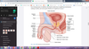
Décrire les fractures de Le Fort

A

A Le Fort I fracture involves a transverse fracture through the maxilla above the roots of the teeth and may be unilateral or bilateral. Patients may report malocclusion, and the maxilla may be mobile when the upper teeth are grasped and rocked. A Le Fort II fracture is typically bilateral and pyramidal in shape. It extends superiorly in the midface to include fractures of the nasal bridge, maxilla, lacrimal bones, orbital floor, and rim. In these cases, the nasal complex moves as a unit with the maxilla when the teeth are grasped and rocked. In the current age of CT scanning, in which the full extent of comminution can be appreciated, simple Le Fort III fractures are rare but essentially involve fracturing of the connections between the elements of the skull and face (craniofacial dysjunction). These fractures start at the bridge of the nose, extend posteriorly along the medial wall of the orbit (ethmoids), along the floor of the orbit (maxilla), and through the lateral orbital wall, and finally break through the zygomatic arch. Intranasally, they extend through all the lesser bones to the base of the sphenoid and frequently are associated with a CSF leak.

24
Q

Qu’est-ce qu’un foetus viable?

A

plus de 500 g et 24 sem

25
Nommer les changements hémodynamiques de la femme enceinte selon les trimestres
26
Nommer des FR de mort périnatale en trauma Quelle est la cause de décès principale du foetus en trauma blunt?
Risk factors significantly predictive of fetal death include ejection, motorcycle and pedestrian collisions, maternal death, maternal tachycardia, abnormal fetal heart rate, lack of restraints, and an injury severity score greater than 9. DPPNI - signe le plus sensible de DPPNI est la détresse foetale. Risque important de mort-né et accouchement pré-terme
27
Avoir une idée des doses de radiation en grossesse
Pas d'évidence d'augmentation du risque foetal si exposition \< 50 mGy
28
Nommer les indicateurs préhospitaliers d'Ohio en trauma gériatrique
Trauma gériatrique ≥70 years Anatomie: 2 régions ou plus atteintes Physiologie: GCS \<15 ave c TCC, TA 100 mm Hg Mécanisme: Fx 1 ou plusieurs os longs dans MVC, piéton/auto, chute avec TCC
29
Nommer des FR de chute en gériatrie
Faiblesse Trouble d'équilibre / mobilité Trouble cognitif Trouble visuel Médication Hypotension orthostatique
30
Nommer les facteurs modifiant la prise en charge d'un trauma gériatrique
Airway: risque d'intubation difficile augmenté ( mobilité cervicale limitée, ouverture TMJ limitée, crico difficile par ATCD chx/RT/ A/C ) Breathing: réserve pulmonaire limitée, détérioration et fatigue + rapide Circulation: réponses à l'hypovolémie diminuées Disability: augmentation du risque de fracture vertébrale et de lésion neurologique. Augmentation des SCIWORA (2nd sténose cervicale + kyphosis) Exposure: risque d'hypothermie augmentée (moins tissus/graisse et mécanismes de thermorégulation moins efficaces) 3 types de lésions cervicales + fréquentes: Syndrome central de la moelle Lésion extension-distraction Fx odontoide
31
Expliquer la classification de Dennis
The three parallel vertical column model proposed by Denis2 depicts the anterior column as being formed by alternating vertebral bodies and intervertebral disks surrounded by the annulus fibrosus capsule and anterior longitudinal ligament. The middle column consists of the posterior part of the annulus fibrosus and posterior vertebral wall, posterior longitudinal ligament, spinal cord, paired laminae and pedicles, articulating facets, transverse processes, nerve roots, and vertebral arteries and veins. The posterior column consists of the spinous processes, nuchal ligament, interspinous and supraspinous ligaments, and ligamentum flavum. Disruption of a single column usually preserves stability but does not preclude an SCI from displaced fracture fragments. Disruption of two columns results in an injury that is stable in one direction but unstable in another
32
Classer les fx de la colonne selon le mécanisme et la stabilité
Clay shoveler: fracture par avulsion du processus épineux d'une vertèbre cervicale basse ## Footnote Hangman: fractures bilatérales des pédicules de C2 Extension teardrop: fracture par avulsion du coin antéroinférieur d'une vertèbre. Syndrome central de la moelle possible par buckling du ligament jaune a/n moelle Jefferson: rupture de l'arc antérieur + postérieur de C1, distance dent - masses latérales \> 7mm
33
Décrire le ratio de Power et la distance normale dent-basion
34
wedge vs flexion teardrop
Flexion teardrop: Déplacement antérieur du fragment antérieur, indique rupture des éléments postérieurs donc instable
35
Nommer 3 mécanismes grossiers de blessure de la moelle
1. trauma pénétrant ou blunt massive avec lésion directe à la moelle 2. syndrome central de la moelle 2nd lésions ostéophytiques et ligament jaune comprimant moelle 3. Lésion vasculaire ( par compression via hématome ou lésion artères vertébrales / adamkiewicz
36
décrire la classification des lésions whiplash
0Whiplash injury but **no** pain, symptoms, or signs 1**Delayed** neck pain, minor stiffness, nonfocal tenderness only, **no** physical signs 2**Early** onset of neck pain, focal neck tenderness, spasm, stiffness, **radiating** symptoms 3**Early** onset of neck pain, focal neck tenderness, spasm, stiffness, radiating symptoms and signs of **neurologic** deficit 4Neck complaint (grade 2 or 3 above) and **fracture** dislocation
37
Ne pas oublier classification ASIA
38
Nommer des signes d'épargne sacrée
Sensation péri-anale Tonus rectal Flexion orteils
39
Nommer et détailler les 2 règles cliniques en imagerie cervicale
Nexus Absence de douleur centrale, absence sx neuro, absence d'intoxication, état conscience N, pas dlr distractrice CCR 1. facteurs de haut risque ? (plus de 65 ans, mécanisme dangereux, paresthésies) 2. facteurs de bas risque? (ambulant, sitting ED, simple collision arrière, dlr retardée, absence dlr centrale) 3. Capacité à tourner 45 deg
40
Décrire l'utilité de la ligne de Swisschuk et de l'espace prédental
Ligne Swisschuk (spinolamellaire): pseudosubluxation C2 sur C3 si ligne C1-C3 et processus épineux C2 à moins de 2 mm Espace prédental N 5 mm enfants et 3 mm adultes, sinon suspecter Jefferson
41
Nommer des conséquences néfastes des colliers cervicaux et planches dorsales
Plaies pression Douleur Diminution CRF Peut masquer lacérations et hématomes Peut aggraver une lésion existante
42
Nommer des ss/sx du choc spinal
Paralysie flasque Aréflexie Hypotension / bradycardie / hypothermie Rétention urinaire et iléus Absence bulbo-caverneux
43
Prédicteurs ou non du whiplash outcome
44
Nommer les zones du cou et les structures pouvant être lésées
**Zone I creux sternal ad cricoide** Carotide / vertébrales / sous-clavières / vaisseaux médiastin supérieur Apex pulmonaires / oesophage / trachée / moelle épinière Thyroide Canal thoracique **Zone II cricoide ad angle machoire** Carotide / vertébrales / jugulaires Larynx/ phaynx / trachée / oesophage / moelle épinière N. vague et récurrents laryngés **Zone III angle machoire ad base du crâne** Carotide / vertébrale / jugulaire Glandes salivaires et parotides NC 9 à 12 Moelle épinière
45
Management trauma pénétrant du cou
Instabilité HD = OR Hard sign avec ou sans instabilité = OR ou parfois imagerie avant OR Soft sign = imagerie Asx = parfois imagerie Hard sign: THE RATS Thrill, hémorragie / hémoptysie massive, hématome expansif, pouls radial absent, compromis airway/stridor/voix rauque, bulles d'air à la peau, TA basse, stroke Soft signs: Hémoptysie mineur/hématémèse, hématome non expansif, sgmt oropharyngé, sx neurologique, dysphonie, dysphagie, emphysème sous-cutané/médiastinal,
46
Nommer les critères de Denver
* 1,2,3, FLASH BANG * Fracture C1, C2, C3 ou foramen en transverse * Facial fracture (lefort 2-3), fracture mandibule * Luxation cervicale/sublux * Augmentation hématome cervical * Souffle * Hanging * Base skull fracture * Arterial bleed * Neuro exam not corresponding to CT, déficits focaux, stroke * GCS\<6
47
Nommer les grades de lésions vasculaires en trauma blunt cervical
Injuries are typically graded as 1 (intimal irregularity with \<25% narrowing), 2 (dissection or intramural hematoma with ≥25% narrowing), 3 (pseudoaneurysm), 4 (occlusion), and 5 (transection with extravasation).
48
Expliquer la règle NEXUS chest CT
Pouvons omettre TDM thorax si absence des éléments suivants - RX pulmonaire anormal - Décélération importante, \> 60 km/hr ou chute 20' - Douleur distractrice - Dlr paroi thoracique / sternum / colonne / scapula
49
Indications de fixation costale et méthodes d'analgésie
Indications: Sevrage ventilateur limité par le volet thoracique, douleur persistance, instabilité thoracique importante, aggravation de la fonction pulmonaire Analgésie: PO, blocs intercostaux, épidurale
50
Nommer 9 complications des fx costales
PTX, hémothorax Contusion pulmonaire Atélectasie, pneumonie Empyème Neurome post trauma Non union Séparation costochondrale
51
Indications de drain thoracique et complications possibles
PTX modéré à large, sx respiratoires PTX traumatique (sauf asx, apex) Augmentation post observation, récurrence post retrait Bilatéral, sous tension Hémothorax associé Besoin VM (sauf occulte) , anesthésie générale Complications: hémothorax, lacération a. intercostales, fistule bronchopleural, fuite pleural, PTX controlatéral, infection, empyème, lacération pulmonaire, emphysème sous-cutané
52
Indications de thoracotomie en trauma thoracique
Initial thoracostomy tube drainage is more than 20 mL of blood per kilogram. Persistent bleeding at a rate greater than 7 mL/kg/hr is present. Increasing hemothorax seen on chest x-ray films. Patient remains hypotensive despite adequate blood replacement, and other sites of blood loss have been ruled out. Patient decompensates after initial response to resuscitation.
53
Nommer 10 complications d'un trauma cardiaque blunt
Arythmie, trouble de conduction Tamponnade, péricardite constrictive Insuffisance cardiaque/choc cardiogénique Thrombus IV, occlusion a. coronaires, phénomene thrombo-embolique Contusion myocardique, rupture myocardique, rupture valve Anévrysme ventriculaire Commotio cordis
54
Nommer 6 mécanismes de rupture cardiaque en trauma blunt
1. Compression des cavités cardiaques lorsqu'elles sont remplies et fermées (ex oreillette en systole tardive ou ventricule en diastole/systole précoce) 2. Décélération importante avec ancrages fixes a/n VCS et VCI 3. Déplacement brusque du sang et des organes intra-abdo augmentant la pression intra-cardiaque 4. Compression directe du coeur entre sternum et vertèbres 5. Lacération par fx costale ou sternale 6. Complication d'une nécrose ou contusion myocardique avec rupture 2nd
55
Critères EDT
Critères EAST Autres Rosen: - choc réfractaire (ex TA\< 50) malgré réanimation après trauma pénétrant thoracique - choc sévère avec évidence de tamponnade ( trauma pénétrant thoracique)
56
Nommer les principaux sites du rupture aortique en trauma et sx associés
80% to 90% : aorte descendante distale à SC G Aorte ascendante ( + lésions létales cardiaques) Aorte descendante a/n diaphragme Aorte descendante mi thorax A l'origine a. SC G Symptomatologie souvent frustre. - dlr rétrosternale, interscapulaire, voix rauque par atteinte récurrent laryngé, dysphagie par compression, dyspnée par compression, sx ischémiques distaux - hypertension par stimulation récepteurs a/n isthme, masse +/- pulsatile a/n cervical par accumulation sgmt médiastinal, souffle systolique médiastinal, hémothorax massif Management: TA entre 100-120, esmolol +/- nipride
57
Nommer les 3 sites de perfo oesophagienne reliés aux CE
(1) the cricopharyngeal muscle near the esophageal introitus, (2) the level at which the esophagus crosses the left mainstem bronchus and the aortic arch, and (3) the gastroesophageal junction
58
Nommer les 6 causes de perforation oesophagiennes les + fréquentes
Iatrogénique Caustiques Corps étrangers Boerhaave Déhiscence d'anastomose Trauma blunt ou pénétrant
59
Nommer 9 conditions mimant la perforation oesophagienne
- Embolie pulmonaire / pneumonie - Pneumomédiastin / SCA / anévrysme aorte - Ulcère perforé / ischémie mésentérique / pancréatite - Cholécystite
60
définition abdomen antérieur, flancs, dos, thorax bas
. The anterior abdomen is defined as that region between the anterior axillary lines from the costal margins to the groin creases. The low chest begins at the nipple line or fourth intercostal space anteriorly and the inferior scapular tip or seventh intercostal space posteriorly, and then extends down to the inferior costal margins. The flank is between the anterior and posterior axillary lines bilaterally from the inferior scapular tip to the iliac crest. The back is between the posterior axillary lines, beginning at the inferior scapular tip and extending down to the iliac crest.
61
Nommer la catégorisation utilisée pour les plaies par balle selon la distance
Based on distance from the weapon to the victim, type I wounds involve a long range (\>7 yards or 6.4 m) and a penetration of subcutaneous tissue and deep fascia only. Type II wounds occur at a distance of 3 to 7 yards (2.7 to 6.4 m) and may create a large number of perforated structures. Type III wounds occur at point-blank range (\<3 yards or 2.7 m) and involve a massive destruction of tissue. When categorized by pattern, type I wounds produce a spread greater than 25 cm in diameter; type II, 10 to 25 cm in diameter; and type III, less than 10 cm in diameter. Close-range shotgun wounds, in addition to the shot, force external contaminants (eg, clothing and parts of the shell wadding) into the wounds. Type III wounds carry a substantial mortality risk
62
Connaître l'algorithme de la WTA en trauma pénétrant abdominal
Indications de laparotomie d'urgence - Éviscération - Empalement - Instabilité HD - Sgmt GI - Péritonite - Lésion diaphragmatique suspectée - Air libre intra-abdominal
63
Management abdominal GSW
* Basically most intra-abdo GSWs go to OR (if stable, then we’ll scan first) and OR should be your default answer. Whether it violates peritoneum isn’t often an issue since its often just assumed unless I guess it’s a graze injury.
64
Indications de laparatomie après trauma abdo blunt
Instabilité HD avec suspicion trauma intra-abdo Péritonite Pneumopéritoine Lésion diaphragmatique Sgmt GI significatif OR direct - instabilité avec FAST + et absence fx bassin instable - Péritonite franche et fast + - FAST + avec autres lésions nécessitant OR
65
Algorithme prise en charge pt instable et bassin instable
66
Algorithme trauma mixte tête abdo
67
Organe le plus atteint en trauma ped abdo blunt
Reins
68
Anatomie urogénital masculine
69
Anatomie testiculaire
70
Algorithme management trauma rénal blunt
Trauma pénétrant: habituellement chirurgical Antibiothérapie suggérée chez tous les traumas rénaux?
71
72
Ss/sx de trauma vésical intra vs extrapéritonéal Quand faire un CT cystogram
Hématurie Sang a/n méat urinaire Incapacité à uriner Ecchymose a/n périné, OGE, cuisse, abdomen Dlr abdominale Intrapéritonéal: trauma blunt sur vessie distendue (chx) extra: habituellement 2nd pénétrant/ fx bassin(conservateur) CT cystogram si hématurie macro + fx pelvis ou trauma pénétrant du pelvis ou hématurie micro + fx anneau pelvien ou obturateur possibilité de faire cystographie rétrograde (mélanger 30mL contraste iode hydrosoluble dans 500mL NS, insérer 300-400 mL via sonde )
73
Sx/ss trauma urétral
- Sang a/n méat urinaire - Échymose a/n périné (en papillon) - oedème/ hématome au périné - rétention urinaire
74
fracture pénienne
Rupture de la tunique albuginée "pop" associé à rapide détumescence et hématome. Si fascia de Buck atteint - ecchymose en papillon associé Dx clinique +/- échographie
75
anatomie artères MS
76
anatomie artères MI
77
Quand suspecter une lésion vasculaire?
Trauma pénétrant Trauma blunt: crush injuries, lésions avec avulsion tissus mous, déplacement os longs, brûlures, trauma par électrisation/foudre ou électriques, morsures, syndrome compartiment
78
Nommer les 5 signes Hard et soft de lésion vasculaire périphérique
HARD (THE R) ( soft) Thrill (déficit nerveux périphérique) Bruit audible (lésion osseuse ou pénétrante à proximité) Hémorragie pulsatile (hémorragie à la scène) Hématome expansif (hématome non expansif) Pouls distaux absents (diminution pouls distaux ou ABI)