MBD - Biochemistry Flashcards
(66 cards)
What are the two groups of symptoms in MBD?
Metabolis symptoms:
- Hypocalcaemia
- Hypercalcaemia
- Hypo/Hyperphosphataemia
Specific to bone
- Bone pain
- Deformity
Fractures
What is caclium incorporated as in bone?
What type of bone is metabolically active? How much remodelling occurs in bone at any time?
Cancellous bone is metabolically active and 5% is remodelling at any one time. total skeleton could be remodelled over 5 years.
There is also continuous exchange of ECF with bone fluid reserve.
What makes a bone strong?
- Mass
- Material properties (matrix and mineral) - collagen, crosslinking, woven vs lamerllar, microcracks
- Microarchitecture - trabecular thickness, connectivity, cortical porosity.
- Macroarchitecture - hip axis length, diameter
Describe the age related changes in bone mass.
- Peak bone mass in mid 20s
- Stable until around 40
- Men slow loss
- Women fast loss in early menopause

How does exercise increase peak bone mass?
There is change in bone mass density and geometry.

How does the tibia change during bone modelling from child to adult?

- More bone is placed anteriorly and posteriorly - this helps optomise deposition as not to waste mass
- Increase in bending strength ratio
- Periosteal apposition - essentially only when we are young. This is deposition of bone on the periosteal/outside surface. This reduces the elasticity of thebone.
Describe the sexual dimorphism in bone growth.
- This is due to sex hormones
- Estrogens limit periosteal bone expansion but stimulate endosteal bone apposition in females, whereas androgens stimulate radial bone expansion in males.
- Men reach higher peak bone mass, greater bone size, develop a stronger skeleton than women.
- However ther skeleton is not denser as bone mineral acquisition is in proportion to the volume of the bone, so volumetric bone density does NOT differ.

Describe the bone remodelilng cycle.

- Activation occurs - microcrack crosses cannaliculi, severing osteocyte processes causing osteocytic apoptosis.
- Connected surface lining cells (of osteoblastic lineage) sense this and release local factors to attract blood and marrow into the remodelling compartment
- Osteoclasts are generated locally to allow the resorption process to start. They resorb matrix and offending microcrack
- Successive teams of osteoblasts deposit new lamellar bone
- Osteoblats that are trapped in the matrix beocme osteocytes , others die or form new flattened osteoblast lining cells.
What investigations would you typically do if you suspect bone disease?
Serum:
Bone profile
- Caclium
- Corrected calcium (albumin)
- Phosphate
- Alkaline phostaphatase
Renal function: creatinine
- PTH
- 25-hydroxy vitamin D
Urine:
- Clacium/phosphate.
- NTX
Are the levels of Ca, P and Alk P high, normal or low in bone diseases:
- Osteoporosis
- Osteomalacia
- Pagets

Are the levels of Ca, P and Alk P high, normal or low in bone diseases:
- Primary HPT
- Renal dystrophy
- Metastases?

Describe the 3 systems involved in calcium balance.
GUT whats comig in; 1g day recommended intake
Kidney whats going out
BONE flux; your compensatory mechanism. This is not an inert tissue. This is where Ca is most abudant - we havea bout 1kg of Ca in bone.

What is meant by “total calcium”?
- This is when we measure calcium not just “free” in the active form, but also protein bound(to albumin) and complexed to citrate and proteins.
- So the calcium result is corrected in the laboratory to complensate for protein levels. If protein levels are high they compensate down (0.02 for each g/L of albumin).
- In acid-base disturbance/ alkalosis, there is more Ca binding to proteins so free levels drop. Happens when we get tingling.
Done using the following equation:
corrected calcium = [calcium] + 0.02 (45 - [albumin])
What are the percentages of protein-bound, complexed and free calcium in blood normally? What is the normal range for total calcium?
46% protein bound (but more in alkalosis)
47% FREE ionised (less in alkalosis)
7% complexed (more if high protein)
2.15-2.56mmol/L
How does PTH regulate Ca levels?
- Secretion of PTH when Ca levels drop
- This acts on 2 systems:
- BONE - acute release of Ca, due to increased resoprtion of bone by osteoclasts.
- Kidney -
- increased Ca re-absorption in the distal convoluted tubule (only place where Ca is absorbed under hormonal control)
- Increases phosphate excretion by inhibiting the NA-P cotransporter - his increases free Ca as usually P forms insoluble salts with Ca
- Increased conversion of 25-hydroxy Vitamin D into 1,25-hydroxy vitamin D (calcitriol) to increase absorption of Ca in the intestines.

What is the cause of hypocalcaemia in alcoholics?
Low magnesium causing low PTH and hypocalcaemia.
What is the half life of PTH? What does this allow in the lcinical setting?
half life (T1/2) = 8min
allows intraoperative testing - if PTH has been too high due to a tumour then surgically removing the tumour should lower PTH levels. PTH serum levels can be measured instantly during the operation.
Describe the molecular structure of PTH.
84 amino acid compound (1-84)
N1-34 (drug) is used for PTH replacement and is also used to build bone. It is more active.
Which ion apart from Calcium, is PTH dependant on?
Magnesium
What is the PTH receptor also activated by?
PTHrP (which is produced by some, so hypercalcaemia may be the first presenting feature)
How does the parathyroid gland monitor serum Ca?
- Through Ca-sensing receptors
- Even at high Ca there is base-line PTH secretion - e.g. a hypercalcaemia of malignancy there will still be detectable PTH
- But a small change in Ca causes a large change in PTH
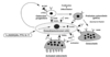
Decribe how PTH drives active Ca absorption in the distal tubule of the kidney.
- Epithelia can absorb Ca2+ by paracellular and transcellular transport. Passive and paracellular Ca2+ transport takes place across the tight junctions and is driven by the electrochemical gradient for Ca2+ (blue arrow).
- The active form of vitamin D (1,25-(OH)2D3) stimulates the individual steps of transcellular Ca2+ transport by increasing the expression levels of the luminal Ca2+channels, calbindins, and the extrusion systems.
- Active and transcellular Ca2+ transport is carried out as a three-step process. After entry of Ca2+ through the (hetero)tetrameric epithelial Ca2+ channels, TRPV5 and TRPV6, Ca2+ bound to calbindin diffuses to the basolateral membrane.
- At the basolateral membrane, Ca2+ is extruded via an ATP-dependent Ca2+-ATPase (PMCA1b) and a Na+/Ca2+ exchanger (NCX1).
- In this way, there is net Ca2+ absorption from the luminal space to the extracellular compartment.

How does PTH cause bone resoprtion through the RANK system?
- PTH stimulate osteoblasts by binding to incease their expression of RANKL and inhibit their secretion of OPg (osteoprotegerin)
- Free OPG usually competitively binds to RANKL as a decoy transport, preventing RANKL from interacting with its receptor “RANK”. Decreased OPG allows RANKL binding to RANK.
- This stimulates osteoclast precursors to fuse forming new osteoclasts which ultimately enhances bone resorption.