MBD - Radiology Flashcards
(41 cards)
Can you see metabolic dysfunction on radiological scans?
No just structural failures such as fractures and ligamentous injuries which could indicate metabolic dyfunction is acting.
Which imaging techniques show bone density? Which scans show biochemical composition and bone turnover?
Density:
- X-rays
- CT
- Bone Densitometry
- MRI - biochemical composition
- Radionuclide bone scans - bone turnover
Where do radionuclide tracers commonly go to?
Radionuclide bone scan - tracer goes to sites of increased osteoblastic activity. Commonly goes to:
- site of joins (wear and tear and degenerative changes)
- site of fractures
- site of tumours
- site of metabolic bone dysfunctions
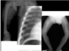
Describe the pathological features of these vertebrae.

- This is a CT - dense things in white and less dense darker.
- Picture 1 : The vertebrae images are T1 MRI- on these the fat shows up as white. When the bone is bright/almost as white as fat, it is because of the bone marrow which consists of a lot of fat. The vertebra above it is much darker which would suggest oedema, soft tissue lesion etc inside the bone. The one below it is completely collapsed.
- Picture 2: The first few vertebrae stand out from the others. This patient had radiotherapy in the past (which causes a lot of changes in the bone marrow). This has made them very well demarcated - they are radiotherapy related changes.
What is the difference between a radiological sign and pathology?
Pathology - disease process that gives rise to symptoms, signs and biochmical ditsurbances and changes in imaging appearance.
Radiological sign - change in imaging appearance, whether structural or functional, that may point towards a pathology.
What si osteoporosis?
- Decreased quantity of bone mass - microstructure is not abnormal, you just have less of it.
- Microstructure normal
Can lead to: fragility fractures, bone deofrmity and pain.
How is osteoporosis diagnosed?
By bone densitometry (aka dual-energy absortiometry, DEXA)
- Measure of BMD
- Compares BMD to normal reference databases (T-score or Z-score)
- T- score -1.5to -2.5=osteopenia; less than -2.5 = osteoporosis.
- Usually BMD is measured in lumbar spine and hips.
What is used to assess fracture risk? How?
FRAX
The BMD T-scores are not very helpful at telling us about risks so we assess risks by typing in results into FRAX
- Age
- Sex
- Weight
- Height
- BMD
Tells you 10 year probability of a major fracture and the probability of a hip fracture.
You can then click onto the nogg guidelines which are linked below.
What are the indications for deciding to do a DEXA scan?
We can’t just do a DEXA scan on all over 55’s so instead we do them when we see signs of osteoporosis:
- Loss of cortical bone/ thinning of cortex
- Loss of trabeculae
- Insufficiency fractures
What is an insufficiency fracture and what are the common sites?
- These are stress fractures due to normal stress on abnormal bones(osteoporosis/osteomalacia)
Common sites:
- Sacrum
- Underside of femoral neck
- verterbral bodies
- pubic rami
What are the radiological signs of osteoporosis? (CT/XR, Bone scan, MRI)
XR/CT
- initially normal
- can get periosteal reaction and callus formation in the later stage.
- more commonly there is increased sclerosis around the fracture lines that never seems to heal.
MRI
- bone oedema i.e. low signal on T1, high signal on T2 and STIR.
Bone scan
- Increased osteoblastic activity i.e. increased uptake, as things attempt to heal.
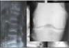
Describe what can be seen on these X-rays and which bone disorder could cause these radiological signs.
This is an example of a femoral fracture. This bone is normal but when compared to the bottom 2 images you can see that they are much darker, signifying osteoporosis. There is also a fracture on the right probably an insufficiency fracture.
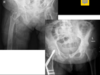
What is the name given to the radiological sign seen on the sacrum ? What other signs of osteoporosis are seen on the CT and MRI?

- HONDA SIGN - looks like a H
- CT - normal bone would have a similar density throughout however on this picture there are areas which have increased density, called “sclerosis”on CT
- MRI - there are also areas of high intensity on this image (similar areas)
Bone scan shows increased bone turnover in the same areas; these points could cause an insufficiency fracture.
What is the Honda sign and why does it occur?
The Honda sign (H sign / H pattern) is a term used to describe the appearances of bilateral sacral insufficiency fractures on a radioisotope bone scan.
Usually due to osteoporosis?
what is the difference between osteomalacia and osteoporosis?
Osteoporosis is reduced amount of normal bone but no abnormalities to the architecture. Osteomalacia is decreased bon mineralisation but amount of bone is still normal.
What is a Looser’s zone?
- They are pseudofractures at high tensile strength areas
- Considered a type of insufficiency fracture
- Have sclerotic irregular margins
- Occur at right angles to the cortex
- Transverse part way through a bone
- Associated with osteomalacia and rickets
What can too much unmineralised osteoid lead to in osteomalacia?
Looser’s Zone
What are the most common sites of Looser’s zone formation?
- Medial proximal femur
- Lateral scapula
- Pubic rami
- Posterior proximal ulna
- Ribs
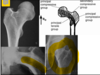
What is shown in this image? Which bone disorder does this correspond to?
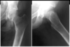
- Looser’s Zone - luscent short line at right angle to cortex with irregular sclerotic margins surrounding it.
- This is a sign of osteomalacia.
The bone would also be bent due to being osteopenic causing the whole femur to be a curved shape

What are cod-fish vertebrae? What conditions are they seen in?
Biconcave deformity of vertebrae
Seen in osteoporosis, osteomalacia.

Differentiate between these 4 vertebral columns.
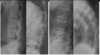
Picture 1 - normal
Picture 2 - wedge fracture, vertebral body collapse.
Picture 3 - cod fish vertebra due to biconcave appearance
Picture 4 - cod fish vertebra due to osteopenia and bone is darker than normal.
List the radiological signs of rickets.
- Indistinct/frayed metaphyseal margin
- Widened growth plate without calcification
- Cupping/splaying metaphyses due to weight bearing
- Enlargement of anterior ribs
- Osteopenia
Describe why there are differences in the radiology of osteomalacia and rickets. What are they?
Differences depend on age and closure of growth plates.
If the patient is an adult with mature skeleton and closed over growth plates then the signs of osteomalacia will be…
- Osteopenia
- Looser’s zones
- Codfish vertebrae
- Bending deformities
In children where growth plates are not closed yet, the signs of rickets will focus mainly on these growth plates.
- Before growth plate closure
- Signs centeres around growth plates
- changs of osteomalacia
Compare the metaphysis of the first and second hand. What condition is this?

First is normal and second is rickets.
Second has a frayed metahyseal margin which is a sign of rickets. There is also cupping.