Rheumatoid Arthritis Flashcards
(41 cards)
What is rheumatoid arthritis?
Chronic autoimmune disease characterised by pain, stiffness and symmetrical synovitis (inflammation of the synovial membrane) of synovial (diarthrodial) joints
Chronic = >6 weeks.
What are the key features of chronic arthritis?
- Polyarthritis - swelling of the small joints of the hand and wrists is common
- Symmetrical
- Early morning stiffness in and around joints – takes a long time to start moving the joints. Indication that treatment works if this improves.
- May lead to joint damage and destruction - ‘joint erosions’ on radiographs
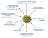
What extra-articular disease can occur in rheumatoid arthritis and what is this associated with?
Extra-articular disease = not just joint features.
Relates to IMMUNE COMPLEXES
- Rheuamtoid nodules
- Other rare: vasculitis, episcleritis.
Which antibody is detected in blood in rheumatoid arthritis?
Rheumatoid factor = IgM autoantibody against IgG
Why is most commonly affected by rheumatoid arthritis?
1% of all population but 3:1 F:M ratio.
What are the genetic and envrionmental risk factors for rheumatoid arthritis?
Genetic:
- Disease concordance rates for twins: 15-30%(monozygotic) and 5%(dizygotic), heritability estimates up to 60%.
- HLA-DRB gene variants; mapping to AAs 70-74 of DRbeta
- Many DRB molecules are associated with RA but what they all have in common is a “shared epitope” in a peptide binding group - a specific amino-acid sequence which is strongly associated with RA
Environmental component:
Smoking - contributes 25% of population attributable risk and interacts with shared epitope to increase risk.
What are the most commonly affected joints in rheumatoid arthritis?
- •Metacarpophalangeal joints (MCP)
- •Proximal interphalangeal joints (PIP)
- •Wrists
- •Knees
- •Ankles
- •Metatarsophalangeal joints (MTP)
Name two types of deformities associated with joint damage and destruction i rheumatoid arthritis.
- Swan-neck deformity
- Boutonniere deformity

Describe the swan-neck deformity.
Swan-neck deformity – there is hyper-extension at the PIP joint and hyper-flexion at the DIP joint
Describe the Boutonniere deformity.
Boutonnière (‘button-like’) deformity – there is hyper-flexion at the PIP joint
What is the primary site of pathology in rhematoid arthritis? Give some examples of pathology.
Synovium: this includes the synovial joints, tenosynovium surrounding tendons, bursa.
Pathology examples:
- proximal IP synovitis,
- extesor tenosynovitis (swelling is between joints, on top of extensors, prevents extension of some fingers as some tendons may become damaged)
- Olecranon bursitis

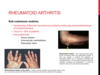
Describe the sub-cutaneous nodules that occur in rheumatoid arthritis. What percentage of patients get them? What is the typical place for such nodules?
- Central area of fibrinoid necrosis surrounded by histiocytes and peripheral layer of connective tissue
- Occur in ~30% of patients
- Associated with:
- Severe disease
- Extra-articular manifestations
- Rheumatoid factor
- Usually occur in ulnar border of the forearm but the hands are also common (e.g. at PIP joints)
What is rheumatoid factor also known as?
IgM anti-IgG antibody
Which portion of the IgG antibody is targeted by rheumatoid factor?
Fc portion is recognised by the rheumatoid factor (IgM - which is pentameric)
This forms a large complex which is through to be the reason why nodules form.

What percentage of rheumatoid arthritis patients are rheumatoid factor positive? How many develop ot later?
Positive in 70% at disease onset and further 10-15% become positive over the first 2 years of diagnosis
Name an antibody which is highly specific for rheumatoid arthritis.
Antibodies to citrullinated protein antigens (ACPA)
- Antibodies to citrullinated peptides are highly specific for rheumatoid arthritis
- AKA anti-cyclic citrullinated peptide antibody ‘anti-CCP antibody’
What enzymes are responsible for the citrullination of peptides?
•Peptidyl arginine deiminases (PADs) —>
Arginine –> Citrulline
Why do antibodies to citrullinated protein antigens (ACPA) form in rheumatoid arthritis?
- Caused by inflammatory cells? Inflammed synovium = high presence of neurophils and monocytes (which contain high concentrations of PADs) –> increased citrullination of autologous peptides i
- Smoking causes citrullination which causes the production of antibodies which are more pathogenic in the presence of a shared epitope–> increases ACPA-positive rheumatoid arthritis risk?
- Changes in microbiota?
- Chronic infections e.g. gingivitis due to poor dental health.
What is the rheumatoid arthritis shared epitope?
Shared epitope= amino acids 70-74 of the HLA-DRb-chains associated with rheumatoid arthritis
How could a shared epitope increase presence of ACPA?
ACPA is strongly associated with (smoking and) HLA ‘shared epitope’
- Shared epitope preferentially binds non-polar aa’s like citrulline/citrulline containing peprtide antigens
- but not positively charged aa’s like arginine
- Citrulline is increased during inflammation
- so ACPA more likely to develop among individuals with citrulinated autoantigens who have the shared eptiope
Why was rheumatoid arthritis found to be associated with multiple HLA serotypes?
Because multiple HLA serotypes - HLA-DR4, -DR1, -DR6, DR10- contained the SHARED EPITOPE
Individuals with HLA-DR4 are not affected if they do not have the shared epitope which preferentially binds non-polar aa’s like citrulline.
Name some common and uncommon extra-articular features of rheumatoid arthritis.
Common
- Fever (due to cytokines), weight loss
- Subcutaneous nodules
Uncommon
- vasculitis
- Ocular inflammation e.g. episcleritis
- Neuropathies
- Amyloidosis
- Lung disease – nodules, fibrosis, pleuritis
- Felty’s syndrome – triad of splenomegaly, leukopenia and rheumatoid arthritis
What are the early, later and latest radiographic abnromalities seen in rheumatoid arthritis?
Early
- Juxta-articular osteopenia
Later
- Joint erosions at margins of the joint
Later still
- Joint deformity and destruction
Where in the synovium is the first place of bone erosion in rheumatoid arthritis?
Erosions occur initially at the margins of the joint where the synovium is in direct contact with bone (the ‘bare’ area)