Part 19-images Flashcards
(28 cards)
Findings? Diagnosis?

cresent rim sign, bite sign
AVN of the femoral head (Chandler’s AVN)
Findings? Diagnosis?

subchondral collapse
AVN of the femoral head
Findings? Diagnosis?

bite sign, snow cap sign
healing phase of AVN of the femoral head
Findings? Diagnosis?

loss of marrow signal
AVN of femoral head bilaterally
Findings? Diagnosis?

obturator fat pad, gluteal fat pad, small epiphysis, lateral displacement of ossification center, flattening and fracture of the ossification center, metaphyseal widening and foreshortened widened irregular physis, irregular Kline’s line, irregular tear drop distance
Legg-Calve-Perthes disease
Findings? Diagnosis?

lesion at the lateral aspect of the medial femoral condyle
Findings? DIagnosis?
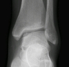
lesion of the medial aspect of the talus
osteochondrosis dessicans
Findings? Diagnosis?

lesion of the capitellum of the elbow
osteochondrosis dessicans
Patient has pain over the tibial tuberosity and this Xray finding. What is the diagnosis?

Osgood Schlatter’s
Patient has pain on the patella. Diagnosis?

Sindig-Larsen-Johannsen disease
Findings? Diagnosis?

AVN of the second metatarsal head
Freiber’s disease
Findings? Diagnosis?

AVN of the lunate, negative ulnar variance
Kienbock’s disease
Patient has pain over the navicular. Findings? Diagnosis?

vascular insufficiency of the navicular (if no pain, could just be normal variant)
Kohler’s disease
Findings? Diagnosis?

anterior body wedging, decreased disc space, increased kyphosis, more than 3 vertebra involved
Scheuermann’s disease
Findings? Diagnosis?

decreased disc space of 1 disc
Juvenile Discogenic Disease
Findings? Diangosis?

calcaneal apophysitis (not AVN), sclerosis and fragmentation of the calcaneal apophysis represents normal anatomy
Severs disease
Differential diagnosis?

chondrosarcoma
enchondroma
calcified medullary infarct
Findings? Most likely diagnosis?

hair on end skull
thalassemia
Findings? Diagnosis?

H vertebra
sickle cell anemia
Findings? Likely diagnosis?

dense hemoarthrosis, squared off patella
hemophilia
Patient has a WBC count of 40,000. Findings? Diagnosis?

submetaphyseal lucent bands, osteopenia, periosteal reaction
leukemia
Finding?

Involucrum
Pain is worse at night, relieved by aspirin. There is no microorgansims in the biopsy. Findings? Diagnosis?

localized, aborted form of suppurative osteomyelitis, oval, elliptical, or serpiginous radiolucency with no visible matrix surrounded by heavily reactive sclerosis
Brodie’s abscess
Findings? Diagnosis?

endplate destruction, disc destruction
spinal infection


