Skin & Systemic Disease Flashcards
(45 cards)
In Diabetes Mellitus, what condition is this called?
Describe it.

Diabetic Dermopathy
Atrophic macules and patches on the shins.
(Possibly due to trauma)
In Diabetes Mellitus, what condition is this called?

Diabetic Bullae
Tense non-inlammatory bullae on the lower limbs.
Unknown why it happens.
In Diabetes Mellitus, what condition is this called?
Describe it.
Necrobiosis Lipoidica
- Very common. Yellow atrophic plaques on the anterior shins.
- Collagen degeneration with a granulomatous response.
- SCC can develop from chronic lesions.
- Can Ulcerate.
- TREATMENT: Very resistant. Can try potent topical steroids, intralesional steroids, topical PUVA or narrow band UVB. Occasionally tacrolimus.
In Diabetes Mellitus, what condition is this called?
Describe it.

Acanthosis Nigricans
- Common in high BMI and insulin resistance.
- Pathology: IGF propagates epidermal growth.
- More common in pigmented skin.
- Treatment: Pigmanorm or topical retinoids.
- Pigmanorm( hydroquinone 5%, tretinoin 0.1g, hydrocortisone 1g)
In Diabetes, What condition is this?

Partial Lipodystrophy
Atrophy of subcutaneous tissue secondary to insulin use.
In Diabetes, what condition is this called?

Scleredema of Buschke
- Only seen in diabetes
- Induration of the skin in the upper back and nape of neck.
- Due to deposits of glycosaminoglycans.
- Skin feels woody and hard to touch.
In thyroid disease, what is this called?
Is it common?
What form of thyroid disease is it associated with?

Thyroid Acropachy
- Not Common
- Grave’s Disease
In Thyroid disease, What is this?
What form of thyroid disease is it associated with?
Does it reverse with treatment?

Pretibial Myxoedema
- Associated with Grave’s Disease
- Peau dórange appearance
- Treatment: intralesional steroids or steroids under occlusion.
- It does not reverse with treatment.
What can happen to eyebrows in thyroid disease?
The lateral third of the eyebrow may be lost.

What cutaneous features do we see in Cushings Disease?
- Subcutaneous fat redistribution - mood face, buffalo hump.
- Skin atrophy - global atrophy of epidermis and dermis.
- Cutaneous infections - candidiasis, pityriasis versicolor.
- Appendageal effects - steroid-related acne, hirsutism.
What are some cutaneous manifestations in addison’s disease?
The Excess ACTH stimulates melanin production by melanocytes
- Pigmentation in mucosal surfaces, palmar creases, nail beds and in scars.
- Vitiligo

In IBD, what is this?
What are some other non-IBD causes for it?
Erythema Nodosum
- idiopathic - most common
- Streptococcal infections - upper respiratory tract.
- Drugs - oestrogens, COCP, pinicillin, iodides, sulphonamides.
- Sarcoidosis
- Behcet’s disease
- Sweet’s syndrome
- Pregnancy
What investigations should be done in Erythema Nodosum?
- Thorough drug history
- Infection screen - viral and bacterial cultures, stool cultures, urine, Sputum, Serum ACE levels & Calcium, CXR and Heaf test.
- Skin Biopsy - needs ot be a deep incisional biopsy - not a punch biopsy - subcutis needs to be obtained where a panniculitis is seen.
What is the treatment for Erythema Nodosum?
- Treat the underlying cause.
- Rest and NSAIDs
What is this?

Leucocytoclastic Vasculitis
(Seen in both UC and Crohns)
What condition is this in IBD and where on the body is it usually found?
What investigations should you do?

Pyoderma gangrenosum
- Lower limbs
- Investigations: look for inflammatory bowel disease, rheumatoid arthritis or malignancy.
- Biopsy - primarily to exclude other causes.
- DO NOT HAVE SURGEONS DEBRIDE - it will only make it worse.
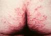
What is the most common skin manifestation in Coeliac disease?
Dermatitis Herpetiformis
- Intensly itchy
- Most common on buttocks, scalp and extensor surfaces.
-
Biopsy shows neutrophil microabscesses in the dermal papilla.
- IMF has granular IgA deposition.
Whats a cutaneous sign of liver cirrhosis?
Palmar erythema

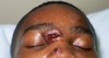
What are some signs of Hepatitis C infection?

- Lichen Planus
- Small vessel vasculitis
- Cryoglobuinaemia
- Pruritus
- Porhyria Cutanea Tarda
(Picture on the other page is lichen planus)
(This picture - Porphyria Cutanea Tarda - blisters and erosions and milia on sun-exposed areas.)

In patients with sarcoidosis, what fraction have affected skin?
1/3
What organ is most commonly affected in sarcoidosis?
Lungs
What are the cutaneous patterns sarcoidosis can manifest like?
- Papules
- Plaques
- Hypopigmented patches
- Subcutaneous Nodules (including Erythema Nodosum)
- Annular
- Ulcerative
- Scar sarcoid
What pattern of lupus is this?
Tell me a bit about it

Lupus Pernio
- Nodules & plaques
- Form on the nose, ears, cheeks.
- 75% have chronic lung involvement
- It is resistant to treatment
- Scarring
What pattern of sarcoid is this?

Annular Sarcoid