Lower Limb Muscles Flashcards

1
Q

What are the 2 groups of muscles in the gluteal region?

What are their functions?

A

Superficial muscles:

  • they abduct and extend the femur

Deep muscles:

  • they laterally rotate the femur
2
Q

What are the superficial muscles in the gluteal region?

A
  1. Gluteus maximus
  2. Gluteus medius
  3. Gluteus minimus
  4. Tensor fascia lata

They act to abduct and extend the lower limb at the hip joint

3
Q

What are the attachments, actions and innervation of gluteus maximus?

A

origin:

  • gluteal surface of ileum, sacrum and coccyx

attachment:

  • iliotibial tract and gluteal tuberosity of femur

actions:

  • main extensor of the thigh
  • assists in lateral rotation

innervation:

  • inferior gluteal nerve
4
Q

What are the attachments, actions and innervation of gluteus medius?

A

Origin:

  • gluteal surface of the ilium

Insertion:

  • greater trochanter of femur

actions:

  • ​abducts and medically rotates the lower limb
  • secures the pelvis and prevents pelvic drop during walking

innervation:

  • superior gluteal nerve
5
Q

What are the attachments, actions and innervation of gluteus minimus?

A

origin:

  • ilium

insertion:

  • greater trochanter

actions:

  • abducts and medically rotates the limb
  • prevents pelvic drop of the opposite limb during walking

innervation:

  • superior gluteal nerve
6
Q

What are the attachments, actions and innervation of the tensor fascia lata?

A

Origin:

  • anterior superior iliac spine

Insertion:

  • iliotibial tract, which attaches to the lateral condyle of the tibia

action:

  • assists gluteus medius and gluteus minimus in abduction and medial rotation of the lower limb

innervation:

  • superior gluteal nerve
7
Q

What are the deep muscles of the gluteal region?

A
  1. Piriformis
  2. Superior and inferior gemelli
  3. Quadratus femoris
  4. Obturator internus

They laterally rotate the lower limb

8
Q

What are the attachments, actions and innervation of piriformis?

A

Origin:

  • anterior surface of the sacrum

insertion:

  • travels through greater sciatic foramen
  • Inserts into greater trochanter of femur

actions:

  • lateral rotation and abduction

innervation:

  • nerve to piriformis
9
Q

What are the attachments, actions and innervation of obturator internus?

A

Origin:

  • pubis and ischium at obturator foramen

insertion:

  • travels through lesser sciatic foramen
  • attaches to greater trochanter of femur

actions:

  • lateral rotation and abduction

innervation:

  • nerve to obturator internus
10
Q

What are the attachments, actions and innervations of the gemelli?

A

origin:

  • superior gemellus - ischial spine
  • inferior gemellus - ischial tuberosity

insertion:

  • greater trochanter of femur

actions:

  • lateral rotation and abduction

innervation:

  • superior gemellus - nerve to obturator internus
  • inferior gemellus - nerve to quadratus femoris
11
Q

What are the attachments, actions and innervation of quadratus femoris?

A

Origin:

  • ischial tuberosity

insertion:

  • quadrate tuberosity on the intertrochanteric crest

actions:

  • lateral rotation

innervation:

  • nerve to quadratus femoris
12
Q

Label the muscles of the gluteal region

A
13
Q

What is the innervation and general action of the muscles in the anterior compartment of the thigh?

A

Innervation:

  • femoral nerve (L2-L4)

Action:

  • extension of the leg at the knee joint
14
Q

What are the muscles within the anterior compartment of the thigh?

A
  1. Pectineus
  2. Sartorius
  3. Quadriceps femoris
  4. Iliopsoas (the end passes into the anterior compartment)
15
Q

What are the origins and insertions of iliopsoas?

A

Origin:

  • Psoas major originates from the lumbar vertebrae
  • iliacus originates from the iliac fossa

Insertion:

  • lesser trochanter of femur
16
Q

What are the actions and innervation of iliopsoas?

A

Actions:

  • flexes the thigh at the hip joint

innervation:

  • psoas major - anterior rami of L1-L3
  • iliacus - femoral nerve
17
Q

Label the muscles of the anterior thigh

A
18
Q

What are the 4 muscles that make up quadriceps femoris?

A
  1. Rectus femoris
  2. Vastus lateralis
  3. Vastus medialis
  4. Vastus intermedius
19
Q

What are the attachments, actions and innervation of vastus lateralis?

A

Origin:

  • ​greater trochanter and lateral lip of linea aspera

Insertion:

  • Quadriceps tendon, which attaches to the patella

Action:

  • extends the knee joint and stabilises the patella

Innervation:

  • femoral nerve
20
Q

What are the attachments, actions and innervation of vastus medialis?

A

Origin:

  • intertrochanteric line and medial lip of linea aspera

Actions:

  • extends the knee joint and stabilises the patella

Innervation:

  • femoral nerve
21
Q

What are the attachments, actions and innervation of vastus intermedius?

A

Origin:

  • anterior and lateral surfaces of femoral shaft

Actions:

  • extends the knee joint and stabilises the patella

Innervation:

  • femoral nerve
22
Q

What are the attachments, actions and innervation of rectus femoris?

A

Origin:

  • Ilium (just superior to acetabulum)

actions:

  • flexes the thigh at the hip joint
  • extends the leg at the knee joint

Innervation:

  • femoral nerve
23
Q

What are the attachments, actions and innervation of sartorius?

A

Origin:

  • anterior superior iliac spine

Insertion:

  • superior, medial surface of the tibia

Actions:

  • flexes, abducts and laterally rotates the hip
  • flexes the knee joint

Innervation:

  • femoral nerve
24
Q

Label the muscles

A
25
Q

What is meant by pectineus being a “transitional muscle”?

A

it has a dual innervation

it is a transitional muscle between the anterior and medial thigh compartments

26
Q

What is the passage of sartorius like?

A

it is long and thin and runs across the thigh in an inferomedial direction

it is more superficial than the other muscles in the leg

27
Q

What are the attachments, actions and innervation of pectineus?

A

Origin:

  • pectineal line on anterior surface of pelvis

Insertion:

  • Pectineal line on posterior femur (just inferior to lesser trochanter)

Actions:

  • adduction and flexion at the hip joint

Innervation:

  • femoral nerve
  • also receives a branch from the obturator nerve
28
Q

Label the prosection image

A
29
Q

What are the 5 medial thigh muscles?

What is their artery and nerve supply?

A

The ‘hip adductors’

  1. Gracilis
  2. Obturator externus
  3. Adductor brevis
  4. Adductor longus
  5. Adductor Magnus

They are supplied by the obturator nerve and obturator artery

30
Q

Label the muscles of the medial thigh

A
31
Q

What are the attachments of the 2 parts of the adductor Magnus muscle?

A

Adductor part:

  • originates from inferior rami of pubis and rami of ischium
  • attaches to linea aspera

Hamstring part:

  • orginates from ischial tuberosity
  • attaches to adductor tubercle and medial supracondylar line of the femur
32
Q

What are the actions and innervation of adductor magnus?

A

Actions:

  • adduction of the thigh
  • the adductor part also flexes the thigh
  • the hamstring portion also extends the thigh

Innervation:

  • ‘adductor part - obturator nerve (L2-L4)
  • hamstring part - sciatic nerve (L4-S3)
33
Q

What are the attachments, innervation and actions of adductor longus?

A

Origin:

  • originates from the pubis and expands into a fan shape

Insertion:

  • linea aspera of femur

Innervation:

  • obturator nerve (L2-L4)
34
Q

Why can the adductor brevis be used as an anatomical landmark?

A

it lies between the anterior and posterior divisions of the obturator nerve

35
Q

What are the attachments, actions and innervation of the adductor brevis?

A

Origin:

  • body of pubis and inferior pubic rami

Insertion:

  • Linea aspera of femur
  • proximal to adductor longus

actions:

  • Adduction of the thigh

Innervation:

  • obturator nerve (L2-L4)
36
Q

What are the attachments, innervation and actions of obturator externus?

A

Origin:

  • membrane of obturator foramen and adjacent bone

Insertion:

  • posterior aspect of greater trochanter

Actions:

  • adduction and lateral rotation of the thigh

Innervation:

  • Obturator nerve (L2-L4)
37
Q

What are the attachments and passage of gracilis?

A

Originates from the inferior ramus of the pubis and body of the pubis

descends vertically down the leg to attach to the medial surface of the tibia

it attaches between the tendons of sartorius (anteriorly) and semitendinosus (posteriorly)

38
Q

What are the actions and innervation of gracilis?

A

Actions:

  • adduction of the thigh at the hip
  • flexion of the leg at the knee

Innervation:

  • obturator nerve (L2-L4)
39
Q

Label the medial thigh muscles

A
40
Q

What are the muscles in the posterior compartment of the thigh?

A

The Hamstrings

  1. Biceps femoris
  2. Semitendinosus
  3. Semimembranosus
41
Q

What are the actions and innervation of the posterior thigh muscles?

A

they extend at the hip and flex at the knee

they are innervation by the sciatic nerve (L4-S3)

42
Q

Label the muscles of the posterior thigh

A
43
Q

What are the attachments of the biceps femoris muscle?

A

Long head:

  • originates from the ischial tuberosity of the pelvis

short head:

  • originates from the linea aspera on the posterior surface of the femur

insertion:

  • the two heads form a tendon, which inserts into the head of the fibula
44
Q

What are the actions and innervation of biceps femoris?

A

Actions:

  • flexion at the knee
  • also extends the thigh at the hip
  • laterally rotates the hip and knee

Innervation:

  • long head - tibial part of sciatic nerve
  • short head - common fibular part of the sciatic nerve
45
Q

What are the attachments, actions and innervation of semitendinosus?

A

Origin:

  • ischial tuberosity of pelvis

Insertion:

  • medial surface of the tibia

Actions:

  • flexion of the leg at the knee joint
  • extension of the thigh at the hip
  • medially rotates the thigh and leg
46
Q

What are the attachments, actions and innervation of semimembranosus?

A

Origin:

  • ischial tuberosity of pelvis
  • more superiorly than the semitendinosus and biceps femoris

Insertion:

  • medial tibial condyle

Actions:

  • flexion of the leg at the knee joint
  • extension of the thigh at the hip joint
  • medially rotates the thigh and leg

Innervation:

  • tibial part of sciatic nerve
47
Q

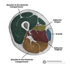
Label the cross section of the thigh

A
48
Q

What are the 4 muscles in the anterior compartment of the leg?

A
  1. Tibialis anterior
  2. Extensor digitorum longus
  3. Extensor hallucis longus
  4. Fibularis tertius
49
Q

What is the general action of the anterior leg muscles?

What is their blood supply and innervation?

A

They dorsiflex and invert the foot at the ankle joint

they are innervated by the deep fibular nerve (L4 - S1)

they are supplied by the anterior tibial artery

50
Q

What are the attachments, actions and innervation of tibialis anterior?

A

Origin:

  • lateral surface of the tibia

Insertion:

  • medial cuneiform and the base of metatarsal I

Actions:

  • dorsiflexion and inversion of the foot

Innervation:

  • deep fibular nerve
51
Q

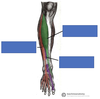
Label the muscles of the anterior leg

A
52
Q

What are the attachments, actions and innervation of extensor digitorum longus?

A

Origin:

  • lateral condyle of the tibia and medial surface of fibula

Insertion:

  • the tendons insert onto the dorsal surface of the 4 toes

Actions:

  • extension of the lateral four toes
  • dorsiflexion of the foot

Innervation:

  • deep fibular nerve
53
Q

What are the attachments, actions and innervation of extensor hallucis longus?

A

Origin:

  • medial surface of the fibular shaft

Insertion:

  • the tendon attaches to the base of the distal phalanx of the big toe

Action:

  • extension of the big toe
  • dorsiflexion of the foot

Innervation:

  • deep fibular nerve
54
Q

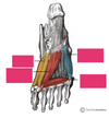
Label the tendons of the foot

A
55
Q

What is significant about fibularis tertius?

A

It arises from the most inferior part of the EDL

It is not present in all individuals

56
Q

What are the attachments, actions and innervation of fibularis tertius?

A

Origin:

  • originates with the EDL from the medial surface of the fibula

Insertion:

  • the tendon attaches to metatarsal V

Actions:

  • Eversion and dorsiflexion of the foot

innervation:

  • deep fibular nerve
57
Q

What is footdrop a clinical sign of?

What is it usually a consequence of?

A

It indicates paralysis of the muscles in the anterior compartment of the leg

It occurs due to damage to the common fibular (peroneal) nerve (from which the deep fibular nerve arises)

58
Q

What is the appearance of the leg like in foot drop and why?

A

The muscles in the anterior compartment are paralysed

the unopposed pull of the muscles in the posterior leg produce permanent plantarflexion

59
Q

What are the 2 muscles in the lateral compartment of the leg?

What is their innervation and main function?

A
  1. Fibularis brevis
  2. Fibularis longus

They are innervated by the superficial fibular nerve

Their main function is eversion (turning the sole of the foot outwards)

60
Q

Label the muscles of the lateral leg?

A
61
Q

What are the attachments and path of the tendon of fibularis longus?

A

Origin:

  • superior and lateral surface of the fibula and lateral tibial condyle

Path of the tendon:

  • the tendon descends into the foot, posterior to the lateral malleolus
  • it crosses under the foot

Insertion:

  • bones on the medial side of the foot - mainly medial cuneiform and base of metatarsal I
62
Q

What are the actions and innervation of fibularis longus?

A

Actions:

  • eversion and plantarflexion of the foot
  • supports the lateral and transverse arches of the foot

Innervation:

  • superficial fibular (peroneal) nerve (L4-S1)
63
Q

What are the attachments, actions and innervation of fibularis brevis?

A

Origin:

  • inferolateral surface of fibular shaft

Insertion:

  • tendon passes posterior to the lateral malleolus and over the calcaneus and cuboidal bones
  • attaches to a tubercle on metatarsal V

Actions:

  • eversion of the foot

Innervation:

  • superficial fibular (peroneal) nerve
64
Q

What is the general action and innervation of the muscles in the posterior compartment of the leg?

A

the posterior compartment muscles are organised into a superficial and deep layer

they act to plantarflex and invert the foot

they are innervated by the tibial nerve

65
Q

What is the insertion of the superficial muscles in the posterior leg?

A

They all insert into the calcaneus of the foot via the calcaneal tendon

66
Q

What are the two bursae associated with the calcaneal tendon?

What is their role?

A

Subcutaneous calcaneal bursa:

  • lies between the skin and calcaneal tendon

Deep bursa of the calcaneal tendon:

  • lies between the tendon and the calcaneus

The bursae work to minimise friction during movement

67
Q

What are the muscles in the superficial layer of the posterior leg?

A
  1. Plantaris
  2. Gastrocnemius
  3. Soleus
68
Q

What are the attachments of gastrocnemius?

A

Origin:

  • lateral head originates from lateral femoral condyle
  • medial head originates from medial femoral condyle
  • the fibres converge to form a single muscle belly

Insertion:

  • The muscle belly combines with soleus to form the calcaneal tendon
  • this inserts onto the calcaneus
69
Q

What are the actions and innervation of gastrocnemius?

A

Actions:

  • plantarflexion of the ankle
  • flexion of the knee

Innervation:

  • tibial nerve
70
Q

Label the superficial layer of posterior leg muscles

A
71
Q

What are the attachments, actions and innervation of plantaris?

A

Origin:

  • lateral supracondylar line of femur

Insertion:

  • ​the tendon blends with the calcaneal tendon

Actions:

  • weak plantarflexor of the ankle
  • weak flexor of the knee

Innervation:

  • tibial nerve
72
Q

What are the attachments, actions and innervation of soleus?

A

Origin:

  • soleal line of the tibia and proximal fibular area

Insertion:

  • joins the calcaneal tendon

Actions:

  • plantarflexion of the ankle

Innervation:

  • tibial nerve
73
Q

What are the 4 muscles in the deep compartment of the posterior leg?

A
  1. Popliteus
  2. Tibialis posterior
  3. Flexor digitorum longus
  4. Flexor hallucis longus
74
Q

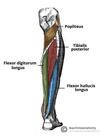
Label the muscles in the deep layer of the posterior thigh

A
75
Q

What are the attachments, actions and innervation of popliteus?

A

Origin:

  • lateral condyle of femur and posterior horn of lateral meniscus

Insertion:

  • it inserts above the origin of the soleus muscle

Actions:

  • laterally rotates the femur on the tibia
  • this is “unlocking” the knee joint so that flexion can occur

Innervation:

  • ​tibial nerve
76
Q

What are the attachments, actions and innervation of tibialis posterior?

A

Origin:

  • interosseous membrane between tibia and fibula

Insertion:

  • the tendon enters the foot posterior to the medial malleolus
  • it inserts onto the plantar surface of the medial tarsal bones

Actions:

  • inversion and plantarflexion of the foot
  • maintains the medial arch of the foot

Innervation:

  • tibial nerve
77
Q

What are the attachments, actions and innervation of flexor digitorum longus?

A

Origin:

  • medial surface of tibia

Insertion:

  • plantar surfaces of the lateral four digits

Actions:

  • flexes the lateral four toes

Innervation:

  • Tibial nerve
78
Q

What are the attachments, actions and innervation of flexor hallucis longus?

A

origin:

  • posterior surface of the fibula

Insertion:

  • plantar surface of the phalanx of the big toe

Actions:

  • flexes the big toe

Innervation:

  • tibial nerve
79
Q

Label the tendons of the posterior leg

A
80
Q

What are the 2 intrinsic muscles on the dorsal aspect of the foot?

What is their innervation?

A
  1. Extensor digitorum brevis
  2. Extensor hallucis brevis

They are both innervated by the deep fibular nerve

81
Q

What are the attachments, actions and innervation of extensor digitorum brevis?

A

Origin:

  • calcaneus, interosseous talocalcaneal ligament and inferior extensor retinaculum

Insertion:

  • attaches to long extensor tendons of the 4 lateral digits

Actions:

  • aids EDL in extending lateral four toes at metatarsophalangeal and interphalangeal joints

Innervation:

  • deep fibular nerve
82
Q

What are the attachments, actions and innervation of extensor hallucis brevis?

A

Origin:

  • calcaneus, interosseous talocalcaneal ligament and inferior extensor retinaculum

Insertion:

  • base of the proximal phalanx of the big toe

Action:

  • aids EHL in extending the big toe at the metatarsophalangeal joint

Innervation:

  • deep fibular nerve
83
Q

What is the collective action of the intrinsic muscles in the sole of the foot?

What is their innervation?

A

they stabilise the arches of the foot and control movement of the digits

they are all innervated by either the medial plantar nerve or the lateral plantar nerve

these are both branches of the tibial nerve

84
Q

What are the 3 muscles in the first layer of plantar muscles?

A

The first layer is located immediately underneath the plantar fascia

  1. Abductor hallucis
  2. Flexor digitorum brevis
  3. Abductor digiti minimi
85
Q

What are the attachments, actions and innervation of abductor hallucis?

A

Origin:

  • medial tubercle of calcaneus, flexor retinaculum and the plantar aponeurosis

Insertion:

  • medial base of proximal phalanx of the big toe

Action:

  • abducts and flexes the big toe

Innervation:

  • medial plantar nerve
86
Q

Label the first layer of plantar muscles

A
87
Q

What are the attachments, actions and innervation of flexor digitorum brevis?

A

Origin:

  • medial tubercle of calcaneus and plantar aponeurosis

Insertion:

  • middle phalanges of lateral 4 digits

Actions:

  • flexes the lateral 4 digits at the proximal interphalangeal joints

Innervation:

  • medial plantar nerve
88
Q

What are the attachments, actions and innervation of abductor digiti minimi?

A

Origin:

  • medial and lateral tubercles of the calcaneus and the plantar aponeurosis

Insertion:

  • lateral base of the proximal phalanx of the 5th digit

Actions:

  • abducts and flexes the 5th digit

Innervation:

  • lateral plantar nerve
89
Q

What are the muscles that make up the second layer of plantar muscles?

A
  1. Quadratus plantae
  2. Lumbricals
  3. Tendons of flexor digitorum longus
90
Q

Label the second layer of plantar muscles

A
91
Q

What are the attachments, actions and innervation of quadratus plantae?

A

Origin:

  • medial and lateral plantar surface of the calcaneus

Insertion:

  • Attaches to the tendons of flexor digitorum longus

Actions:

  • Assists FDL in flexing the lateral four digits

Innervation:

  • lateral plantar nerve
92
Q

What are the attachments, actions and innervation of the four lumbrical muscles of the foot?

A

Origin:

  • tendons of flexor digitorum longus

Insertion:

  • extensor hoods of the lateral 4 digits

Actions:

  • flexes the metatarsophalangeal joints, whilst extending the interphalangeal joints

Innervation:

  • the most medial lumbrical is innervated by the medial plantar nerve
  • the remaining 3 are innervated by the lateral plantar nerve
93
Q

What muscles make up the third layer of plantar muscles?

A
  1. Flexor hallucis brevis
  2. Adductor hallucis
  3. Flexor digiti minimi brevis
94
Q

Label the third layer of plantar muscles

A
95
Q

What are the attachments, actions and innervation of flexor hallucis brevis?

A

Origin:

  • plantar surfaces of the cuboid and lateral cuneiforms

Insertion:

  • base of the proximal phalanx of the big toe

Actions:

  • flexes the proximal phalanx of the big toe at the metatarsophalangeal joint

Innervation:

  • medial plantar nerve
96
Q

What are the attachments, actions and innervation of adductor hallucis?

A

Origin:

  • oblique head - bases of 2nd, 3rd and 4th metatarsals
  • transverse head - plantar ligaments of the metatarsophalangeal joints

Insertion:

  • lateral base of proximal phalanx of the big toe

Actions:

  • adduction of the big toe
  • assists in forming the transverse arch of the foot

Innervation:

  • deep branch of lateral plantar nerve
97
Q

What are the attachments, actions and innervation of flexor digiti minimi brevis?

A

Origin:

  • base of fifth metatarsal

Insertion:

  • base of proximal phalanx of fifth digit

Actions:

  • flexes proximal phalanx of fifth digit

Innervation:

  • superficial branch of lateral plantar nerve
98
Q

What makes up the fourth layer of plantar muscles?

A

the plantar interossei - which are unipennate

and

the dorsal interossei - which are bipennate

99
Q

What are the attachments, actions and innervation of the 3 plantar interossei?

A

origin:

  • the medial side of metatarsals 3 to 5

insertion:

  • medial sides of phalanges of digits 3 to 5

Actions:

  • abducts digits 3 to 5
  • flexes the metatarsophalangeal joints

innervation:

  • lateral plantar nerve
100
Q

What are the attachments, actions and innervation of the 4 dorsal interossei?

A

Origin:

  • sides of metatarsals 1 to 5

Insertion:

  • first muscle attaches to medial side of proximal phalanx of the second digit
  • second to fourth muscles attach to lateral sides of the proximal phalanges of digits 2 - 4

Actions:

  • abducts digits 2 to 4
  • flexes the metatarsophalangeal joints

Innervation:

  • lateral plantar nerve