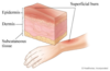
Skin Histology Flashcards
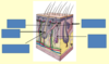
What are the 3 layers of the skin?
- epidermis
- dermis
- subcutis

What are the types of cells present in the epidermis?
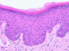
contains continuously proliferating stratified squamous epithelium
it produces keratin
it is in direct contact with the external environment
it is constantly shed and contains no blood vessels
What is present in the dermis?
fibrous and fibroadipose tissue that supports the epidermis, both physically and metabolically
it contains blood vessels, nerves and sensory receptors
What is present in the subcutis?
it contains adipose tissue with supporting fibrous bands (septa)
it contains larger blood vessels
What type of epithelium is the epidermis?
keratinised stratified squamous epithelium
What type of skin is present on the foot?
glabrous skin
this is non-hair bearing and thick skin

What are the 5 layers of the epidermis that are present in thick skin?
- stratum basale - basal cell layer
- stratum spinosum - prickle cell layer
- stratum granulosum - granular layer
- stratum lucidum
- stratum corneum - keratin layer
What layer of the skin is not present in thin skin?
stratum lucidum
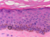
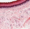
What is shown by letters a-e in this picture?
a - stratum basale
b - stratum spinosum
c - stratum granulosum
d - stratum lucidum
e - stratum corneum

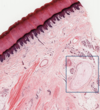
What is shown by the letters in the high magnification of keratinised squamous epithelium?

b - stratum spinosum
c - stratum granulosum
e - stratum corneum
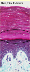
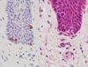
What is shown in this image?

resin section of stratified squamous epithelium
What is shown in the image?
What is the composition of this layer like?
stratum lucidum of the sole of the foot
this consists of several layers of flattened dead cells
nuclei already begin to degenerate in the outer part of the stratum granulosum
in the stratum lucidum, faint nuclear outlines are visible in only a few of the cells
What 3 types of cells are found in the epidermis?
- keratinocytes
- melanocytes
- langerhans cells
What type of cell is shown here?
What is their role?
melanocytes
these produce melanin (skin and hair colour)
they are transferred to the keratinocytes through a network of melanocyte cytoplasmic processes
What is shown in this image?

Langerhans cells
these are intra-epidermal antigen presenting cells
they are present in all layers of the epidermis but are most easily recognised in the prickle cell layer
How can Langerhans cells be recognised?
they are pale-staining in the epidermis
they have irregularly lobulated nuclei and almost clear cytoplasm
cytoplasmic processes (CP) extend from the cells and insinuate between keratinocytes of all layers

Where are keratinocytes present?
they are present in all layers of the epidermis, but are most easily recognised in the prickle cell layer
they are present in the upper dermis, particularly around small blood vessels
when stimulated, they migrate to the dermis and then via lymphatics to the lymph nodes
What structures are labelled in the diagram?

What is shown in this image?

hair bearing skin
What is shown in this image?
pilosebaceous unit
this consists of hair, sebaceous gland and arrector pili muscle
this image is from the scalp
What is shown by a, b and c?

a - arrector pili
b - hair follicle
c - sebaceous gland
What is shown here?
fingertip
What is shown in this image of the fingertip?
each lamella is composed of flattened Schwann cells and endoneurial fibroblasts
there is fluid between each layer
delicate collagen fibres may be present as well as occasional capillaries
What cells are shown here and what type of staining is used to see them?
CK20 staining is used for Merkel cells
these are intra-epidermal receptors for touch
they are rounded cells without dendritic processes found in the basal layer of the epidermis
they are associated with a myelinated nerve forming a Merkel cell-neurite complex
What is shown in this image?
Meissner’s corpuscles
these are an encapsulated body in the papillary dermis
they are fast-adapting discriminatory touch and vibration receptors

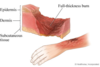
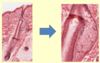
What is meant by a first degree burn?
an injury that affects only the outer layer of skin - the epidermis
symptoms of redness, minor inflammation and swelling but no blistering
What is a second degree burn?
this affects both the epidermis and dermis
there is blistering and some thickening of the skin

What is a third degree burn?
this type of burn destroys the whole of the epidermis and the dermis
the skin has a widespread thickness and a white, leathery appearance