Brainstem & Cerebellum Flashcards
What are the 3 general functions of the brainstem?
- Conduit
- Integrative
- Cranial nerve
How does the brainstem act as a conduit?
It allows ascending and descending pathways to reach the thalamus and cerebellum from the spinal cord
It contains relay nuclei
These are a collection of grey matter (nerve cell bodies) within the CNS

How does the brainstem have an integrative function?
It is involved in the control of cardiovascular, respiratory and consciousness
Consciousness is controlled by the reticular formation
It is involved in complex motor patterns (e.g. balance)
What does locked in syndrome result from?
An infraction of vessels in the ventral pons
as the reticular formation is intact, the patient is conscious but is unable to move
What is the ‘cranial nerve” function of the brainstem?
These are the head’s equivalent to spinal nerves
They are involved in sight, hearing, equilibrium and gustation
There are cranial nerve nuclei and reflex centres within the brainstem
What are the 3 structures within the brainstem?
- Midbrain
- Pons
- Medulla oblongata
What are the rostral, caudal, ventral and dorsal relationships of the brainstem?
Rostrally:
- midbrain is continuous with the diencephalon
Caudally:
- medulla is continuous with the spinal cord at the level of the foramen magnum
Ventrally:
- clivus of occipital bone
Dorsally:
- cerebellum

What components of the ventricular system run through the brainstem?
- IVth ventricle (IVth)
- Cerebral aqueduct (Caq)
This is continuous with the central canal of the spinal cord
These are cavities containing cerebral spinal fluid
Label the components of the ventricular system and brainstem


Which cranial nerves arise from the brainstem?
10 out of 12 cranial nerves arise from the brainstem
These are part of the PNS
They have sensory and motor parts

At any level of the brainstem, which 3 areas can be identified in cross section?
Tectum:
- the tectum is only present in the midbrain
Tegmentum:
- this contains cranial nerve nuclei and the reticular formation
Basal:
- descending motor fibres travel through here
Label the different areas in cross section of the brainstem


Where is the tectum found?
It is posterior to the ventricular system
The only region with substantial tectum (roof) is the midbrain (superior and inferior colliculi)

Where is the tegmentum found?
What does it contain?
Found anterior to the ventricular system
it contains cranial nerve nuclei and tracts, reticular formation and some ascending/descending pathways

Where is the basal found?
What does it contain?
It is found most anteriorly
it contains descending fibres from the cerebral cortex (pyramids, cerebral peduncles)

Which junction is labelled?

Pontomedullary junction (PMJ)
this is between the medulla of the brainstem and the pons

Where is the rhomboid fossa?
What is this?

It forms the floor of the IVth ventricle
It is exposed when the cerebellum is removed

Label the ventral view of the medulla


What is the passage of the cranial nerves visible on the ventral view of the medulla like?
IX and X are lateral to the olives in the anterolateral sulcus
XI is below X
IX, X and XI all leave through the jugular foramen
XII emerges between the pyramids and olives and leaves through the foramen magnum
What is behind the pyramids?
Descending motor fibres - the pyramidal tract
What happens at the decussation of pyramids?
Fibres travelling from the cerebrum cross over and travel on the opposite side of the spinal cord
Label the dorsal view of the medulla


What is the obex?
The point at which the IVth ventricle becomes continuous with the central canal of the spinal cord
What is the difference between the fasciculus cuneatus and fasciculus gracilis?
Fascicles contain a tract of white matter (axons)
Fasciculus cuneatus:
- contains ascending sensory information from the upper limb
Fasciculus gracilis:
- contains sensory information from the lower limbs
What is the difference between the open and closed medulla?
If you cut below the obex, this section is the closed medulla as there is brain tissue dorsally and centrally
If you cut above the obex, this is the open medulla as the IVth ventricle is anterior
What are the sections through the midbrain?


Label the closed and open medulla sections

White matter is shown as black due to the staining
the nucleus within the olive (shown in rostral medulla) is part of the visual system

What 2 junctions are labelled?


Label the ventral view of the pons


Label the dorsal view of the pons


Label the cross sections of the caudal and rostral pons


What junction is shown?

Pontomesencephalic junction of the midbrain

Label the ventral view of the midbrain


Label the ventral view of the midbrain


Label the midbrain at the level of the inferior colliculi?


What is the reticular formation?
A complex, multisynaptic network of neurones within the tegmentum of the brainstem

Label the blood supply to the brainstem


Label the blood supply to the brainstem


Label the cranial nerves
Which ones are missing and where do they originate from?
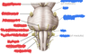
I and II are extensions of the forebrain
IV comes from the dorsal aspect of the midbrain

Label the cerebellar peduncles


What are the views of the cerebellum?


Label the superior/dorsal surface of the cerebellum

The vermis is the middle portion between the 2 hemispheres

Label the inferior/ventral surfaces of the cerebellum

