SDL - Vertebral Column & Spinal Cord Flashcards

1
Q

What are the 7 components that make up the back?

A

the back is the posterior aspect of the trunk and includes:

  1. skin
  2. subcutaneous tissue
  3. vertebral column
  4. spinal cord and meninges in the vertebral column
  5. ribs
  6. nerves and vessels
  7. muscles
2
Q

What makes up the vertebral column?

How many vertebrae are in the typical adult vertebral column?

A

the vertebral column consists of vertebrae separated by intervertebral discs that are bound together by ligaments

the typical adult vertebral column has 33 vertebrae

3
Q

What are the different types of vertebrae that make up the vertebral column?

A

7 cervical (C1 - C7 in the neck)

12 thoracic (T1 - 12) articulate with the ribs

5 lumbar (L1 - 5) in the lower back

5 sacral (S1 - S5) fused into the sacrum

4 coccygeal (Co) fused into the coccyx

4
Q

What are the features of a typical vertebra?

A

a body anteriorly which supports the vertebral column and is connected to the intervertebral discs

a vertebral arch posteriorly

5
Q

what are the different parts of the vertebral arch?

A

pedicle:

  • this attaches the transverse process to the body of the vertebra

lamina:

  • attaches transverse process to spinous process

paired superior and inferior articular processes:

  • protrude posterior to the vertebral notches

spinous and transverse processes:

  • for attachment of muscles and ligaments
6
Q

Label the diagram

A
7
Q

What are the smallest vertebrae?

A

cervical vertebrae

they are the smallest moveable vertebrae and form the bony skeleton of the neck

8
Q

What is the distinctive feature of the cervical vertebrae?

A

transverse foramen

these foramina are smaller in C7 than in other cervical vertebrae

they are occasionally absent

9
Q

What structures pass through the transverse foramina?

A
  • vertebral artery
  • vertebral vein
  • sympathetic nerves from the inferior cervical ganglion
10
Q

Which cervical vertebrae have a different structure?

A

C1 and C2

11
Q

What is significant about the spinous processes of C3 to C6?

A

they are short and bifid (divided into 2 parts)

12
Q

What is significant about the spinous process of C7?

A

it is known as the vertebra prominens and is very long

it is used as a bony landmark from which to count vertebrae

13
Q

Label the typical cervical vertebra

A
14
Q

What are the unique features of C1 (the atlas) and C2 (the axis)?

A
  • it has NO vertebral body and NO spinous process
  • it is ring-like and has an anterior arch, posterior arch and 2 lateral masses
  • the anterior arch has a facet for articulation with the dens of the axis
  • the posterior arch has a groove for the vertebral artery and C1 spinal nerve
15
Q

What is the role of the ligament of the atlas?

A

the anterior arch has a facet for articulation with the dens of the axis

this is secured by the ligament of the atlas that attaches to the lateral masses

16
Q

What are the unique features of C2 (the axis)?

A
  • the dens rises perpendicularly from the upper surface of the body
  • it articulates with anterior arch of the atlas, creating the medial atlanto-axial joint
  • they are specialised to allow a greater range of motion
17
Q

Complete the labelling on the diagrams of C1 and C2

A
18
Q

What are the distinguishing features of the thoracic vertebrae?

A
  • vertebral body is heart-shaped
  • demi-facets on the sides of each vertebral body articulate with the heads of the ribs
  • costal facets on the transverse processes articulate with the tubercles of the ribs (T1 - T10 only)
  • spinous processes are long and slant inferiorly
19
Q

Label the thoracic vertebrae

A
20
Q

Label the lumbar vertebra

A
21
Q

What are the distinguishing features of the lumbar vertebrae?

A
  • transverse processes are long and slender
  • articular processes have nearly vertical facets
  • spinous processes are short and broad
22
Q

What is the purpose of accessory and mammillary processes of the lumbar vertebrae?

A

accessory processes:

  • found on posterior aspect of the base of each transverse process
  • site of attachment for deep back muscles

mammillary processes:

  • found on the posterior surface of each superior articular process
  • site of attachment for deep back muscles
23
Q

What is significant about the fifth lumbar vertebra?

A

it has a large vertebral body and transverse processes as it carries the weight of the entire upper body

24
Q

Label the features of the sacrum and coccyx

A
25
Q

What are vertebrae held together by?

A
  1. facet joints
  2. intervertebral discs
  3. ligaments
26
Q

What is the purpose of facet joints?

A

(also called zygapophyseal joints)

they connect the superior and inferior articular processes of adjacent vertebrae

27
Q

What is the role of intervertebral discs?

A

intervertebral discs between all non-fused vertebrae provide flexibility to the spine and act as shock absorbers

28
Q

what is the purpose of the ligaments of the vertebral column?

A

ligaments bind vertebrae together and give stability to the vertebral column

29
Q

What are the 2 components of the intervertebral disc?

A
  1. annulus fibrosus
  2. nucleus pulposus
30
Q

Label the components of the intervertebral disc

A
31
Q

What are the functions of the annulus fibrosus and nucleus pulposus?

A

annulus fibrosus:

  • enhances spine’s rotational stability
  • helps to resist compressive stress

nucleus pulposus:

  • transmits stress and weight from vertebra to vertebra
32
Q

What are the 5 types of ligaments associated with the vertebral column?

A
  1. anterior longitudinal ligament
  2. posterior longitudinal ligament
  3. ligamentum flavum
  4. ligamentum nuchae
  5. interspinous ligaments
33
Q

What are the attachments and function of the anterior longitudinal ligament?

A

attachment/location:

  • covers the anterior aspects of the vertebral bodies and intervertebral discs
  • attaches to the upper and lower edges of each vertebra, body

function:

  • limits extension of the vertebral column and reinforces the intervertebral disc
34
Q

What are the attachments and functions of the posterior longitudinal ligament?

A

posterior longitudinal ligament:

  • arises from the body of the axis (C2)
  • travels downward and posterior to the vertebral bodies and intervertebral discs
  • attaches to the back of the sacrum

function:

  • limits flexion of the vertebral column
  • reinforces intervertebral disc
35
Q

What is the attachment and function of the ligamentum flavum?

A

attachments:

  • connects the laminae of adjacent vertebrae from C2 to S1

functions:

  • resumes the shape of the vertebral column after flexion
  • preserves upright posture
  • resists excessive separation of the adjacent vertebral laminae
  • prevents canal compression
36
Q

What are the functions and attachments of the ligamentum nuchae?

A

attachments:

  • continuous with the supraspinous ligament
  • spans from the occiput to the spine of C7

functions:

  • limits flexion
37
Q

What are the attachments and functions of the interspinous ligaments?

A

attachments:

  • connects the spinous processes (from roots to apexes) from C1 to S1 one segment at a time
  • fibres connect with the ligamentum flavum anteriorly
  • fibres connect with the supraspinous ligament posteriorly

functions:

  • the role of the interspinous ligament is to limit flexion (bending fowards)
  • it restricts separation of the spinous processes of the vertebral column
38
Q

What are the movements possible at the vertebral column?

How do these vary?

A

the arrangement of the vertebral column, with many small bones separated by intervertebral discs, permits a wide range of movements of the back

  • flexion
  • lateral flexion
  • extension
  • rotation

these movements are possible to different extents in different regions of the vertebral column

39
Q

What are the 2 groups of muscles of the back?

A

superficial (or extrinsic)

deep (or intrinsic)

40
Q

What are the extrinsic (superficial) back muscles?

What is their innervation?

A
  1. rhomboid major and minor
  2. levator scapulae
  3. trapezius
  4. latissimus dorsi

they are innervated by the ventral rami of spinal nerves

trapezius is innervated by the accessory nerve

41
Q

What is the innervation of the intrinsic (deep) back muscles?

How can they be divided?

A

dorsal (posterior) rami of spinal nerves

they can be divided into superficial, intermediate and deep

42
Q

What are the muscles in the superficial group of intrinsic back muscles?

A
  1. splenius muscles on the posterolateral surfaces of the neck
  2. splenius capitis
  3. splenius cervicis
43
Q

what are the following superficial intrinsic back muscles?

A
44
Q

What are the functions of splenius capitis?

A

acting bilaterally:

  • extension of the head and cervical spine

acting unilaterally:

  • lateral flexion of the head and neck
  • rotation of the head to the same side
  • it also assists in supporting the head in the erect position
45
Q

What are the functions of splenius cervicis?

A

acting bilaterally:

  • they extend the neck

acting unilaterally:

  • they laterally flex and rotate the head and neck to the same side
  • they also assist in supporting the head in the erect position
46
Q

What muscle is part of the intermediate group of deep back muscles?

What is its composition like?

A

the large erector spinae muscles that lie on either side of the vertebral column

it is composed of 3 separate muscles and acts as the chief extensor of the vertebral column

47
Q

What are the 3 muscles that make up erector spinae?

A

from midline to most lateral:

  • vertebral column
  • spinalis
  • longissimus
  • iliocostalis
48
Q

Label the constituents of erector spinae

A
49
Q

What muscles form part of the deep group of intrisic back muscles?

Where do they originate from?

A

deep to erector spinae there is a group of much smaller muscles that originate from the transverse processes of the vertebrae

and insert onto the spinous process of more superior vertebrae

this is the transversospinalis group

it consists of semispinalis, multifidus and rotatores

50
Q

What are the primary contents of the vertebral canal?

A
  1. spinal cord
  2. spinal nerve roots
  3. their meningeal coverings
51
Q

At which vertebral level does the spinal cord end in a newborn and an adult?

What does this mean for the lower spinal nerve roots in adults?

A

the vertebral column ends at L1 - L2

at birth, the vertebral column ends at L3

the lower spinal nerve roots in adults form the cauda equina

they travel down the vertebral column to exit below their respective vertebrae

52
Q

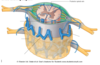
Label the diagram of the spinal cord

A
53
Q

What is contained within the subdural and subarachnoid space?

A

subdural space:

  • potential space between arachnoid and dura mater
  • does not contain any bridging veins (unlike cranial subdural space) so haemorrhage in this area is rare

subarachnoid space:

  • space between the arachnoid and the pia mater
  • filled with cerebrospinal fluid
54
Q

What is contained within the epidural space in the spinal cord?

A

this is a space between the dura and vertebra

it contains fat

55
Q

What is the arterial supply to the spinal cord?

A

the spinal cord receives its arterial supply from three small, longitudinally running arteries

these are one anterior and two posterior spinal arteries

56
Q

Where do the anterior and posterior spinal arteries originate?

A

anterior spinal artery:

  • originates from vertebral artery
  • travel in the anterior median fissure

posterior spinal artery:

  • originate from the vertebral artery or posteroinferior cerebellar artery
  • they anastamose with each other in the pia mater
57
Q

Which areas of the spinal cord are supplied by the anterior and posterior spinal arteries?

A

anterior spinal artery:

  • supplies anterior portion of spinal cord

posterior spinal artery:

  • supplies grey and white posterior columns of the spinal cord
58
Q

What arteries reinforce the anterior and posterior spinal arteries?

A

the anterior and posterior spinal arteries are small and are reinforced by radicular branches of the intercostal and lumbar arteries

these enter the vertebral canal through the intervertebral foramina

59
Q

Complete the labelling of the blood supply to the spinal cord

A
60
Q

Label the venous drainage of the spinal cord

A
61
Q

What is the venous drainage of the spinal cord like?

A

venous drainage is via three anterior and three posterior spinal veins

these are valveless and form an anastomosing network along the surface of the spinal cord

they also receive venous blood from the radicular veins

62
Q

What do the spinal veins drain into?

A

they drain into the internal and external vertebral plexuses

these empty into the systemic segmental veins

the internal vertebral plexus also empties into the dural venous sinuses superiorly

63
Q

At each vertebral level, what venous connections are present?

A

at each vertebral level there are connections with thoracic, abdominal and intercostal veins and the external vertebral venous plexus

64
Q

Where does blood pass into after flowing through a spinal vein?

A

spinal veins have no valves

blood passes directly into the systemic venous system

65
Q

What is significant about the continuity of the vertebral venous plexuses with the prostatic plexus?

A

the continuity of the vertebral venous plexuses with the prostatic plexus is probably the path along which prostatic neoplastic cells metastasize to the central nervous system

66
Q

label the components of the venous drainage of the spinal cord

A
67
Q

what is the passage of the spinal nerve like in the upper part of the vertebral canal?

A

in the upper part of the vertebral canal, each spinal nerve has a short course before emerging through the intervertebral foramen as the anterior primary ramus and posterior primary ramus

68
Q

what happens to spinal nerves in the lower part of the spinal canal?

A

the roots of the sacral nerves begin inside the vertebral column at the level of the L1 vertebra

they descend into the sacrum

this is where the cauda equina begins

69
Q

Label the diagram

A
70
Q
A