Localisation Flashcards
What are the cerebral hemispheres?
Dense core of white matter with an overlying thin layer of grey matter (cortex)
What is the thickness of the cerebral cortex?
What is the surface of it like?
It varies in thickness from 2-4 mm
it is heavily folded to increase surface area
over half of the total surface area is hidden by the walls of the sulci
What is the arrangement of the cerebral cortex like?
It has a laminar arrangement (I - VI)
it consists of 6 layers of cells, with different types of cells being present in each layer
specific sulci are used to divide the cerebral hemispheres into lobes and specific gyri
What is the main difference between the precentral and postcentral gyrus?
Precentral gyrus:
- this is the primary motor cortex
- it is involved sending out efferent fibres
postcentral gyrus:
- this is the somatic sensory cortex
- it receives afferent (sensory) information

Label the following sulci and gyri


Label the following sulci and gyri

- The collateral sulcus is found in the temporal lobe
- the parahippocampal gyrus hooks upon itself to form the uncus
- the cingulate sulcus and surrounding gyrus are above the corpus callosum

What is significant about the cingulate gyrus and parahippocampal gyrus?
The parahippocampal gyrus, cingulate gyrus and uncus form the limbic lobe
this is a C-shaped rim of grey matter surrounding the diencephalon

What is the role of the limbic lobe?
It is part of the limbic system
this is involved in emotion and memory
What is the role of the olfactory bulb?
It is the primary olfactory area
It connects with the olfactory tract
it eventually brings information into the area around the uncus
this is the only sensation that does not pass through the thalamus
Label the lobes of the brain


What is meant by Brodmann’s areas?
It involves a cortical map that divides the cortex into 46 areas

What does the mapping of Brodmann’s areas show about the different regions within the brain?
It is based on the cellular organisation of the cerebral cortex
specific areas of the cortex are believed to carry out specific functions
What are the primary projection areas?
What do they do?
They receive sensory information or send out motor information FIRST
sensory areas:
- specific sensory pathways terminate here
motor areas:
- specific motor pathways originate here
In which primary projection area are the following sensations located?


What are the primary projection areas involved in motor functions?

The primary motor cortex is found in the pre-central gyrus
this sends signals that initiate movement

Label the following primary projection areas


What is the role of the association (secondary) areas?
They interpret information and give it meaning and an understanding
What are the roles of the secondary sensory and motor areas?
Secondary sensory areas:
- receive input from primary sensory area
- involved in interpretation and understanding
secondary motor areas:
- send output to primary motor area
- organise patterns of movement
Where are the following secondary sensory areas located?


Where are the following secondary motor areas located?


Label the following association (secondary) areas


Where is the primary motor cortex located?
What is its role?
Pre-central gyrus
it controls voluntary contraction of specific muscles
it sends out descending motor information

What is meant by the primary motor cortex being somatotopically organised?
Specific areas of the gyrus control muscle contraction in specific areas of the body
Which association motor areas are indicated by the Brodmann’s numbers?

6 (pm) and 6 (sm):
- represents the supplementary motor (sm) and pre-motor (pm) areas
8:
- frontal eye field
44 and 45:
- Broca’s area

Where is the primary somatosensory cortex located?
What is its function?
Post-central gyrus
it receives sensory pathways for touch, temperature, vibration, pain and proprioception

What are the two fundamental principles around somatotopic organisation?
Regions:
- specific regions of the brain analyse information from and control specific areas on the body
Size of the regions:
- some areas are more sensitive/have more motor control
- more information is processed in this area of the cortex, so the area is larger

What is meant by the somatosensory and motor homunculi?
The size of each region of the homuniculi is related to its importance in sensory or motor function, resulting in a distorted appearance

Where are the association somatosensory areas located?
What is their function?
Superior parietal lobe
they are involved in interpretation, understanding and recognition
and spatial analysis

Where is the primary auditory cortex located?
What is its role?
Superior temporal gyrus (Heschl’s gyrus)
it is involved in the conscious perception of sound

How is the primary auditory cortex organised?
Tonotopically organised
this means that the spectrum of audible frequencies is mapped onto the primary auditory cortex
low frequency sounds are analysed by the most anterior regions of the gyrus

Where is the association auditory cortex located?
It surrounds the primary auditory area
It also involves Wernicke’s area which is located in the dominant hemisphere

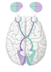
Where is the primary visual cortex located?
Where does it receive information from?
It is the area around the calcarine sulcus and occipital pole
it receives information from the retina
(LG = lateral geniculate nucleus of thalamus)

How is the primary visual cortex organised?
Retinotopically organised
information is projected onto the retina in a specific way
right and left visual fields are sent to specific areas of the cortex
Where is the association visual cortex found?
What is its function?
It surrounds the primary visual cortex
it is involved in interpretation of visual information
e.g. Recognising faces, objects, etc.

Where is the taste cortex located?
It extends from the inferior margin of the post-central gyrus
It is known as the insula
the lateral fissure must be opened up in order to see the insula

What is the role of the general association cortex?
How is it divided?
It is involved in more complex aspects of behavioural and intellectual functioning
It is divided into the prefrontal cortex and parieto-temporal cortex

What are the 2 language areas within the dominant hemisphere?
What does damage to these areas result in?
Broca’s area and Wernicke’s area - connected by the arcuate fasciculus
Damage to either of these areas leads to aphasia - an impairment of language
