Descending Motor Pathways Flashcards
How are motor pathways divided?
Into upper and lower motor neurone regions
They involve a 2 neurone pathway
What are the characteristics of upper motor neurones?
Where do they originate from?
Originate in the cerebrum and subcortical structures
they influence lower motor neurone activity
they modify local reflex activity
they superimpose more complex patterns of movement
What are the roles of lower motor neurones?
Where do they originate from?
They originate from the brainstem and the ventral grey horn of the spinal cord
they are peripheral nerves which travel to motor end plates / neuromuscular junctions
they are in direct contact with the muscle
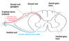
Label the diagram showing a typical lower motor neurone

What type of information is carried by the lower motor neurones?
Afferent nerve:
- visceral sensory and somatic
- these travel in the dorsal root
Efferent nerve:
- somatic motor
- these travel in the ventral root
- the cell body is in the ventral grey horn
What are the 3 main categories of descending motor pathways?
Corticospinal:
- the cell body is in the cortex and it runs to the spinal cord
Corticobulbar / corticonuclear:
- corticobulbar is from the cortex to the brainstem
- corticonuclear is from the cortex to the cranial nerve nuclei in the brainstem
Extrapyramidal:
- these originate in other regions outside of the cerebrum

What are examples of extrapyramidal descending motor pathways?
- reticulospinal - from reticular formation
- rubrospinal
- tectospinal
- vestibulospinal
These are mainly involved in modification of the main pathways elicited in the cortex
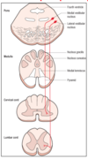
In general, what is the pathway like of the corticospinal / corticonuclear descending motor pathway?
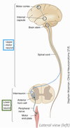
- cerebral cortex
- cell bodies in the precentral gyrus
- UMN descends via the internal capsule
- it passes between the cerebral peduncles and through the medullary pyramids
- crosses the midline at the decussation of pyramids
What is the difference between corticonuclear and corticospinal pathways?
corticonuclear:
- from the cortex to the brainstem
- lower motor neurone located in the cranial nerve nuclei for a specific function
corticospinal:
- from the cortex to the spine
- involves spinal nerves
Label the components involved in the corticospinal / corticonuclear descending motor pathway

What areas of the somatotopic organisation of descending fibres are missing?


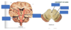
Through which regions of the internal capsule do the corticospinal/nuclear fibres pass through?
the fibres retain somatotopic organisation as they pass through the internal capsule
those travelling to the face travel in the genu
those travelling to the arm, trunk and leg travel in the posterior limb
Label the components of the internal capsule

the posterior limb is located between the thalamus and the lentiform nucleus
Which
What is the difference between the fibres contained within the internal capsule and the crus cerebri (cerebral peduncles)?
the internal capsule also contains ascending sensory fibres that connect to the thalamus
the internal capsule connects to the crus cerebri
the peduncles contain descending fibres only
Label the features of the cerebral peduncles

What is significant about the descending motor fibres within the crus cerebri?
somatotopic representation is still present
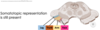
Label the location of descending fibres in different parts of the brain

- fibres move from the peduncles, through the basal pons and pyramids
- in the pons, the fibres are interrupted by transpontine fibres
- the fibres recollect to travel in the pyramids of the medulla
- some fibres will cross the midline at the decussation of pyramids

Label the components of the corticospinal tract
Do all the fibres cross at the decussation of pyramids?
What kind of innervation does this produce?

85% of fibres cross at the decussation of pyramids and then enter the lateral corticospinal tract
these produce contralateral innervation
15% of UMNs descend the cord ipsilaterally in the anterior corticospinal tract
these produce bilateral innervation

What types of muscles receive bilateral and contralateral innervation?
bilateral innervation:
- comes from the anterior corticospinal tract
- for axial musculature (in the midline)
- at the appropriate SC level, some fibres will cross and some remain ipsilateral
contralateral innervation:
- comes from the lateral corticospinal tract
- for limb musculature
- fibres cross at the decussation of pyramids

What test can be used to determine whether there is an upper motor neurone lesion?
Babinski test
a sharp object is run along the surface of the foot and the toes should become flexed
In UMN lesion, the big toe will be extended and the other toes will fan out

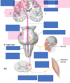
What is the exception to the bilateral innervation of the head and neck muscles?
the lower facial and extrinsic tongue muscles are under contralateral control

Label the branches of the facial nerve
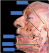

What injury would be associated with the following facial symptoms?

lower motor neurone lesion:
- Bell’s palsy
- painless unilateral weakness of the facial muscles
- weakness is present on the upper and lower face of the ipsilateral side
upper motor neurone lesion:
- weakness of the lower face only
- lesion is contralateral to lower facial muscles

What is the innervation to the extrinsic muscles of the tongue like?
innervation of the extrinsic muscles of the tongue is contralateral
these change the shape and direction of the tongue

If there is a lesion of the left hypoglossal nerve, where will the tongue deviate to?
the tongue will deviate to the left
this is ipsilateral to the lesion

If there is a lesion of fibres coming from the right side of the cortex, where will the tongue deviate to?
the tongue will deviate to the left
this is contralateral to the lesion

What structures, other than the cerebral cortex, can be involved in control of muscles?
- basal ganglia
- tectum and red nucleus
- reticular formation
- vestibular system
What are some of the roles of the reticulospinal pathway?
this runs from the reticular formation (in pons and medulla) to the spinal cord
it is invovled in voluntary movement, breathing and consciousness

What are some of the roles of the vestibulospinal pathway?
this runs from the vestibular nuclei (in pons and rostral medulla) to the spinal cord
it is invovled in controlling posture
What are some of the roles of the rubrospinal pathway?
this runs from the red nucleus (in the midbrain) to the spinal cord
it is invovled in controlling muscle tone
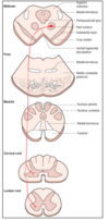
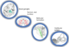
Label the descending tracts


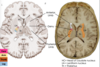
Label the locations of the major ascending and descending tracts
What sensory modalities are they concerned with?

dorsal column:
- fine touch
- pressure
- vibration
- joint position sense
spinothalamic tract:
- crude touch
- pain
- temperature
lateral & ventral corticospinal tract:
- voluntary movement
