Cerebellum (Metencephalon) Flashcards

1
Q

Where is the Cerebellum located?

A
  • largest part of the hindbrain
  • located in the posterior cranial fossa
  • behind the pons and medulla oblongata
  • separated by the 4th ventricle
2
Q

What is tentorium cerebelli?

A

an extension of the dura mater that separates the cerebellum from the inferior portion of the occipital lobes.

3
Q

Role of cerebellum

A
  • Maintenance of Equilibrium - balance, posture, eye movement

posture, gait

  • Adjustment of Muscle Tone
  • coordination of voluntary movements
  • Motor Leaning – Motor Skills
  • Cognitive Function
  • does not initiate movement
4
Q

Does the cerebellum relay directly to motor neurons

A
  • no
  • exerts action indirectly via circuits which terminate onto the UMN or the LMN
  • major component of extrapyramidal motor system
5
Q

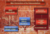
How does the cerebellum communicate with other structures

A

via superior, middle and inferior cerebellar peduncles

  • afferent and efferent connections run between the cerebellum, brainstem and SC travel through cerebellar peduncles
6
Q

What are cerebellar peduncles

A
  • a nerve tract that permits communication between the cerebellum and the other parts of the central nervous system
  • Three pairs of cerebellar peduncles conduct this communication
7
Q

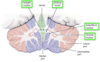
Describe the structure of the cerebellum

A
  • consists of two large cerebral hemispheres united in the middle by the vermis
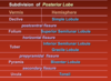
  • numerous transverse fissures divide the cerebellum into three lobes - anterior, posterior and flocculonodular
  • longitudinal divisons - vermis, paravermal region and cerebellar hemisphere
  • contain 8 lobules
8
Q

Superior Cerebellar Peduncle - function, what borders do they form and what do they connect?

A
  • brachia conjuctiva
  • major efferent fibers of cerebellum
  • emerge from the upper and medial part of the white matter of the cerebellum
  • they form the upper and lateral boundaries of the fourth ventricle
  • they connect the cerebellar nuclei, mainly the dentate nuclei of the cerebellum, with the midbrain structures and the thalamus
9
Q

Inferior Cerebellar Peduncle -

A
  • sometimes named restiform bodies
  • formed by fibers of the posterior spinocerebellar tract and the axons of the inferior olivary nucleus
  • they lie between the lower part of the fourth ventricle and the roots of the glossopharyngeal and vagus nerves
  • contain mainly afferent fibers
  • from SC and MO
10
Q

Middle Cerebellar Peduncles

A
  • brachia pontis
  • largest peduncle
  • contain only afferent fibers
  • fibers of these peduncles come from the pontine nuclei of the opposite side, so, the middle cerebellar peduncles connect the cerebellum with the pons
  • pontine nuclei receive input from the cerebral cortex, the stimuli arrive via the middle cerebellar peduncles in the cortex of the cerebellar hemispheres
  • these fibers that arrive at the cerebellum from the pathway are part of the so-called mossy fiber system
11
Q

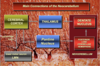
How the peduncles bring about movement

A
  • The inferior peduncles bring sensory information about the actual position of body parts such as limbs and joints
  • The middle peduncles transmit information about the desired position of these parts.
  • After integrating and analyzing, the cerebellum sends impulses through the superior peduncles to the midbrain
  • In response, motor impulses are transmitted down through the pons, medulla oblongata, and spinal cord to stimulate or inhibit skeletal muscles at appropriate times and cause movements of body parts into the desired positions
  • This activity makes rapid and complex muscular movements possible.
12
Q

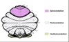
Longitudinal divisons of cerebellum

A
13
Q

Transverse divisions of cerebellum

A
14
Q

Function of Anterior lobe

A

responsible for mediating unconscious proprioception and receives input mainly from the spinal cord.

15
Q

Function of Posterior Lobe

A

important role in fine motor coordination specifically in the inhibition of involuntary movement via inhibitory neurotransmitters especially GABA

  • The posterior lobe receives input mainly from the brainstem and the cerebral cortex
16
Q

Subdivisions of flocculonodular lobe

A

Nodulus and Flocculus

17
Q

Subdivisions of anterior lobe

A
18
Q

Subdivision of posterior lobe

A
19
Q

What is cerebellum composed of?

A
  • composed of grey cerebellar cortex, medullary core of white matter and 4 parts of intrinsic nuclei
  • corpus medullare - white matter
  • deep nuclei - fastigial, globose, emboliform and dentate - main centers of communication
  • globose + emboliform = interposed nucleus
20
Q

How is the white matter arranged in the cerebellum?

A

assumes a laminar organisation and on sagittal sections has a branching appearance

21
Q

Dentate Nucleus

A
  • center of corpus medullare
  • convex, indented lamina of grey matter with a hillum open anteromedially
  • interior if filled with efferent white fibers - leave the nucleus and form the major part of the superior cerebella peduncle
22
Q

Emboliform Nucleus (interposed)

A
  • lies at the hillum of the dentate nucleus
  • youngest of the cerebellar nuclei (w/ dentate)
  • receives input from cerebrocerebeullum (lateral parts/hemispheres)
  • projects axons to the contralateral red nucleus and thalamus via SCP
23
Q

Globose Nucleus

A
  • includes several small cellular groups found medially to the emboliform nucleus
  • associated with spinocerebellum
24
Q

Fastigial nucleus

A
  • it is close to the midline
  • oldes nucleus
  • associated with vestibulocerebellum
  • receives afferents from it and projects it to the vestibular nuclei in the pons and RF
25
Q

Cerebellar Cortex

A
  • modulate information flowing through the deep nuclei
  • 3 layers - molecular, purkinje cell layer and granular layer
26
Q

Describe the molecular layer of the cortex

A
  • contains two types of inhibitor neurons - (outer) stellate and (inner) basket cells
  • external layer
  • deep to pia mater and contain very few cells
27
Q

Describe stellate and basket cells

A
  • molecular layer

Stellate cells

  • small interneurons; their processes do not extend beyond the external layer
  • their axons form inhibitor synapses with the dendrites of the purkinje cells
  • NT: taurine

Basket Cells

  • larger, with numerous dendrites
  • axon of each cell forms a basket-like plexus around the bodies of a group of purkinje cells, establishing axo-somatic inhibitory synapses
  • NT: GABA
28
Q

Describe the purkinje layer of the cortex - function, arrangement, pathway, collateral branches

A
  • primary neurons of cortex and provides its sole output
  • made up of bodies of the purkinje cells
  • arranged in a singular layer, but the processes penetrate other layers
  • they are relatively large
  • the dendrites branch heavily with the molecular layer
  • their axons arise at the base of the cell bodies and pass through the granular layer
  • when they enter white matter, they get myelin sheaths and terminate at the cerebellar nuclei with inhibitor synapses (GABA)
  • collateral branches of the axons form inhibitor synapses with other purkinje, basket and golgi cells (afferent?)
29
Q

What do the few purkinje cells, who bypass the cerebellar nuclei, synapse with?

A
  • vestibular nuclei in the pons
30
Q

The excitation of purkinje cells is regulated by what?

A

feed-back mechanisms through closed circuit chains of two or more neurons, confined within the cerebellar cortex

31
Q

Describe the granular layer of the cortex

A
  • deepest, superficial to white matter
  • packed with small cells which are characterised with densely strained nuclei and scarce cytoplasm
  • these are granule and golgi cells (and unipolar bush cells)
32
Q

Describe granular cells

A
  • granular layer

Granule cells

  • scarce cytoplasm, short heavily branched dendrites and a long non-myelinated axon
  • dendrites do not reach beyond the granular layer, but the axon reaches the molecular layer (runs perpendicular to the purkinje cells)
  • establishes numerous axodendritic inhibitor synapses with them
  • synapse with stellate, basket and dendrites of golgi
33
Q

Describe Golgi cells

A
  • granular layer
  • distributed in the upper part of the granular layer, near the purkinje cells
  • their dendrites branch in the molecular layer in their axons - in the granular layer itself
  • they form axodendritic inhibitor synapses with the granule cells at the site of the glomeruli
34
Q

Granular and Golgi afferent and efferent

A
  • afferent: mossy fiber
  • efferent: purkinje cell dendrite, basket, stellate, golgi
  • afferent: parallel fiber, mossy fiber rosette
  • efferent: granule cell dendrite
35
Q

Where does the cerebellar cortex receive its input from?

A
  • massive excitatory input from the spinal cord and brainstem
  • two types of afferent fibers: climbing and mossy
36
Q

Climbing fibers - where it starts form and its path to the molecular layer

A
  • terminal fibers of the olivocerebellar tract
  • originate at the inferior olivary nucleus in the medulla oblongata
  • pass through the ICP and reach the cortex
  • ascend through the layers of the cortex (in a manner resembling a vine on a tree)
  • before reaching molecular layer, it sends collateral branches to and synapse with the cerebellar nuclei to the interneurons of the cortex - granule, golgi, stellate, basket
  • in the molecular layer, each climbing fiber wraps around and makes a large number of synaptic contacts with the dendritic complexes of the purkinje cells
37
Q

Mossy fibers

A
  • other afferent cerebellar tracts (exc. olivocerebellar) terminate at the cerebellar cortex via mossy fibers
  • thick axons, branch heavily
  • in granular layer, their thin terminations synapse with the dendrites of the granule cells, forming globular structures (cerebellar glomeruli)
  • stimulatory effect of mossy fibers is then transferred by the granule cells to the purkinje cells
  • through these numerous connections, they can stimulate thousands or purkinje cells - more diffuse excitatory effect on purkinje cells
38
Q

Describe the synaptic glomerulus

A
  • small intertwined mass of nerve fiber (afferent) terminals in granular layer
  • consists of post-synaptic granule cell dendrites and pre-synaptic golgi cell axon terminals surrounding the pre-synaptic terminals of mossy fibers
  • processing station for afferent nerve fibers entering the cerebellum
39
Q

Describe the detail of the glomerulus

A
  • input comes from mossy fibers (afferent) - (except inferior olivary input) which terminate there and synapse with golgi and granule
  • granular cell dendrite - main afferent input
  • golgi cell axon - synapse on granule cell dendrite, GABA, surrounded by astrocyte foot process
40
Q

Classification of phylogenetic and ontogenetic development, afferent connection and efferent connection of cerebellum respectively

A

Archicerebellum - Vestibulocerebellum - Vermis

Paleocerebellum - Spinocerebellum - Paravermal Region

Neocerebellum, Pontocerebellum, Cerebellar Hemisphere

41
Q

Vestibulocerebellum

A
  • archeocerebellum
  • represented by nodulus and flocculus and fastigeal nucleus
  • bilaterally connected to the vestibular nuclei
  • involves consecutive input from the vestibular nuclei in the pons (vestibulocerebellar tract)
  • rise to vestibulospinal tract - descends to LMN
  • influence of the ipsilateral extensor muscle tone associated with maintenance of eqm.
42
Q

Main connections of vestibulocerebellum

A
43
Q

Spinocerebellum

A
  • paleocerebellum
  • vermis and medial hemisphere
  • emboliform and (part of fastigeal which has connections with SC)
  • trunco-cerebellar citcuitry - red nucleus and inferior olivary nucleus
  • rubroolivary tract and olivocerebellar tract
  • maintenance of posture, muscle tone, movements of the axial and proxomal musculature
44
Q

Main connections of Paleocerebellum

A
45
Q

Cerebrocerebellum

A
  • neocerebellum
  • dentate
  • linked to basilar part of pons
  • most advanced module
  • involved in motor precision, learning and memory
  • corticopontine tract - pontocerebellar tract
46
Q

Main connections of neocerebellum

A
47
Q

Afferent connections of Cerebellum

A
  • comprise of different pathways form different parts of the CNS that terminate at the cerebeller cortex
  • pass through all 3 peduncles
48
Q

Afferent connections of inferior cerebellar peduncle - 3 nuclei and 2 bodies

A
  • transmits afferents from:
    1. nucleus dorsalis of the spinal medulla (posterior spinocerebellar tract)
    2. nucleus cuneatus and olivaris inferior of med.obl. (cuneocerebellar and olivocerebellar tracts)
    3. vestibular nuclei of pons (vestibulocerebella tract)
  • Restiform body - This bundle contains mainly fibers that arise in the spinal cord or medulla
  • Juxtarestiform body - subdivison of inferior cerebellar peduncle
49
Q

Tracts in restiform body

A

Posterior Spinocerebellar Tract

Olivocerebellar tract

Cuneocerebellar Tract

Reticulocerebellar Tract

50
Q

Tracts in juxtarestiform body

A

Vestibulocerebellar Tract

Primary Vestibular Fiber

51
Q

Afferent connections of middle cerebellar peduncle

A

Pontocerebellar fiber

Corticopontocerebellar Fiber

Reticulocerebellar Fiber

  • made only of the pontocerebellar tract - from the pontine nuclei to the cerebro-cerebellar cortex, where they temrinate as mossy fibers
52
Q

Afferent connections of superior cerebellar peduncle

A
  • form spinal cord to the cerebellum - Anterior (ventral) Spinocerebellar Tract

from RF - reticulocerebellar tract

  • from the locus cerleus of the pontine tegmentum

Cerulocerebellar fiber

Raphecerebellar fiber

Rubrocerebellar fiber

Hypothalamocerebellar fiber

53
Q

Where do the afferent tracts of the superior peduncle terminate into?

A

in the spinocerebellar cortex as mossy fibers

54
Q

Efferent connections of the cerebellum

A
  • most from SCP
  • originate in the cerebellar nuclei (vide supra)
  • begin from cerebellar nuclei
  • in the midbrain, they cross the midline and terminte at three different domains:
  • red nucleus - cerebellorubral tract
  • thalamus - cerebellothalamic tract (NVA)
  • RF nuclei - cerebelloreticular fibers
55
Q

Efferent connections in superior cerebellar peduncle

A

Cerebellothalamic fiber

  • from 3 deep nuclei to ventrolateral preoptic nucleus (VPLo), ventral lateral nucleus (VLc) and central lateral nucleus (CL)

Cerebellorubral fiber

  • from nucleus interpositus and dentate nucleus

ascending portion of

uncinate fasciculus of Russell

56
Q

Efferent connections of inferior cerebellar peduncle

A
  • the cerebelloolivary tract to the inferior olivary nucleus
  • cerebellovestibular tract - from the fastigeal nucleus to the lateral vestibular nucleus

Fastigiovestibular fiber

descending portion of uncinate fasciculus of Russell

57
Q

Where do the tracts from the inferior cerebellar peduncle terminate in?

A

cortex of the vestibulo and spino-cerebellum

58
Q

Pyramidal tract and associated circuits

A
59
Q

Cerebellum and Automatic Motor Control

A
60
Q

Corticonuclear connections

A

A zone - fastigial and medial vestibular nucleus

B zone - lateral vestibular nucleus

C1, C3 zone - emboliform nucleus

C2 zone - globose nucleus

D1 - parvocellular portion of dentate nucleus

D2 - magnocellular portion of dentate nucleus

61
Q

Olivocerebellar connections

A
  • caudal (post.) portion of medial and dorsal accessory olivary nucleus, vermis of cortex (A,B), fastigial and vestibular nucleus
  • rostral (near nose) portion of medial and dorsal accessory olivary nucleus, paravermal region (C1,2,3) and nucleus interpositus
  • principal inferior olviary nucleus - cerebellar hemisphere (D1,2) and dentate nucleus
62
Q

Cerebellar Ataxia

A
  • incoordination of movement
  • decomposition of movement
  • dysmetria, past-pointing - lack of coordination of movement
  • dysdiadochokinesia - cannot perform rapid movements
  • rebound phenomenon of Holmes reflex that occurs when one attempts to move a limb against resistance that is suddenly removed

- gait ataxia, truncal ataxia, titubation - deviation from normal walking

  • Intention Tremor, Hypotonia and Nystagmus
63
Q

Different lesions on the cerebellum which cause a form of ataxia

A

Archicerebellar (vestibul-) Lesion: medulloblastoma

Paleocerebellar (spino-) Lesion: gait disturbance

Neocerebellar (cerebro-) Lesion: hypotonia, ataxia, tremor

64
Q

Describe Cerebellar Medulloblastoma

A

Cerebellar tumors on vermis

  • Truncal Ataxia
  • Frequent Falling
65
Q

Function of Flocculus

A

the flocculus receives input from the inner ears’ vestibular system and regulates then balance

  • In addition, many floccular projections connect the motor nuclei involved in control of eye movement.