Telencephalon (Cerebrum) Flashcards
(43 cards)
Telencephalon
- large part of the brain containing the cerebral cortex (of the two cerebral hemispheres)
- as well as several subcortical structures, including the hippocampus, basal ganglia, and olfactory bulb
- the cerebrum is the uppermost region of the central nervous system
Where does the telencephalon develop from?
The prosencephalon or forebrain is the embryonic structure from which the cerebrum develops prenatally
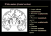
Telencephalic white matter - projection fibers
- projection fibers consist of efferent and afferent fibers uniting the cortex with the lower/other parts of the brain and with the spinal cord
e. g. Corona radiata - white matter sheet of both ascending and descending axons carries most of the neural traffic from and to the cerebral cortex - The corona radiata is associated with the corticopontine tract, the corticobulbar tract, and the corticospinal tract.

Telencephalic white matter - commissural fibers
- or transverse fibers
- axons that connect the two hemispheres of the brain
Corpus Callosum
- It is the largest white matter structure in the human brain
- wide, thick nerve tract, consisting of a flat bundle of commissural fibers
- beneath the cerebral cortex in the brain
- only found in placental mammals
- It spans part of the longitudinal fissure, connecting the left and right cerebral hemispheres, enabling communication between them
- four main parts; individual nerve tracts that connect different parts of the hemispheres. These are the rostrum, the genu, the trunk or body, and the splenium
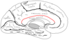
Relations of Corpus Callosum
- fibers radiate in the white matter and pass to the various parts of the cerebral cortex
- those curving forward from the genu into the frontal lobes - forceps minor
- those curving backward from the splenium into the occipital lobes - forceps major (also forceps posterior)
tapetum - main body of fibers between these two parts
Commissura anterior
- commissural fiber, pars anterior/post.
- white matter tract (a bundle of axons) connecting the two temporal lobes of the cerebral hemispheres across the midline
- placed in front of the columns of the fornix
- The great majority of fibers connecting the two hemispheres travel through the corpus callosum, which is over 10 times larger
- key role in pain sensation, more specifically sharp, acute pain, contains decussating fibers from olfactory tracts
Commissura fornicis
- commissural fiber
- a C-shaped bundle of nerve fibers in the brain that acts as the major output tract of the hippocampus
- also carries some afferent fibres to the hippocampus from structures in the diencephalon and basal forebrain
- The fornix is part of the limbic system
Commissura posterior
- left and right parts of tectum and tegmentum of midbrain
- important in the bilateral pupillary light reflex.
Association fibers
- axons that connect cortical areas within the same cerebral hemisphere
- short fibers - Many of the short association fibers (also called arcuate or “U”-fibers) lie immediately beneath the gray substance of the cortex of the hemispheres, and connect together adjacent gyri.
- Some pass from one wall of the sulcus to the other
Long association fibers
- The long association fibers connect the more widely separated gyri and are grouped into bundles
Fasciculus longitudinalis superior
- ass. f.
- frontal lobe to occipital lobe
- ass. f.

Fasciculus longitudinalis inferior
- ass. f.
occipital to temporal lobe

Cingulum
- ass. f.
- cingulate gyrus to entorhinal cortex
- ass. f.

Fasciculus uncinatus
4.
- ass. f.
frontal lobe to temporal lobe
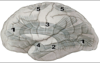
Speech areas
Lateralisation of functions

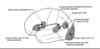
Split brain syndrome
- the corpus callosum connecting the two hemispheres of the brain is severed to some degree
dominant hemisphere - mentions the stimulus
nondominant hemisphere - points the stimulus
anomia - can not name stimuli on the left hand
alexia in the left visual field
test - hybrid face

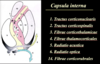
Internal Capsule
- white matter structure situated in the inferomedial part of each cerebral hemisphere of the brain
- It carries information past the basal ganglia
- contains both ascending and descending axons, going to and coming from the cerebral cortex
- also separates the caudate nucleus and the putamen in the dorsal striatum, a brain region involved in motor and reward pathways.
- corticospinal tract is a large part of the internal capsule, carrying motor information from the primary motor cortex to the lower motor neurons in the spinal cord
- Above the basal ganglia the corticospinal tract is a part of the corona radiata, below the basal ganglia the tract is called cerebral crus(a part of the cerebral peduncle) and below the pons it is referred to as the corticospinal tract.
Structure of Internal Capsule
- consists of three parts and is V-shaped when cut horizontally, in a transverse plane
- the bend in the V is called the genu
the anterior limb or crus anterius is the part in front of the genu, between the head of the caudate nucleus and the lenticular nucleus
- the posterior limb or crus posterius is the part behind the genu, between the thalamus and lenticular nucleus
- the retrolenticular portion is caudal to the lenticular nucleus and carries the optic radiation (from medial part of lateral geniculate nucleus) also known as the geniculocalcarine tract
- the sublenticular portion is beneath the lenticular nucleus and are tracts involved in the auditory pathway from the medial geniculate nucleus to the primary auditory cortex (Brodmann areas 41 and 42)
- thalamolenticular portion - between thalamus and lenticular nucleus
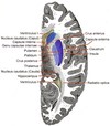
Internal Capsule tracts and fibers
Tracts on frontal section of cerebrum
Basal Ganglia - what it is, what it contains, connections and functions
- masses of grey matter situated within each hemisphere
- include caudate and lentiform nucleus
- substantia nigra and subthalamic nucleus is also present
- strongly interconnected with the cerebral cortex, thalamus, and brainstem, as well as several other brain areas - major relay station
- associated with a variety of functions, including control of voluntary motor movements, procedural learning, habit learning, eye movements, cognition, and emotion.
- surrounded by white matter (internal, external and extrema capsule)

Components of the Basal Ganglia
- lentiform nucleus has two parts - lateral (putamen), medial (globus pallidus)
- GP (also called pallidum) is further divides into external (GPe) and internal (GPi)
- putamen and caudate nucleus united by origin, structure, function - known as striatum


