Upper Limb Flashcards
Articulation of Clavicle
- medially with sternum
- laterally with acromion process
- forms pectoral girdle
Function of clavicle
- connects axial skeleton to UL
- allowing weight to be transferred from the upper limb to the axial skeleton
- protect neurovascular structures as they travel between the trunk and the upper limb.
What joints does the clavicle form?
- sternoclavicular joint
- acromioclavicular joint - keep movements of the scapula separated from the trunk when the arm is moved in various ways.
Impression for costoclavicular ligament
clavicle
attachment of costaclavicular ligament which connects clavicle to first rib
Subclavian groove
clavicle
-attachment for subclavius muscle
conoid tubercle
- attachment for conoid ligament - involved in coracoclavicular ligament
- The conoid ligament of the coracoclavicular ligament runs from the inferior surface of the clavicle, the conoid tubercle to the superior aspect of the coracoid process of the scapula
trapezoid line
clavicle
- attachment site for trapezoid ligament
lateral part of coracoclavicular ligament
- runs from the inferior aspect of the clavicle to the coracoid process of the scapula.
intertubercular sulcus
- humerus
- bicipital groove
- because this is where you will find the proximal tendon of the long head of the biceps brachii muscle as it courses distally towards its muscular belly in the arm
anterior divisons of the surface of humerus
anteromedial, anterolateral
- anterior border runs from crest of greater tubercle
deltoid tuberosity
humerus
- lateral midshaft
- point of insertion or distal attachment for the deltoid muscle.
fossae
humerus
radial fossa which is located laterally
- medial side, coronoid fossa.
medial epicondyle
humerus
attachment for several of the anterior or flexor muscles of the forearm
lateral epicondyle
humerus
attachment to the posterior muscles or extensor muscles of the forearm
radial groove
humerus
- posterior
- radial nerve runs through
- same for ulnar groove on distal end
subscapular fossa
scapula
gives origin to the subscapularis muscles which extends laterally to the humerus
scapulothoracic joint
physiological joint as opposed to a true anatomical joint due to the fact that there is no bony articulation here.
coracoid process
- scapula
- attachment site for several muscles such as the short head of the biceps brachii, pectoralis minor, and coracobrachialis muscles.
supraspinous and infraspinous fossa
scapula, posterior
- provide attachments for muscle of the rotator cuff which are the supraspinatus and infraspinatus muscles respectively
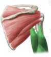
supraglenoid tubercle
- scapula
- proximal attachment point or origin of the long head of the biceps brachii muscle
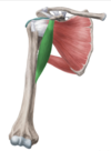
infraglenoid tubercle
- scapula
provides attachment for the long head of the triceps brachii muscle
coracoclavicular ligament
more medial is known as the conoid ligament which originates from the knuckle of the coracoid process
- trapezoid ligament which runs from the trapezoid edge of the coracoid process
- One primary function of this ligament is to hold the acromial end of the clavicle in place preventing it dislocating from the acromioclavicular joint.
coracoacromial ligament
extends from the coracoid process to the acromion of the scapula
forms what is known as the coracoacromial arch
Acromioclavicular joint
- synovial joint
- connects the acromion of the scapula with the clavicle at its lateral end
- This joint is stabilized by two sets of ligaments – the coracoacromial ligament and the coracoclavicular ligaments
- functions to allow anterior and posterior movement of the acromion during the protraction and retraction of the scapula, rotation during adduction and abduction of the shoulder, and the tilting of the acromion during adduction and abduction of the arm.

glenohumeral ligaments, Flood’s ligament
- superior - supraglenoid tubercle of the scapula to the medial ridge of the intertubercular groove of the humerus
middle - originating inferior to the superior glenohumeral ligament and inserting on the lesser tubercle of the humerus
inferior - originates mostly along the inferior edge of the glenoid margin and attaches onto the neck of the humerus
- all the ligaments blend with the capsule and are not clearly distinguishable.
- function to reinforce the articular capsule of the glenohumeral joint