Ch1 Anatomy Flashcards

(118 cards)

1
Q

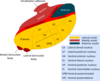
What are the ligaments of the atlanto-occipital junction from anterior to posterior?

A

Anterior atlanto-occipital membrane (continuation of the ALL) Apical ligament Cruciate ligament Tectorial membrane (continuation of the PLL) Dura Spinal cord Dura Ligamentum flavum Posterior atlanto-occipital membrane (continuation of the interspinous ligament)

2
Q

What structure lies between the cruciate ligament and the dura at the craniocervical junction?

A

The tectorial membrane (which is a continuation of the PLL)

3
Q

Where does the tectorial membrane attach?

A

The posterior aspects of the VBs of C2/3 to the basion of the foramen magnum. An accessory portion connects the C2 to the occipital condyles laterally.

4
Q

What is the dentate ligament?

A

Separates ventral and dorsal roots

5
Q

What is the lambda?

A

Junction of the lambdoid suture and the sup sag suture

6
Q

What is the Stephanion?

A

Junction of the coronal suture and the superior temporal line

7
Q

Which bones form the pterion?

A

Frontal, parietal, temporal and sphenoid joined by the coronal suture and the squamosal suture

8
Q

What markings can be used to identify the central sulcus on the skull surface?

A

Taylor-Haughton lines: 1/2 way between nasion and inion, 2 cm behind marks the top of the central sulcus. (also marked by the posterio ear line)

Orbito-zygomatic suture to 3/4 point marks the sylvian fissue

The bottom of the central sulcus is the condylar line (vertical line from the condylar process)

9
Q

What is the frankfurt plane?

A

Line from inferior orbit to the upper part of EAM

10
Q

Which skull suture marks the sylvian fissure?

A

The squamosal suture

11
Q

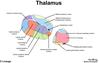
What is the length of the third ventricle?

A

28 mm

12
Q

What level is the scapula spine?

A

T2/3

13
Q

What level is the inferior scapula?

A

T6

14
Q

What is the intercristal line?

A

Line between the iliac crests - marks L4/5

15
Q

What is Bill’s bar?

A

Vertical crest - separates the 7UP (facial) and Coke Down (Cochlear) nerves anteriorly from the superior and inferior vestibular nerves posteriorly

16
Q

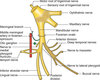
What are the branches of V1?

A

Frontal

Lacrimal

Nasocillary

17
Q

What are the contents of the Sup Orbital Fissure?

A

CN3, 4, V1, 6

Superior opthalmic vein

18
Q

What passes through the Inf Orbital Fissure?

A

Infraorbital veins and arteries and inferior ophthalmic veins

Infraorbital nerve V2

Zygomatic nerve V2

19
Q

What passes through F. Rotundum?

A

V2

20
Q

What passes through F. Ovale?

A

V3

21
Q

What passes through F. Spinosum?

A

Middle meningeal

22
Q

Where does the facial nerve exit the skull?

A

Stylomastoid foramen

23
Q

What passes through the mastoid foramen?

A

Mastoid emissary vein

24
Q

What fibres lie in the anterior limb of the internal capsule?

A

Corticopontine fibres

(forms the anterior thalamic radiation)

25
What fibres lie in the posterior limb of the internal capsule?
Corticospinal tracts (superior thalamic radiation)
26
What fibres lie in the posterior thalamic radiaton?
Parieto- and occipito-thalamic connections
27
What is the audtory thalamic radiation?
Sublenticular part of the internal capsule connecting the medial geniculate nucleus to the A1 (Heschl's gyrus)
28
What is the retrolenticular radiation?
The optic pathway from the LGN to occpitial lobe
29
What is the blood supply to the internal capsule?
Lateral lenticulostriates to the anterior limb ICA branches to the genu Anterior choroidal to the and posterior limb sub- and retrolenticular parts
30
What structure lies above the 7 and 8 th complex as it exits the brainstem?
The flocculus
31
What are the 5 thalamic radiations?
Anterior (frontopontine), Superior (corticospinal), Posterior (parieto-occipital), Sublenticular (auditory) and retrolenticular (optic)
32
Where is the tectorial membrane?
Superficial component: Anterior to the ventral dura (superior continuation of the PLL) Deep/accessory component: connects C2 to the occipital condyle
33
Where does the transverse ligament attach?
C1 tubercle (medial to the C1 lateral mass)
34
Which ligaments are most important for atlanto-occipital stability?
Tectorial membrane and alar ligaments
35
What is the dentate ligament?
Separates dorsal and ventral nerve roots
36
What is the function of the rubrospinal tract?
Flexor tone
37
Where is Lissauer's tract?
Entry into the dorsal entry (dorsolateral tract)
38
Describe the pain pathway
Spinothalamic pathway: Nerve to substantia gelatinosa \> crosses in anteiror white matter commissure after ascending 2-3 levels \> Latearl spinothalamic tract \> VPL \>postcentral gyrus
39
Describe the fine touch and proprioception pathway
Meissner's / Pacinian / Merkel's discs \> DRG \> Nucleus proprius (Rexed 3/4) \> dorsal columns \> gracile / cuneate nuclei \> decussation in lower medulla \> medial lemniscus \> VPL thalamus \> post. central gyrus
40
Where is the C2 dermatome?
Back of the head
41
Where is the C3 dermatome?
Front and back of the neck
42
Where is the C4 dermatome?
Above the clavicle anteriorly and above the trapezius muscle posteriorly
43
What passes through Foramen Rotundum?
V2 (Maxillary nerve) to the pterygopalatine fossa. F. Rotundum lies immediately below the SOF.
44
What passes through F. Ovale?
V3, Lesser petrosal nerve (from CN IX) and the accessory meningeal artery Emissary vein between the cavernous sinus and the pterygoid plexus
45
Where is F. Spinosum?
Posterior and lateral to foramen ovale. Formed at the junction of the Gr. Wing of sphenoid and the petrous temporal bone. The meningeal branch of V3 and the middle meningeal artery pass through this.
46
Where is F. Lacerum?
Medial to F. Ovale. Located between the petrous apex and the Gr. Wing of Sphenoid. Transmits the ICA (lacerum segment = C3), greater petrosal nerve + deep petrosal nerve = vidian nerve and sympathetics
47
Which foramen lies at the junction of the petrous temporal and occipital bones?
Jugular foramen Contains CN9, 10, 11 and the inferior petrosal sinus and sigmoid sinuses that form the IJV.
48
Between which bones is the orbit can the inferior orbital fissure be found?
The zygomatic bone laterally and the maxillary bone medially.
49
Where is the orbital process of the palatine bone in the orbit?
On the floor, posterior and medial to the inferior orbital fissure
50
What lies within the lacrimal fossa of the orbit?
The nasolacrimal duct
51
What forms the boundaries of the SOF?
The greater and lesser wings of the sphenoid bone
52
Which bone forms part of the anterior and middle cranial fossa?
The sphenoid bone
53
What fissure lies anterior to the EAM?
Petrotympanic fissure (separating the tympanic part of the temporal bone and the glenoid fossa)
54
What structures pass through the F. magnum?
Medulla, CN11, vertebral arteries, Anterior and posterior spinal arteries and basilar venous plexus
55
Where is the quadrigeminal cistern?
Between the splenium and dorsal midbrain
56
Where is the ambient cistern?
Between the lateral midbrain and the parahippocampal gyrus
57
How many Brodmann areas are there?
47
58
What are the layers of the cortex?
From outside to inside: Molecular Ext granular Ext pyramidal Int granular Int pyramidal Multiform
59
What structures compose the diencephalon?
The thalamus, hypothalamus, subthalamus and epithalamus.
60
Where is the hypothalamus?
In the ventrolateral wall and floor of the 3rd ventricle
61
What is the boundary between the thalamus and hypothalamus within the 3rd ventricle?
The hypothalamic sulcus
62
What structure separates the thalamus from the posterior limb of the internal capsule?
The external medullary lamina (invests the lateral surface of the thalamus)
63
What white matter structure within the thalamus subserves connections between the thalamic nuclei?
The internal medullary lamina
64
What is the name of the white matter that covers the dorsal surface of the thalamus?
The stratum zonale
65
What is the function of the thalamus?
Integrative center and relay for motor, sensory and limbic pathways
66
Where is the anterior nuclear group of the thalamus?
Dorsal medial surface of the thalamus
67
What structure within the thalamus forms the caudal border of the foramen of monroe?
The anterior tubercle of the thalamus
68
What lies immediately lateral to the anterior nuclear group?
The internal medullary lamina
69
What structures are damages in thiamine deficiency leading to Korsakoff's syndrome?
Anterior nuclei group, mamillothalamic tract and mamillary bodies
70
Where are the lateral group of thalamic nuclei?
Lateral to the internal medullary lamina and is subdivided into dorsal and ventral portions. The ventral lateral nuclei are sensory (ventral posterior) and motor inputs (ventral anterior and ventral lateral)
71
Where is the motor thalamus region?
Ventral anterior and ventrolateral nuclei (involved in motor planning and regulation)
72
Which part of the thalamus projects to the motor cortex?
The ventral lateral nucleus
73
What are the subsegments of the ventral posterior nucleus?
Ventral intermediate (VIM) Ventral posteromedial (VPM) Ventral posterolateral (VPL)
74
What is the role of the VPL?
Receives input from the dorsal column-medial lemniscus pathway transmitting head and body sensation (pain, temperature, fine touch and vibration)
75
What information is relayed through the ventral posterior medial (VPM)?
Sensation from the contralateral head, face and oral cavity via the trigeminothalamic tracts as well as gustatory inputs from CN7, CN9 and CN10 (nucleus solitarius)
76
Where do VPM fibres project to?
The primary sensory cortex for taste
77
Where is the medial geniculate nucleus?
Ventral caudal surface of the pulvinar. It receives inputs from the inferior colliculus (via the brachium) carrying auditory fibres. The fibres from the MGN project to Heschl's gyrus.
78
What is the role of the superior colliculus?
Afferent limb of the pupillary light and accommodation reflexes
79
What is the term given to the lateral and medial thalamic nuclei?
Metathalamus
80
Where is the medial dorsal nucleus?
Medial to the internal medullary lamina. It has limbic connections with widespread visceral and somatic inputs. Bilateral damage results in memory loss and inappropriate behaviour.
81
Where are the intralaminar nuclei?
These are enclosed by the internal medullary lamina. They are involved with wakeful, aroused and vigilant states.
82
What forms the lateral border of the hypothalamus?
The optic tracts
83
What delineates the tuber cinereum?
The supraoptic recess anteriorly and the mamillary bodies posteriorly. The infundibulum arises from a circular ridge of neural tissue in the midline called the median eminance.
84
What are the divisions of the thalamus?
Anterior (supraoptic) Middle (tuberal) Posterior (mammillary)
85
Where is the epithalamus?
In the posterior lateral wall of the third ventricle. Consists of the medial and lateral habenular nuclei and the pineal gland,
86
Which commissures form part of the epithalamus?
The habenular commissure above the pineal gland and the posterior commissure below the pineal gland.
87
Where is the subcommissural organ?
Just above the entral to the cerebral aqueduct. It is one of the circumventricular organs.
88
What is the role of the posterior commissure?
Interconnects nuclei of the pretectal and CN3 nuclei. Involved with light reflexes and eye movements.
89
What connects the thalamus to the habenular?
The stria medullaris thalami connect the habenular with the thalamus, hypothalamus and pallidum.
90
What is the output of the habenular nuclei?
Output is via the habenularpeduncular tract (fasciculus retroflexus of Meynert) to the limbic, hypothalamic, thalamic and extrapyramidal motor regions. The habenular is important interactions with olfaction, mating, reward and autonomics.
91
What cells compose the pineal gland?
Pineocytes and astrocyte-like glial cells (there are no neurons)
92
What substance is produced by the pineal gland?
Melatonin for sleep-night cycles
93
What structures compose the subthalamic region?
Subthalamic nucleus Zona incerta Forel's field H (Motor and sensory tracts to the thalamus also pass through)
94
Where is the STN?
It is an oval shaped structure between the Zona Incerta and the Substantia Nigra. Lies medial to the internal capsule and lateral to the hypothalamus.
95
What is the main input to STN?
The GPe (part of the indirect pathway) via the subthalamic fasciculus which traverses the posterior limb of the internal capsule.
96
What are the connections of the GPi to the thalamus?
Ansa lenticularis (loops under the STN and post. limb of the internal capsule) Lenticular fasciculus (Forels H2 - runs through the internal capsule on top of the STN)
97
Where do the efferents from the GPi go?
They activate the GPi which inhibits the thalamus and therefore inhibits movement (indirect pathway)
98
What does lesions of the STN cause?
Contralateral voluntary movement disturbances (chorea / hemiballismus). It is a DBS target for PD.
99
What is the zona incerta?
A GM region between the thalamus and STN that is encapsulated by the Forels fields.
100
Where is the H1 field of Forel?
Between the thalamus and dorsal zona incerta. Designated as the thalamic fasciculus it contains cerebellothalamic and pallidothalamic tracts.
101
Where is the H2 field of forel?
Aka lenticular fasciculus, runs under the zona incerta above the STN. Contains pallidothalamic and pallidotegmental tracts from the GPi as well as dentothalamic fibres.
102
Which gyri form the subcallosal area?
The paraolfactory and paraterminal gyri
103
What type of cortex is found in the hippocampus and dentate gyri?
Allocortex = 3 layers. (The subiculum contains transitional cortex separating the hippocampus from parahippocampal gyrus). Layers are molecular, pyramidal and polymorphic.
104
What covers the ventricular aspect of the pes hippocampus?
The alveus
105
What is the inferior rolandic point?
Where the central sulcus projects to the sylvian fissure.
106
What lies beneath the anterior sylvian point?
The apex of the insula (with the uncinate and IFOF deep to it)
107
What sulcus is continuous with the intraparietal sulcus?
The posterior central sulcus anteriorly
108
Which sulcus separates the supramarginal and angular gyri?
The intermediate sulcus of Jensen
109
What does the fusiform gyrus relate to?
The temporal horn anteriorly and floor of the atrium posteriorly
110
What lies immediately deep to the anterior perforated substance
Ventral striatum / subst. inominata
111
What is the connection between the caudate and putamen?
Nucleus accumbens
112
Where is the septal region?
Posterior to the subgenual cingulate (paraterminal gyrus)
113
Where does the anterior commissure run laterally?
Under the GPi in the canal of grateolet
114
What fibre tracts are in the sagittal striatum?
Tapetum IFOF SLF / AF Anterior commissure Optic radiation
115
What cortex is medial to the amygdala?
The semilunar gyrus
116
What are the segments of the fimbria / fornix?
(Alveus) \> Fimbria \> Fornix Crus \> Body \> Column
117
Where is the anterior commissure in relation to the fornix?
Anterior
118