Ch90+ Flashcards

(89 cards)

1
Q

What spinal level does the iliac crest demarcate?

A

L4/5 interspace (L4 spinous process)

2
Q

Label the structures:

A

1 = S2 VB

2 = SI joint (synovial portion)

3 = SI joint (non-synovial portion)

4 = Sacral ala

5 = Iliac bone

6 = S2 root

7 = S3 root

8 = Multifidus

9 = Glut. maximus

10 = Glut. medius

3
Q

Label the structures on the CT myelogram:

A
  1. Odontoid process
  2. C1 lateral mass
  3. Lateral neural arch
  4. Posterior neural arch
  5. CSF
  6. Spinal cord
  7. Anterior midline tubercle
  8. Foramen transversarium
  9. Dorsal rootlet
  10. Rectus capitus posterior major
  11. Semispinalis capitus
  12. Tubercle of the transverse ligament
4
Q

What are the muscles of the suboccipital triangle?

A

Trapezius is more superficial. SCM being lateral. Both attach to the superior nuchal line

Then Semispinalis (medial) and splenius capitus (lateral)

Rectus capitus post. major - C2 spinous process to the inferior nuchal line

Inferior oblique - C2 spinous process to C1 lateral tubercle

Superior oblique - C1 lateral tubercle to inferior nuchal line

(The rectus capitus posterior minor attaches from the C1 lamina to the inferior nuchal line)

5
Q

What are the muscle attachment at the back of the skull?

A
6
Q

Label the structures:

A
  1. C4 VB
  2. Spinal cord
  3. C5 sup articular process
  4. C4 inferior articular process
  5. Lamina
  6. Spinous process
  7. Vertebral artery
  8. Facet joint
  9. C5 DRG
  10. Dorsal rootlet
  11. Ventral rootlet
  12. Longus coli
  13. Multifidus
  14. Semispinalis capitus
  15. Splenius captius
  16. IJV

17 ICA

7
Q

What nerves supply the scalp and face?

A
  1. Supratrochlear (from V1)
  2. Supraorbital (from V1)
  3. Infratrochlear (from V1)
  4. External nasal (from V1)
  5. Infraorbital nerve (from V2)
  6. Lacrimal (from V1)
  7. Zygomaticotemporal (from V2)
  8. Zygomaticofacial (from V2)
  9. Auriculotemporal (from V3)
  10. Buccal (from V3)
  11. Mental (from V3)
  12. Greater auricular (C2/3)
  13. Transverse cervical (C4)
  14. Supraclavicular (C4)
  15. Greater occipital (C2)
  16. Lesser occipital (C2)
8
Q

Where is the hypoglossal nerve during high ACDF approach?

A

Beneath the posterior digastric running between the ICA and IJV. Runs deep to mylohyoid on the surface of hyoglossus

9
Q

How do you assess bony fusion in the spine?

A

In the absence of instrumentation lateral flexion-extension Xrays show lack of movement at that level

If there is instrumentation then CT is needed

10
Q

What is the correlation between fusion and clinical outcome?

A

Not strong

11
Q

What is osteoinduction?

A

The process of mesenchymal recruitment to develop osteoblasts and osteoclasts

12
Q

What is osteogenesis?

A

The formation of new bone

13
Q

What is osteoconduction?

A

A scaffold upon which new bone can form

14
Q

Where is autograft taken from?

A

Bone taken during decompression

Iliac crest

Rib

Fibula

Risk of donor site morbidity (~30%)

Can be cancellous / cortical / both (tricortical) or marrow

15
Q

What are the different properties of cortical vs cancellous autograft?

A

Cortical provides mechanical stability

Cancellous is osteogenic, osteoinductive and osteoconductive

16
Q

What is the most osteoinductive substance?

A

BMP

17
Q

What are the risks of using fibula autograft?

A

Common peroneal nerve palsy

Ankle instability

18
Q

Which sutures are braided and non-absorbable?

A

Silk and Ethibond

19
Q

Which sutures are braided and absorbable?

A

Vicryl

20
Q

Which monofilaments are non-absorbable?

A

Ethilon / Prolene

21
Q

Which monofilaments are absorbable

A

Monocryl, PDS and cat gut

22
Q

Which windows are tcds performed through?

A

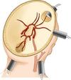
Temporal, orbital, suboccipital and submandibular
23
Q

How does the Lindegaard ratio differentiate vasospasm from hyperaemia?

A

Spasm ratio severe >6 (3-6 is mild)

Hyperaemia ratio <3

Most reliable for MCA and basilar A vasospasm.

An MCA Vmean >180 cm/s suggests spasm or an increase in 20%

24
Q

What range do fluorescein, 5ALA and ICG emit light?

A

520 nM Fluorescein

625 nM 5ALA

700-850 nM ICG

25
What is 5-ALA?
5 aminolevulinc acid A precursor of protoporphyrin 9 in the heme synthesis pathway within mitochondria (so not seen in RBCs!) Photobleaching occurs at an average of 1% per minute under UV light
26
How do you read a Vernier scale?
Align the 0 on the Vernier scale with the rule scan to get the mm distance. Then whichever of the Venier lines that lines up with the rule scale lines tells you the next decimal value. In the picture, the answer is 2.1 mm as the 0 on the vernier is at 2 on the rule scale and the 1 on the vernier is in line with the rule scale.
27
What is Joule's first law?
Heat = current density squared x resistance x time
28
What are cutting and coagulation diathermy setting?
Cutting = Pure continuous sine wave of low voltage that produces high temperature that vaporizes the tissue. Coagulation = intermitted sine wave with a longer off-time i.e. low duty cycle and a higher voltage that produces a coagulation effect. Fulguration is when the coagulation voltage is high enough to create a zone of thermal damage.
29
What is a blend diathermy setting?
A waveform similar to coagulation with a higher duty cycle producing more cut and less coagulation.
30
How do you manage patients with pacemakers pre-operatively?
Pacemakers should be checked and reprogrammed to monitor mode prior to surgery so that they do not inadvertently discharge during surgery with diathermy use. Placing a magnet over the pacemaker can do this.
31
What is the difference between Floseal and Surgiflo?
Floseal has human thrombin and bovine gelatin Surgiflo is porcine gelatin (spongistan is a solid version)
32
What is DuraGuard?
Bovine pericardium with glutaraldehyde
33
Why is partial aspiration of a cystic lesion undertaken?
To decompress the lesion, similar to debulking prior to dissection of the wall. Complete aspiration is avoided to prevent being unable to find the lesion!
34
What causes severe brain swelling during surgery?
Extra-axial bleeding ICH Venous outflow obstruction Vasodilation (check the pCO2!) Diffuse cerebral oedema from stroke
35
How do you manage severe brain swelling?
Alert the anaesthetist: Elevate the head Prevent neck vein kinking - place head in neutral position Reduce the CO2 to between 30-35 mmHg Drain CSF Mannitol bolus (1g/Kg IV) Deepen the sedation to induce burst suppression Intraoperative US to identify an ICH that can be evacuated Decompressive craniectomy
36
What is the risk of a post-op haematoma following craniotomy?
1%
37
What are the causes of a post-operative decline following craniotomy?
Cerebral oedema Haematoma Hydrocephalus (esp. if intraventricular/intracisternal) Infarction Pneumocephalus Seizure
38
Why is intraoperative mapping performed?
To identify eloquent regions of brain such as motor and language cortices. Mapping can be performed awake or asleep.
39
What methods are used to identify the motor cortex?
Phase reversal (performed asleep) - a strip is placed traversing the central sulcus covering precentral and postcentral cortices. SSEPs are undertake with peripheral stimulation. Phase reversal of N20 to become P20 indicates the location of the central sulcus. Direct cortical / subcortical stimulation (awake surgery)
40
Which nerves are anaesthetised with a scalp block?
Supraorbital / supratrochlear Auriculotemporal Posterior auricular branch of the greater occipital Greater and lesser occipital nerves
41
What stimulation settings do you use for cortical mapping?
Using a bipolar electrode: 50Hz Biphasic square wave (2ms peak to peak) Current 1-10 mA A recording strip is also placed on the brain surface. Start at a low current and stimulate for 3-5 seconds and observe for after discharges on the recording strip. Increase current until after discharges occur. Use this as the threshold for speech mapping looking for speech arrest, dysnomia, semantic and phonemic paraphasias
42
Where does cortical stimulation result in speech arrest?
Pars opercularis / precentral gyrus
43
Where does cortical stimulation result in dysnomia?
Frontal and parietal operculum of the dominant hemisphere (and IFOF subcortically)
44
Where does cortical stimulation result in semantic paraphasias?
Inferior frontal, supramarginal and posterior temporal regions
45
Where does cortical stimulation result in phonemic paraphasias?
Superior temporal gyrus (and arcuate fasciculs)
46
What positioning options are there for posterior fossa approaches?
Prone / concorde Park bench Sitting
47
Advantages and disadvantages of the sitting position?
Advantage - CSF and blood drain from surgical site, gravitation retraction and enhanced venous drainage. Disadvantage - Air embolism, fatigue, air embolism, pneumocephalus, sciatic nerve injury and reduced cerebral blood flow.
48
Why do venous emboli occur?
As the venous sinuses are non-collapsible and the negative pressure entrains air. This accumulates in the right atrium and creates an airlock preventing venous return to the heart. Note a patent foramen ovale can cause a paradoxical air embolism resulting in a stroke!
49
How do you treat a venous air embolism?
Occlude the opening by placing a soaking wet swab over the field and flood the field with saline. Lower the head initially Jugular venous compression if accessible Rotate the patient left side down (Durant's manoeuvre) Aspirate air from the right atrium via CVP Ventilate with 100% O2 Use pressors and fluid to maintain BP
50
What is the earliest sign of venous air embolism?
Reduction in end-tidal CO2
51
How can you monitor for air embolism?
Precordial doppler
52
How do you place a patient in the park bench position?
Position the patient at the top of the table so the arm can extend down over the top. Place a roll under the chest wall (not pushed into the axilla, as this may damage the brachial plexus). Upper arm / shoulder are taped downward and forwards to open the working space. Head placed in mayfield clamp looking 30 degrees to the floor. Elevate the head 15 degrees. Round bolster placed at the ASIS and flat bolster on the back. Pillow in between the knees
53
Where do you place the incision for a retrosigmoid craniectomy?
5 mm behind the digastric groove of the mastoid
54
What incision do you use to access a unilateral cerebellar hemisphere?
Mitre/hockey-stick incision. Starting from C2 up to the inion and across (below the transverse sinus) then curve down to the mastoid
55
Where is the asterion?
Junction of the lambdoid, parieto-mastoid and occipito-mastoid sutures. It is an inconsistent marker for the transverse-sigmoid junction.
56
How do you seal mastoid air cells?
Pack them with bone dust mixed with bone wax
57
Where are Frazier and Dandy burr holes for ventriculostomy?
Frazier - 6 cm above and 3 cm lateral Dandy - 3cm above and 3cm lateral (used in paediatrics)
58
How do you retract the cerebellum following retrosigmoid craniectomy?
Inferior retraction at the petrotentorial junction gives access to CN5 Medial retraction of the lateral cerebellar hemisphere gives access to CN7/8 Superior retraction gives access to the CN9/10/11
59
What forms the floor of the 4th ventricle?
The upper 2/3 are formed by the pons and lower 1/3 is formed by the medulla.
60
What are the approaches to the 4th ventricle?
Transvermian - through the inferior medullary velum to the fastigium. The superior medullary velum is not split. Risk of mutism, truncal ataxia and dysequilibrium. Telovelar - through the cerebellomedullary fissure with elevation of the tonsil. Narrow corridor with limited rostal reach.
61
How do you perform a telovelar approach?
Suboccipital craniotomy and U-shaped dural opening. The uvula is separated from the tonsils bilaterally. The tonsils are retracted superiorly and laterally. The PICA is visualised and protected. The tela choroidae and inferior medullary velum are opened as the uvula is retracted. The floor is protected by inserting a pattie.
62
What are the main considerations following posterior fossa surgery?
Cranial nerve function (CN 7 & 10) Respiratory rate and pattern Hypertension must be avoided to prevent rebleed Obstructive hydrocephalus CSF leak
63
Describe the anatomical location of structures following a pterional approach with proximal splitting of the sylvian fissure.
Optic nerve Carotid Anterior choroidal PCom ACom complex CN3 Carotid-oculomotor approach to basilar tip
64
What is the name of the bicoronal incision?
Souttar incision
65
What are the approaches to lesions within the frontal horn or body of the lateral ventricle?
Interhemispheric transcallosal Trans-middle frontal gyrus
66
What are the approaches to lesions within the temporal horn of the lateral ventricle?
Trans-middle temporal gyrus Trans-temporal horn following temporal pole resection
67
What are the approaches to the third ventricle?
Transcortical (middle frontal gyrus for anterior 1/3 ventricle) - requires dilated ventricles Transcallosal - anterior and posterior Subfrontal - trans lamina terminalis through subfrontal or pterional approaches and transphenoidal for suprasellar lesions extending into third ventricle Supracerebellar infratentorial Suboccipital transtentorial
68
Describe the anatomy of the ventricles from an interhemispheric transcallosal approach
Orientation by identifying the thalamostriate vein and following it anteriorly to the foramen of Monroe.
69
How can the window through the foramen of Monroe be expanded?
Transchoroidal (medial to the choroid plexus through the tenia fornices) Or Interforniceal (between the left and right fornices)
70
How do you resect a choroid plexus papilloma?
The ventricles are enlarged due to overproduction of CSF. The lateral ventricle is approached through a transcortical route. The vascular pedicle arising from the choroidal arteries is coagulated and cut (Vineyard technique)
71
Describe how you would perform an interhemispheric transcallosal approach for a colloid cyst
In an appropriately consented and anaesthetised patient. I would apply the Mayfield clamp and position then supine with slight head elevation (10-15 deg). Registration of neuronavigation. Linear bicoronal incision (or box flap) at the level of the coronal suture 6 cm AP (2/3 anterior to coronal suture and 1/3 behind) x 4 cm medial-lateral craniotomy straddling the midline to give control over the SSS. Position is planned to avoid sacrificing large cortical veins. Dura flapped over the SSS Interhemipheric dissection following the falx Once the falx ends you see the cingulate gyri Dissect between the cingulate gyri to visualised the pericallosal arteries Dissection between the pericallosals through the corpus callosum from within 2cm of the genu ensuring not to enter a cavum septum pellucidum.
72
During colloid cyst resection how do you prevent traction on the fornix?
The colloid cyst should be aspirated / debulked. The cysts wall should be delivered into the lateral ventricle through the foramen of monroe. The stalk attachment to the roof of the third ventricle should be coagulated and cut.
73
How do you perform a ventriculostomy following a pterional craniotomy?
Through Paine's point: 2.5cm above and behind the sylvian fissure. Passed directly parallel to the floor of the anterior fossa. This is used when there is significant brain swelling to allow CSF egress. \*\*RISK of hitting the caudate and damaging Broca's area on the left. In which case modify the entry to be more anterior.
74
What is 'Syndrome of the trephined'?
Headache and neurological deficit (incl motor, speech and executive function) associated with the brain below usually 6 months after craniectomy due to atmospheric pressure on the brain. This is thought to cause venous compression and altered CSF dynamics.
75
During transoral odontoidectomy, do you decompress or fuse from the back first?
Decompress first and do OC fixation later as the positioning for OC fixation can cause neurological compression.
76
What specific risk with MEPs should be consented for?
Seizures!
77
What screws are used with occipital plates?
Cortical screws (narrow pitch) with blunt flat tips to prevent dural penetration
78
What procedures are performed stereotactically?
Biopsy Catheter placement Electrode placement Lesioning Cyst/haematoma aspiration SRS
79
What is the yield from stereotactic brain biopsy?
80-95% (lower rates with AIDS)
80
What are the indications for DBS?
Movement disorders (PD, ET and dystonia) Epilepsy (ANT, Centromedian, Hippocampus) Pain (Thalamus) Psychiatric (OCD, Tourette's, depression)
81
Which PD patients are eligible for DBS?
1. Retractory to medical therapy 2. Levodopa-induced dyskinesia 3. Gait and postural instability (PPN)
82
What are the contraindications to DBS for PD?
Dementia Risk of ICH (coagulopathy / uncontrolled hypertension) Ipsilateral hemianopsia (due risk of complete blindness) Age \>85 years Secondary Parkinson's (MSA / PSP / olivopontocerebellar atrophy)
83
Which symptoms improve most following DBS for PD?
Dyskinesia (due to drug reduction) 90% Bradykinesia 85% Rigidity 75% Tremor 60%
84
What are the side effects of VIM stimulation?
Paraesthesia (too posterior - stimulation of lemniscal fibres entering the ventralis caudalis nucleus) Dysequilibrium (due to ZI stim) Dysarthria (too medial - stimulation of the medial Vim) or too lateral if also associated with muscle contractions - stimulation of the internal capsule) Ataxia (too ventral and medial - stimulation fo the brachium conjunctivum) High voltages needed for tremor suppression (too anterior due to stimulation of the ventralis oralis posterior) Phosphenes (too deep - optic tract stimulation)
85
What side effects are expected with the following electrode locations for Vim stimulation?
a - electrode is too anterior and within the VOP - requires high voltages to induce tremor suppression b - electrode is too short within the Vim - will likely not to result in any tremor suppression. c - electrode too posteroir and within the ventralis caudalis nucleus - will likely results in paraesthesias due to leminiscal fibre stimulation
86
What side effects are expected with the following electrode locations for Vim stimulation?
a - medial location within the Vim. Likely to result in dyarthria. b - deep location stimulating the ZI and brachium conjunctivum. Likely to cause ataxia. c - lateral location so within the internal capsule. Likely to cause dysarthria and facial pulling movements/dystonia.
87
What electrodes are used for DBS?
Platinum iridium contacts 1.5 mm length spaced by 1.5 mm. Diameter 1.2 mm. Stimulation parameters: pulse width 75 μs, frequency 150 Hz. With a well-placed electrode, tremor arrest can be achieved at 0.5 to 2.0 volts. Suppression thresholds of 4 volts or greater suggest that repositioning of the electrode may be necessary.
88
Label the adjacent structures to the STN (axial and coronal images shown)
Superior = ventralis oralis anterior / ZI (stim results in tremor suppression) Inferior = Substantia nigra (stim results in akinesia) Medial and deep = midbrain / CN3 nuclus (double vision and gaze deviation) Anterior or Lateral = internal capsule (dysarthria and muscle contraction) Posterior = (perspiration and mydriasis)
89
What are the side effects of incorrectly placed DBS electrodes?
Based on the target: STN / VIM / GPi