Ch32 Neuro-opthalmology Flashcards
(91 cards)
What is nystagmus?
Involuntary rhythmic oscillation of the eyes. Described by the direction of the fast (cortical) component, which is not the abnormal component.
What commonly causes horizontal nystagmus?
Sedatives and AEDs
What commonly causes vertical nystagmus?
Posterior fossa pathologies
What is see-saw nystagmus?
Intorting eye moves up whilst the extorting eye moves down. Associated with diencephalic (thalamic) lesions. Usually accompanied by a bitemporal hemianopia due to chiasmal compression.
What is convergence-retraction nystagmus?
Associated with Parinaud’s syndrome. Nystagmus when the patient accommodate.
What is down-beat nystagmus indicative of?
Pathology at the craniocervical junction - such as Chiari malformations and syringobulbia. Classically also occurs with alcohol, phenytoin and carbamazepine intoxication.
What causes upbeat nystagmus?
Lesions in the medulla
What causes abducting nystagmus?
Internuclear opthalmoplegias (associated with MLF lesion). The abducting eye shows horizontal nystagmus.
What is Brun’s nystagmus?
Large amplitude low frequency ipsilateral and low amplitude fast frequency contralateral nystagmus. Also have upbeat torsional nystagmus. Associated with CP / pontomedullary junction lesions.
What is vestibular nystagmus?
Lesion in the pontomedullary junction or inner ear causing horizontal nystagmus with lateral gaze (worse in the left eye - called Alexander’s law)
What is ocular myoclonus?
Rapid uncontrolled eye movements. Associated with lesion in myoclonic triangle aka Mollaret’s triangle (dentato-rubro-olivary pathway)
What is Mollaret’s triangle?
Red nucleus > Sup cerebellar peduncle > Dentate nucleus > Inferior cerebllar peduncle > Inf. olivary nucleus > Central tegmental tract > Red nucleus. Lesions classically cause hypertrophic olivary degeneration and a Holme’s tremor
What is periodic alternating nystagmus aka pin-pong gaze?
During forward gaze there is alternating left and right-sided nystagmus. Associated with lesions at the foramen magnum and cerebellum
What are square wave jerks?
Inappropriate saccades that take the eye off target when fixating. Suggests a cerebellar lesion.
What is ocular bobbing associated with?
Lesion in the pontine tegmentum
What types of nystagmus occur with foramen magnum pathology?
Down-beat nystgmus (remember up-beat suggests medullary pathology)
Periodic alternating nystagmus
What causes papilloedema?
Axoplasmic stasis - usually due to raised ICP transmitted to the optic disc.
During fundoscopy what is the differential diagnosis of papilloedema?
Optic neuritis and pseudopapilloedema (Drusen)
How long does papilloedema take to develop after a sustained rise in ICP>
24-48 hours
What are the features of papilloedema?
Venous engorgement
Loss of venous pulsations
Blurring of the optic disc margins
Elevation of the optic disc
Other features include retinal haemorrhages and venous tortuosity
What is the grading scale for papilloedema?
Frisen grading: 0 - normal 1 - Minimal - normal temporal margin but blurring of the others 2 - Low degree - elevation of the nasal margin with disc swelling. 3 - Moderate degree - elevation of entire disc with obscuration of a segment of a major vessel at margin 4 - Marked degree - all vessels obscured at the margin but not disc surface 5 - Severe - all vessels obscured on disc surface and margin

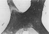
What grade of papilloedema is this?

Grade 1 papilledma with blurring of the margins but sharp in the temporal region (C-shaped halo). Note the cup is still discernible.
What grade of papilloedema is this?

Grade 2 - low degree. The disc margins are blurred all the way around. Cup visible. No obscuration of the vessels at the margin
What grade papilloedema is this?

Grade 3 - moderate
Circumferential blurring and elevation of the disc
Blurring of a single vessel at the margin (arrow) but not on the disc