Photo Exam Flashcards

1
Q
A

Acanthosis Nigricans

This is a skin condition characterized by areas of dark, velvety discoloration in body folds and creases–esp. armpits, groin, & neck.

2
Q
A

Acne vulgaris

This is a common chronic skin disease involving blockage and/or inflammation of pilosebaceous units (hair follicles and their accompanying sebaceous gland)

3
Q
A

Actinic keratosis

This is a skin condition caused by sun damage that causes scaly, rough, or bumpy spots on the skin. Common locations include sun exposed areas i.e. scalp, face, neck…etc.

4
Q
A

Acute Otitis Media

A) Early w/ inflammatin

B) Purulent effusion with air fluid level

C) Bulging purulent effusion filling the middle ear
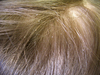
5
Q
A

Alopecia areata

This is a chronic immune-mediated disorder that targets anagen hair follicles and causes nonscarring hair loss.

6
Q
A

Anal Fissure

This is a tear in the anoderm distal to the dentate line.

7
Q
A

Angioedema

This is a self-limited, localized subcutaneous (or submucosal) swelling, which results from extravasation of fluid into interstitial tissues.

*****Commonly caused by ACE-inhibitors

8
Q
A

Angular Cheilitis

This is acute or chronic inflammation of the skin and contiguous labial mucosa located at the lateral commissures of the mouth.

9
Q
A

Ankyloglossia

This is also known as a “tongue-tie,” a congenital anomaly in which a short, lingual frenulum or a highly-attached genioglossus muscle restricts tongue movement.

10
Q
A

AV Nicking

This is a conditions where small artery (arteriole) is seen crossing a small vein (venule), which results in the compression of the vein with bulging on either side of the crossing.

*****Most commonly caused by HTN i.e. hypertensive retinopathy

11
Q
A

Bartholin Cyst

This occurs when a Bartholin’s gland is blocked and the gland becomes inflamed

12
Q
A

Blepharitis

This is a chronic inflammation of the eyelid, generally the part where eyelashes grow. It generally presents when very small oil glands near the base of the eyelashes don’t function properly, resulting in inflamed, irritated, itchy, and reddened eyelids.

13
Q
A

Bulla

This is a large vesicle described as a rounded or irregularly shaped blister containing serous or seropurulent fluid.

14
Q
A

Cafe-au-lait spots

These are flat, uniformly hyperpigmented macules that appear during the first year after birth. The presence of >6 is highly suggestive fo Neurofibromatosis-1

15
Q
A

Cataract

This is a clouding of the lens inside the eyewhich leads to a decrease in vision.

16
Q
A

Cavernous Hemangioma

This a type of blood vessel malformation or hemangioma, where a collection of dilated blood vessels form a tumor.

17
Q
A

Cherry Angioma

This is a common, benign neoplasms made up of clustered capillaries and blood vessels. These usually on the trunk and proximal limbs, also the mons pubis. The lesions increase in number with pregnancy

18
Q
A

Chloasma

This is a hyperpigmentation of the face referred to as the “mask of pregnancy.”

19
Q
A

Chondritis

I.e. “Inflammation of the cartilage”

20
Q
A

Clubbing

This is increased distal finger tip mass and increased longitudinal and transverse nail plate curvature. Commonly associated with pulmonary or cardiovascular disease.

21
Q
A

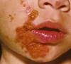
Condyloma acuminatum

These are also known as anogenital warts or venereal warts that are manifestations of human papillomavirus (HPV) infection that typically appear as flesh-colored or hyperpigmented verrucous papules or plaques in the perianal or genital region.

22
Q
A

Conjunctivitis

Literally means “inflammation of the conjunctiva.”

23
Q
A

Corneal arcus

This is a white, grey, or blue opaque ring in the corneal margin (peripheral corneal opacity), or white ring in front of the periphery of the iris.

*****Can be associated with hypercholesterolemia

24
Q
A

Crust

Note that this is often assocaited with impatigo.

25
Q
A

Pathologic cupping

The optic disc is the anatomical location of the eye’s “blind spot”, the area where the optic nerve and blood vessels enter the retina. The optic disc can be flat or it can have a certain amount of normal cupping. But glaucoma, which is due to an increase in intraocular pressure, produces additional pathological cupping of the optic disc.

26
Q
A

Darier’s Disease

This is a rare autosomal dominant genodermatosis. It is characterized by a persistent eruption of greasy hyperkeratotic papules in seborrheic areas, nail abnormalities, and mucosal changes [1]. The disease usually starts around puberty and runs a chronic course with exacerbations induced by sun exposure, heat, friction, or infections.

27
Q
A

Darwin Tubercle

This is a congenital ear condition which often presents as a thickening on the helix at the junction of the upper and middle thirds.

28
Q
A

Dermatochalasis

In elderly patients, redundant eyelid skin and prolapse of the orbital fat (dermatochalasis) can cause the eyelid to overhang the eyelid margin

29
Q
A

Drusen

These are multilobulated, globular concretions composed of mucoprotein matrix with acid mucopolysaccharides and ribonucleic acids that progressively calcify.

*****Associated with Macular Degeneration

30
Q
A

Dupuytren contracture

This is a benign, slowly progressive fibroproliferative disease of the palmar fascia. As the process evolves, nodules progress to form longitudinal bands referred to as cords on the palmar fascia, and the finger gradually loses extension, with contractures that draw one or more fingers into flexion at the metacarpophalangeal (MCP) joint.

31
Q
A

Ectropian

This is a condition in which the lower eyelid turns outwards.

32
Q
A

Eczema

This is inflammation of the skin. It is characterized by itchy, erythematous,vesicular, weeping, and crusting patches

33
Q
A

Enophthalmos

This is posterior displacement of the eyeball within the orbit due to changes in the volume of the orbit (bone) relative to its contents (the eyeball and orbital fat), or loss of function of the orbitalis muscle.

*****DO NOT CONFUSE with exophthalmos

34
Q
A

Entropion

Inward turning of eyelid as the result of chronic inflammation.

35
Q
A

Epstein pearls

Multiple small, white epithelial inclusion cysts found in the midline of the palate in newborn infants.

36
Q
A

Epulis

This is any benign tumor (i.e. lump) situated on the gingival or alveolar mucosa

****Note that the word literally translates into gingiva.

37
Q
A

Erythema multiforme

This is an acute, immune-mediated condition characterized by the appearance of distinctive target-like lesions on the skin. The disorder is most commonly induced by infection, with herpes simplex virus being the most frequent precipitator.

38
Q
A

Esotropia

This is an inward turning of the eye.

39
Q
A

Excoriation

This is a loss of the epidermis with a linear hollowed-out & crusted area.

****Note that this can be psychogenic in etiology i.e. “skin-pciking disorder” or drug induced (photo is methampheatmine)

40
Q
A

Exophthalmos

This a bulging of the eye anteriorly out of the orbit. Exophthalmos can be either bilateral (as is often seen in Graves’ disease) or unilateral (as is often seen in an orbital tumor)

41
Q
A

Exudative Tonsillitis

This is inflammation of the tonsils most commonly caused by viral or bacterial infection.

42
Q
A

Fissured Tongue

This is a benign condition of unknown etiology usually seen in adults. Deep grooves are located on the midline or evenly distributed on the tongue surface

43
Q
A

Flat wart

44
Q
A

Folliculitis

This is the infection and inflammation of one or more hair follicle

45
Q
A

Fordyce Spots

These are ectopic sebaceous glands of the bucceal mucosa appearing as small yellow-whote raised lesions found on the inner surface and vermilion border of the lips.

46
Q
A

Furuncle

This is a deep folliculitis, infection of the hair follicle. It is most commonly caused by infection by the bacterium Staphylococcus aureus, resulting in a painful swollen area on the skin caused by an accumulation of pus and dead tissue.

47
Q
A

Geographic tongue

Geographic tongue (benign migratory glossitis) is a disorder of unknown etiology that affects the epithelium of the tongue [90]. Local loss of filiform papillae leads to ulcer-like lesions that appear as erythematous patches on the dorsal tongue with circumferential white polycyclic borders

48
Q
A

Gingivitis

49
Q
A

Gynecomastia

This is a common endocrine disorder in which there is a benign enlargement o fbreast tissue in males.

50
Q
A

Hairy Tongue

This is a benign condition associated with antibiotic use, candida albicans infection, or poor oral hygiene.

51
Q
A

Halo nevus

This is a melanocytic nevus surrounded by a round or oval, usually symmetric, halo of depigmentation.

52
Q
A

Heberden node

These are hard or bony swellings that can develop in the distal interphalangeal joints (DIP).

*****Associated with osteoarthritis.

53
Q
A

Hemorrhoid

These are normal vascular structures in the anal canal. Approximately 5 percent of the general population is affected by symptoms related to hemorrhoidal disease [1] The cardinal features of hemorrhoidal disease include bleeding, anal pruritus, prolapse, difficulty with hygiene, and pain due to thrombosis.

54
Q
A

Herpes labialis

55
Q
A

Herpes Simplex

This is a viral disease caused by the herpes simplex virus.[1] Infections are categorized based on the part of the body infected. Figure shows primary genital HSV.

56
Q
A

Herpes Zoster

This is commonly known as shingles and also known as zona; a viral disease characterized by a painful skin rash with blisters in a limited area on one side of the body (left or right), often in a dermatomal pattern.

57
Q
A

Hirsutism

This is defined as excessive male-pattern hair growth, affects between 5 and 10 percent of women of reproductive age. It may be the initial, and possibly only, sign of an underlying androgen disorder, the cutaneous manifestations of which may also include acne and male-pattern balding (androgenetic alopecia). The most common cause of hirsutism is polycystic ovary syndrome (PCOS)

58
Q
A

Hordeolum

This is more commonly referred to as a “stye” and is a red and painful lump on the eyelid. It happens when a small gland on the edge of the eyelid gets infected or inflamed.

59
Q
A

Hirdradenitis suppurativa

This a chronic inflammatory skin condition that is also known as acne inversa (AI); it is a chronic follicular occlusive disease involving the follicular portion of folliculopilosebaceous units (FPSUs).

The primary sites of involvement for HS/AI are the intertriginous skin areas of the axillary, groin, perianal, perineal, and inframammary regions. The clinical manifestations vary, ranging from recurrent inflamed nodules and abscesses to draining sinus tracts and bands of severe scar formation. The associated pain, malodor, drainage, and disfigurement that accompany HS/AI contribute to a profound psychosocial impact of the disease on many patients.

60
Q
A

Hydrocele

This denotes a pathological accumulation of serous fluid in a body cavity. A hydrocele testis is the accumulation of fluids around a testicle, and is fairly common.

61
Q
A

Hydrocephalus

This is a condition where there is an abnormal accumulation of cerebrospinal fluid (CSF) in the ventricles of the brain. This causes increased intracranial pressure inside the skull and may cause progressive enlargement of the head if it occurs in childhood.

62
Q
A

Hypospadias

This is a congenital anomaly of the male urethra that results in abnormal ventral placement of the urethral opening.

63
Q
A

Impetigo

This is a contagious superficial bacterial infection observed most frequently in children.

64
Q
A

Kaposi Sarcoma

This is a tumor caused by infection with human herpesvirus 8(HHV8), also known as Kaposi’s sarcoma-associated herpesvirus (KSHV) or KS agent. It can present with cutaneous lesions with or without internal involvement.

65
Q
A

Keloid

These are fibrous growths that extend beyond the original area of injury to involve the adjacent normal skin

66
Q
A

Kyphosis

Excessive kyphosis or overcurvature called roundback or Kelso’s hunchback.

67
Q
A

Leukoplakia

This is a common early finding of HIV infection.

68
Q
A

Lichenification

This is increased skin markings and thickening with induration secondary to chronic inflammation caused by scratching or other irritation

69
Q
A

Linea Nigra

This is a dark vertical line that appears on the abdomen during about three quarters of allpregnancies.

70
Q
A

Lordosis

This is the normal inward curvature of the lumbar and cervical regions of the spine.[1]Excessive or hyperlordosis can happen and lumbar hyperlordosis is commonly referred to as sway back, hollow back or saddle back.

71
Q
A

Lyme Disease

Erythema migrans (EM) is a distinctive skin rash that occurs at the site of the tick bite. The rash is usually salmon to red-colored; the color may cover the entire lesion or may have an area in the center that is flesh-colored. In some cases, the rash consists of multiple rings, which give it a “bull’s eye” appearance

72
Q
A

Macule

A macule is a change in surface color, without elevation or depression and, therefore, nonpalpable, well or ill-defined,[30] variously sized, but generally considered less than either 5[30] or 10 mm in diameter at the widest point.[29]

73
Q
A

Malocclusion

A malocclusion is a misalignment or incorrect relation between the teeth of the two dental arches when they approach each other as the jaws close

74
Q
A

Mastitis

This is an infection of the breast tissue that results in breast pain, swelling, warmth and redness of the breast.

75
Q
A

Measles

The exanthem of measles is a maculopapular, blanching rash beginning on the face and spreading cephalocaudally and centrifugally to involve the neck, upper trunk, lower trunk, and extremities

76
Q
A

Melanoma

77
Q
A

Melasma

This is an acquired hyperpigmentation of the skin that typically affects the sun-exposed areas of the face. It is most common in women with darker complexions who live in areas of intense ultraviolet radiation exposure

78
Q
A

Milk Bottle Teeth

This is a severe decay in the teeth of infants or young children.

79
Q
A

Mongolian Spots

Mongolian spots are bluish-green areas of skin discoloration often seen in African-American, Hispanic, or Asian infants. While typically described as disappearing by one year, many persist, sometimes into adulthood. They are seen most commonly on the buttocks and lower back, but may extend over the entire back and on extremities

80
Q
A

Mucus Retention Cyst

is a cyst caused by an obstruction of a duct, usually belonging to the parotid gland or a minor salivary gland.

81
Q
A

Nasal Polyp

Nasal polyps are soft, painless, noncancerous growths on the lining of your nasal passages or sinuses. They hang down like teardrops or grapes. They result from chronic inflammation due to asthma, recurring infection, allergies, drug sensitivity or certain immune disorders.

82
Q
A

Neovascularization

This is the formation of functional microvascular networks with red blood cell perfusion.

*****This is a hallmark of Diabetic Retinopathy

83
Q
A

Neurofibromatosis

NF1 is due to mutations in the NF1 gene located at chromosome 17q11.2. Hallmarks of NF-1 include nerve tissue grows tumors (neurofibromas) that may be benign and may cause serious damage by compressing nerves and other tissues.

84
Q
A

Nevus

Simply means “mole.”

85
Q
A

Nodule

This is a solid, raised areas in or under the skin that are larger than 0.5 centimeters.

86
Q
A

Notched Teeth

These are also referred to as Hutchinson teeth, which are smaller and more widely spaced than normal and are notched on their biting surfaces. The sides of the teeth taper toward the biting edges.

*****Associated with congenital syphilis

87
Q
A

Onychomycosis

This is characterized by the appearance of dull white spots on the surface of the nail plate of one or several nails. Untreated, the diseased areas spread centrifugally (picture 3). Eventually the whole of the plate may be involved. The white areas are soft and when scraped lightly with a dermal curette yield a chalky scale suitable for laboratory examination.

88
Q
A

Otitis Externa

The term external otitis (also known as otitis externa or swimmer’s ear) refers to inflammation of the external auditory canal. Infectious, allergic, and dermatologic disease may all lead to external otitis. Acute bacterial infection is the most common cause of external otitis [1].

89
Q
A

Paget’s Disease

In 1874, Sir James Paget described 15 women with chronic nipple ulceration who all went on to develop cancer of the involved breast within two years [1]. The ulceration was described as an eczema-like eruption on the nipple and areola with a copious clear yellowish exudate. Ultimately this would become known as Paget disease of the breast (PDB) or mammary Paget disease. The hallmark of PDB is a scaly, raw, vesicular, or ulcerated lesion that begins on the nipple and then spreads to the areola

90
Q
A

Papilloma

Also known as a wart, papilloma refers to a benign epithelial tumor[1] growing exophytically (outwardly projecting) in nipple-like and often finger-like fronds.

****Caused by HPV.

91
Q
A

Papule

This is a circumscribed, solid elevation of skin with no visible fluid, varying in size from a pinhead to 1 cm

92
Q
A

Paronychia

This is a skin infection that happens around the fingernails or toenails.

93
Q
A

Patch

This is a macule that is >2cm.

*****Mongolian spots are an example of a patch.

94
Q
A

Peau d’ orange

Peau d’orange is caused by cutaneous lymphatic edema, which causes swelling; it is a sign of inflammatory breast cancer.

95
Q
A

Pectus carinatum

This is also called pigeon chest, is a deformity of the chest characterized by a protrusion of the sternum and ribs.

96
Q
A

Pectus excavatum

This is the most common congenital deformity of the anterior wall of the chest, in which several ribs and the sternum grow abnormally. This produces a caved-in or sunken appearance of the chest.[2] It can either be present at birth or not develop until puberty.

97
Q
A

Pediculosis capitis

Pediculosis capitis is a common condition caused by infestation of the hair and scalp by Pediculus humanus capitis (the head louse), one of three distinct varieties of lice specifically parasitic for humans

98
Q
A

Pemphigus foliaceus

Pemphigus foliaceus is a superficial variant of pemphigus that presents with cutaneous lesions. The mucous membranes are typically spared [1]. Pemphigus foliaceus usually develops in a seborrheic distribution. The scalp, face, and trunk are common sites of involvement. The skin lesions usually consist of small, scattered superficial blisters that rapidly evolve into scaly, crusted erosions

99
Q
A

Peyronie Disease

This is also known as induratio penis plastica (IPP)[1] orchronic inflammation of the tunica albuginea (CITA), is a connective tissue disorder involving the growth of fibrous plaques[2] in the soft tissue of the penis affecting an estimated 5% of men.[3] Specifically, scar tissue forms in the tunica albuginea, the thick sheath of tissue surrounding the corpora cavernosa causing pain, abnormal curvature, erectile dysfunction, indentation, loss of girth and shortening.[4][5][6][7] A variety of treatments have been used, but none have been especially effective.

100
Q
A

Pinguecula

A pinguecula is a yellowish, slightly raised conjunctival lesion arising at the limbal conjunctiva (picture 7). Unlike a pterygium that arises from the limbus and progresses onto the cornea, a pinguecula arises from the limbus and remains confined to the conjunctiva without corneal involvement.

101
Q
A

Pityriasis rosea

Pityriasis rosea is a harmless skin rash that causes small, itchy spots on the belly, back, chest, arms, and legs. The rash usually lasts about 4 to 6 weeks, but in some people, it can last for months.

102
Q
A

Port Wine Stain

Port wine stains, or nevus flammeus, are low-flow vascular malformations of dermal capillaries and postcapillary venules. They are present at birth as blanchable pink to red patches and may be located anywhere on the body, typically with a unilateral or segmental distribution that respects the midline.

103
Q
A

Psoriasis

Psoriasis is a common chronic skin disorder most commonly characterized by well-demarcated erythematous plaques with silver scale

104
Q
A

Pterygium

A pterygium is a triangular wedge of fibrovascular conjunctival tissue that typically starts medially on the nasal conjunctiva and extends laterally onto the cornea

105
Q
A

Ptosis

Ptosis occurs when the muscles that raise the eyelid (levator and superior tarsal muscles) are not strong enough to do so properly

106
Q
A

Purpura

This is a red to purplish spots on the skin that do not blanch with pressure.

*****Associated with meningitis.

107
Q
A

Pyogenic granuloma

Pyogenic granuloma (PG) or lobular capillary hemangioma [1] is a benign vascular tumor of the skin or mucous membranes characterized by rapid growth and friable surface.

108
Q
A

Pyorrhea

Also known as periodontitis; a set of inflammatory diseases affecting theperiodontium, i.e., the tissues that surround and support the teeth. Periodontitis involves progressive loss of thealveolar bone around the teeth, and if left untreated, can lead to the loosening and subsequent loss of teeth.

109
Q
A

Ranula

Ranulas are pseudocysts associated with the sublingual glands and submandibular ducts. They can be congenital, probably from improper drainage of sublingual glands, or acquired after oral trauma. They appear as blue, fluctuant swellings lateral to the midline in the lower mouth

110
Q
A

Raynaud’s Disease

The Raynaud phenomenon (RP) is an exaggerated vascular response to cold temperature or emotional stress. The phenomenon is manifested clinically by sharply demarcated color changes of the skin of the digits. Abnormal vasoconstriction of digital arteries and cutaneous arterioles due to a local defect in normal vascular responses is thought to underlie the disorder.

111
Q
A

Raynaud’s Disease

The Raynaud phenomenon (RP) is an exaggerated vascular response to cold temperature or emotional stress. The phenomenon is manifested clinically by sharply demarcated color changes of the skin of the digits. Abnormal vasoconstriction of digital arteries and cutaneous arterioles due to a local defect in normal vascular responses is thought to underlie the disorder.

112
Q
A

Rectal prolapse

A complete rectal prolapse is the protrusion of all layers of the rectum through the anus, manifesting as concentric rings of rectal mucosa

113
Q
A

Retinoblastoma

Retinoblastoma, the most common primary intraocular malignancy of childhood, exists in sporadic and germline forms [1-3]. Children with retinoblastoma frequently (but not always) present with leukocoria (picture 1) [4,5]. Prompt referral to ophthalmology and other pediatric specialists is necessary to optimize visual outcome and survival

114
Q
A

Rhinophyma

Rosacea is a common, chronic skin disorder that presents with a variety of clinical manifestations primarily localized on the central face [1,2]. The disorder is divided into four main subtypes: erythematotelangiectatic, papulopustular, phymatous, and ocular rosacea.

*****Phymatous rosacea exhibits tissue hypertrophy manifesting as thickened skin with irregular contours (picture 4). Involvement most commonly occurs on the nose i.e. “rhinophyma”

115
Q
A

Rosacea

This is a chronic skin condition characterized by facial redness, small and superficial dilated blood vessels on facial skin, papules, pustules, and swelling.

116
Q
A

Salmon patch/ Stork bite

This is a common birthmark occurring in up to 80 percent of newborns [1]. It is similar in appearance to capillary malformations but has more indistinct borders and is usually located in the midline. The most common locations are the glabella, upper eyelids, and nape of the neck

117
Q
A

Scoliosis

Scoliosis is a condition that makes the spine (backbone) curve sideways, like the letter “S” or “C.” Often, the spine also twists so the back is not flat. If the spine twists, one side of the back sticks out more than the other

118
Q
A

Seborrheic keratoses

Seborrheic keratoses are common benign keratinocytic tumors presenting as macular or elevated lesions, with variable brown pigmentation and a dull surface (picture 6). On the face, seborrheic keratoses may remain superficial for a long period. Dermoscopic examination is helpful in differentiating seborrheic keratosis from LM. Histologically, there is a proliferation of normal keratinocytes and an increased number of melanocytes

119
Q
A

Serous otitis media

Otitis media with effusion (OME), also called serous otitis media, is defined as middle-ear effusion without acute signs of infection

120
Q
A

Squamous cell carcinoma

SCC can develop on any cutaneous surface, including the head, neck, trunk, extremities, oral mucosa, periungual skin, and anogenital areas (picture 1A-D). In fair-skinned individuals, SCCs most commonly arise in sites frequently exposed to the sun.

121
Q
A

Stomatitis

This is inflammation of the mouth and lips.[1] It refers to any inflammatory process affecting the mucous membranes of the mouth and lips, with or without oral ulceration.

122
Q
A

Strawberry Hemangioma

Superficial hemangiomas have been called “strawberry” or “capillary” hemangiomas, but superficial hemangioma is the preferred term

123
Q
A

Striae

Also known as stretch marks.

124
Q
A

Subconjunctival hemorrhage

Patients with subconjunctival hemorrhage may have a history of trauma or contact use or may report no history of trauma and note the hemorrhage after waking from sleep. A typical subconjunctival hemorrhage appears as a focal, flat, red region on the ocular surface representing a collection of blood between the sclera and the conjunctiva

125
Q
A

Swan neck deformity

Deformity of the figners that is assocaited with RA.

126
Q
A

Syndactyly

Syndactyly (two or more digits fused together) is a characteristic feature of Apert syndrome that permits distinction from other similar syndromes

127
Q
A

Telangiectasia

This is a dilated superficial blood vessel.

128
Q
A

Thrush

Oropharyngeal candidiasis, or thrush, is common in young infants.

129
Q
A

Thyroglossal duct cyst

A thyroglossal cyst is a fibrous cyst that forms from a persistent thyroglossal duct. It usually presents as a midline neck lump (in the region of the hyoid bone) that is usually painless, smooth and cystic, though if infected, pain can occur. There may be difficulty breathing, dysphagia (difficulty swallowing), ordyspepsia (discomfort in the upper abdomen), especially if the lump becomes large.

130
Q
A

Tinea barbae

Dermatophyte infections involving the beard area are referred to as tinea barbae. Patients may present with pustules, inflammatory papules and nodules, or scaly erythematous plaques

131
Q
A

Tinea capitis

Tinea capitis, dermatophyte infection of the scalp, almost always occurs in small children.

132
Q
A

Tinea corporis

Tinea corporis, dermatophyte infection of the body that is also known as “ringworm.”

133
Q
A

Tinea cruris

Tinea cruris (jock itch) is a special form of tinea corporis involving the crural fold.

134
Q
A

Tophi

This is a deposit of uric acid crystals, in the form of monosodium urate crystals, in people with longstanding hyperuricemia (high levels of uric acid in the blood). Tophi are pathognomonic for the disease gout.

135
Q
A

Torus palatinus

Torus palatinus is an exostosis located on the midline of the hard palate. It presents as a bony hard, nodular, lobular, or spindle-shaped mass covered with normal mucosa. The lesion appears during childhood, enlarges slowly over many years, and is asymptomatic. Torus palatinus is typically an incidental finding during routine physical examination.

136
Q
A

Transverse crease (allergic salute)

This iss the characteristic and sometimes habitual gesture of wiping and/or rubbing the nose in an upwards or transverse manner with the fingers, palm, or back of the hand. It is termed a salute because the upward movement of the hand acts as an unintentional gesture.

137
Q
A

Turbinate hypertrophy

138
Q
A

Tympanosclerosis

Tympanosclerosis (myringosclerosis), or whitish plaques of calcium and phosphate crystals deposited on the TM. These plaques are firm, contained within the middle layer of the TM, are typically horseshoe-shaped, and move with the TM during pneumatic otoscopy [31]; however, TM movement may be decreased or absent in patients with tympanosclerosis.

139
Q
A

Varicella

Also known as the “chicken pox.”

140
Q
A

Variocele

A varicocele is a collection of dilated and tortuous veins in the pampiniform plexus surrounding the spermatic cord in the scrotum.

141
Q
A

Vesicle

Elevated, circumscribed, superficical lesion filled with fluid that is less than 1 cm in diameter i.e. a small blister

142
Q
A

Vitiligo

Vitiligo is an acquired skin depigmentation occurring in approximately 1 percent of the population worldwide. It results from an autoimmune process directed against the melanocytes

143
Q
A

Wheals

Wheals (raised areas surrounded by a red base) from urticaria & can appear anywhere on the surface of the skin.

144
Q
A

Xanthelasma

Xanthelasma are cholesterol-filled, soft, yellow plaques that usually appear on the medial aspects of the eyelids bilaterally. They most often occur in middle-aged and older adults.