Cardiovascular mechanics Flashcards
(29 cards)
Explain what is needed in order for a single ventricular cell to contract
Action potential
Calcium ion influx and release
contractile event
Describe the parameters (length and width of a single ventricular cell)
100uM in length
15uM wide
Describe the structure of T tubules and how much do they open up due when theres an action potential? Where do that carry AP to?
Finger like invagination from cell surface
opens up to 200nm in diameter
it is spaced so that T tubule lies alongside each Z line of every myofibril
carries surface depolarisation deep into cell

Describe the 3D component of a single cardiac cell structure in terms of organelles distribution
describe significance of mitochondria
Myofibrils, mitochondria, nucleus, SR
mitochondria - 36%; need large ATP

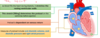
Describe the excitation- contraction coupling in the heart?
- Ca2+ moves into sarcoplasm via L-type calcium channel
- stimulate ryanodine receptor in SR to release Ca ions
- same amount of Ca2+ that entered now leave sarcoplasm via Na+/Ca2+ transporter to avoid bulid up of Ca2+.. this doesnt use ATP; only electrical gradient.

What’s the relationship w=etwwen intracellular Ca ions and force production
There’s a threshold needed but it’s SIGMOIDAL

What is the relationship between length and tension? what other factors increase
The longer the muscle the higher the maximum contractility force (iSOMETRIC contraction)
There’s also more PASSIVE FORCE
Contrast skeletal and cardiac muscle
what causes these differences
- Cardiac muscle more resistant to stretch
- LESS compliant than skeletal muscle
this is due to differences in properties of ECM and cytoskeleton
in the relationship between length and tension, What limb is only important/relavant for cardiac muscle
Ascending limb of graph; heart muscle doesnt pull as it is contained in pericardial sac unlike skeletal muscle

What are two forms of contraction that the heart uses ? which occurs earlier?
- Isometric- muscle fibres doesnt change in length but pressure increases
- Isotonic- shortening of fibres and blood is ejected
theres isometric FIRST then isotonic

What is the law of LaPlace
When pressure within a cylinder is led constant, the tension on it’s walls increases with increasing radius
wall tension = (P X R)/h
Pressure in vessel, R= radius of vessel
what is preload?
Blood fills the heart during diastoles and stertches resting ventricular walls.
this fills ventricles and the stretching determines the pre-load
allows heart to adapt its force to the volum of blood it recieves
Dependent on venous return
what are the measures of pre-load?
- END-DIASTOLIC VOLUME
- END-DIASTOLIC PRESSURE
- RIGHT ATRIAL PRESSURE
what is after load?
what si the signifcance of increase in afterload?
THE PRESSURE in which the ventricles MUST WORK against to OPEN the aortic valve.
high afterload means there’s less amount of isotonic shortening. Also less velocity of shortening

what are the measures of afterload?
Diastolic blood pressure
what does an increase in pre load affect?
increase in ISOMETRIC CONTRACTION
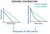
what does an increase in after load affect?
- shorter ISOTONIC contraction
- lower VELOCITY of contraction
note the change in gradient of velocity
describe the Frank-Starling relationship?
Increased diastolic fibre length (due pre-load) will increase ventricular contraction.
hence ventricles pump greater stroke volume; at equilibrium, cardiac output exactly balances augemnted venous return
what are the potential causes of the frank-starling relationship
- Changes in number of myofilament crossbridges formed- the more stretched, the more cross bridges can form
- Changes in Ca2+sensitivity of the myofilaments
descirbe hypothesis 1 for increase in Ca senstivity
at longer sarcomere length, the affinity of troponin C for Ca2+ increases due to change in protein structure

describe the other hypothesis for the starling relationship
With higher length, there’s decreasing myofilament lattice spacing, the probabilty of forming strong bidning cross-bridges increases
what is stroke work?
give equations? what affects each value?
Stroke work= Stroke volume x Pressure at which blood is ejected
SV; affected by pre-load AND after load
Pressure- affetced by cardiac structure
hence stroke work is work done by heart to pump blood under pressure into aorta or pulmonary artery
How does radius of curvature affect Tension using Law of Laplace?
Lower radius of curvature increases pressure as thickness is taken into consideration
hence HIGHER PRESSURE; Tension is the same
those with failing heart have heart with HIGHER radius of curvature; more work and pressure on heart

Does skeletal muscle need influx of extracellular calcium in order to contract?
No; ONLY CARDIAC muscle cells do


