DIT review - Heme 2 Flashcards

1
Q

Where does erythropoeisis occur in early life and later life

A
  • Fetal development - liver
  • After 28 weeks - bone marrow
    • Infancy and childhood:
      • Flat bones
      • Sternum, pelvis, ribs, cranial bones, vertebrae, long bones of leg
    • Later adolescence and adulthood
      • Axial skeleton
      • Vertebrae, sternum, ribs, and pelvis
2
Q

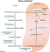
What is the rate limiting enzyme of heme synthesis

A

ALA synthase:

Succinyl CoA + Glycine –> ALA

3
Q

What is the cofactor required for ALA synthase

A

B6

4
Q

What are causes of polycythemia (increased RBC)

A
  • Polycythemia vera – monoclonal proliferation of red cells
  • Chronic hypoxia – need to increase O2 carrying capacity so kidney produces more erythropoietin to make more RBCs
    • Pulmonary disease
    • Cyanotic heart disease
    • High altitudes
  • Inappropriate elevation of EPO – e.g. EPO-producing tumor
  • Trisomy 21
5
Q

What are the EPO-producing tumors?

A

THINK: Potentially Really High Hematocrit

  • Pheochromocytoma
  • Renal cell carcinoma
  • Hepatocellular carcinoma
  • Hemangioblastoma
6
Q

What is the enzyme deficiency in Acute intermittent porphyria

A

Porphobilinogen (PBG) deaminase

THINK:

o Acute intermittent = guys hollering “damn” (deam-inase) intermittently at A CUTE pretty big girl (PBG)

7
Q

Presentation of acute intermittent porphyria

A

§ Symptoms – 5 P’s

· Painful abdomen

· Port wine colored urine (due to increase PGB)

· Polyneuropathy

· Psychological disturbances

· Precipitated by drugs (CYP450 inducers – e.g. Rifampin), alcohol, and starvation

8
Q

Treatment of acute intermittent porphyria

A

· Glucose + heme = inhibition of ALA synthase

9
Q

What is the deficienct enzyme in porphyria cutanea tarda?

A

Uroporphyrinogen carboxylase

THINK of a homeless man living in a cardboard box (carbox)

10
Q

Presentatin of porphyria cutanea tarda

A

§ Symptoms

· Blistering cutaneous photosensitivity

· Hyperpigmentation

· Hyerptrichosis (extra hair)

· Tea colored urine

· Exacerbated with alcohol consumption

· Associated with Hepatitis C

§ (THINK of a stereotypical homeless man)

· Alcoholic, face blistered and dark from sun, facial hair, liver disease

11
Q

Enzyme defiecient in lead poisoning

A

ALA dehydratase and Ferrochelatase

12
Q

Presentation of lead poisoning

A
  • GI (abd pain, constipation, anorexia)
  • Neuro (cognitive defects, peripheral neuropathy, encephalopathy, memory loss, delirium)
  • Hematologic (microcytic anemia with basophilic stippling, ringed sideroblasts in marrow)
  • Burton lines – lead lines in gingiva and gums
  • Hyper dense lines on metaphysis of long bones
  • Renal failure
13
Q

What are the different causes of microcytic anemia

A

Iron deficiency

Anemia of chronic disease (late)

Thalassemia

Lead poisoning

Sideroblastic anemia

14
Q

What is the basic principle behing microcytic anemia

A
  • MCV < 80
  • Due to decreased production of hemoglobin = extra division to maintain Hb concentration
    • Hemoglobin = heme + globin
      • Heme = iron + protoporphyrin
  • Cells are small and hypochromic (pale)
15
Q

What are lab values of iron deficiency anemia (ferritin, serum iron, TIBC, % saturation)

A
  • Low ferritin (low iron stores)
  • Low serum iron
  • Low % saturation (of transferrin – iron transporter)
  • High TIBC (Total iron-binding capacity = # of transferrin molecules in the blood – will be elevated because the liver is pumping out more in a state of low iron in order to replenish iron
    • Ferritin and TIBC are always opposite
16
Q

What is the molecule responsible for anemia of chronic disease

A

Hepcidin sequesters iron into storage sites

17
Q

What are lab values of anemia of chronic disease(ferritin, serum iron, TIBC, % saturation)

A
  • Labs:
    • High ferritin
    • Low TIBC
    • Low serum iron
    • Low % saturation
  • Early disease presents as nonhemolytic, normocytic anemia
  • Late disease presents as microcytic anemia
18
Q

Describe the defect in alpha thalassemia

A
  • Defect in a-globin synthesis
  • Alpha Thalassemia
    • Due to alpha-globin gene deletion = decreased alpha-globin synthesis
    • There are 4 alpha genes on chromosome 16
19
Q

Describe the different types of alpha-thalassemia

A
  • 1 gene deleted = asymptomatic
  • 2 genes deleted = mild anemia
    • Cis = increased risk in offspring – Asians
    • Trans = Africans
  • 3 genes deleted
    • Beta chains form tetramers – B4 = Hemoglobin H (HbH)
  • 4 genes deleted
    • No a-globin at all
    • Gamma (y) chains form tetramer – y4 = Hemoglobin Barts (Hb Barts)
    • Incompatible with life – hydrops fetalis
20
Q

Describe the defect in beta thalassemia

A
  • Due to beta globin gene mutation
  • There are 2 beta genes on chromosome 11
    • Mutations can result in absent (B0) or diminished (B+) production of b-globin
  • Seen in Mediterranean populations
21
Q

Describe beta-thalassemia minor (genes, presentation, types of Hb, histology)

A
  • B-thalassemia minor (B/B+)
    • Decreased amount of B-globin
    • Minimal anemia
    • Increased HbA2 (a2d2) and HbF (a2y2)
    • Will see target cells
22
Q

Describe beta thalassemia major (genes, presentation, histology)

A
  • B-thalassemia major (B0/B0)
    • No B-globin at all
    • Severe anemia requiring blood transfusions
      • Risk of hemochromatosis
    • High HbF at birth is temporarily protective
    • Will see target cells
    • Erythroid hyperplasia – hematopoiesis occurring in unusual places, such as face, skull, liver, spleen
      • “Crewcut” appearance on X-ray (“hair-on-end”)
      • “Chipmunk” facies
      • Hepatosplenomegaly
    • Risk of aplastic crisis with Parvovirus B19 infection of erythroid precursors
23
Q

Describe the defect and causes of sideroblastic anemia

A
  • Defect in heme synthesis
    • Defect in protoporphyrin synthesis leads to iron buildup in the mitochondria = iron-laden mitochondria form a ring around nucleus of erythroid precursors
  • Causes:
    • Congenital:
      • ALA synthetase deficiency
    • Acquired:
      • Lead poisoning
      • Vitamin B6 deficiency (cofactor for ALA synthetase)
      • Alcohol
24
Q

What are the lab values of sideroblastic anemia (ferritin, TIBC, serum iron, % saturation)

A
  • High ferritin
  • Low TIBC
  • High serum iron
  • High % saturation
25
Q

Describe the defect and presentation of lead poisoning

A
  • Lead poisoning inhibits ferrochelatase and ALA dehydratase, thus inhibiting heme synthesis and increasing protoporphyrin
  • Presentation:
    • Lead lines on gingivae (Burton lines) and on metaphyses of long bones
    • Encephalopathy
    • RBC basophilic stippling
    • Abdominal colic
    • Sideroblastic anemia
    • Wrist and foot drop
26
Q

What are the causes of macrocytic anemia

A
  • Megaloblastic anemia
    • Folate deficiency
    • B12 deficiency
    • Orotic aciduria
  • Alcoholism
  • Liver disease
  • Drugs
27
Q

Describe how folate and B12 are involved in the synthesis of DNA precursors

A
  • Folate circulates as methyl THF
  • Methyl is transferred from THF to B12 so that THF can participate in DNA synthesis
  • Methyl is transferred from B12 to homocysteine, creating methionine
28
Q

How can you differentiate folate and B12 deficiency

A

Both will have megaloblastic anemia with hypersegmented neutrophils

  • Folate deficiency:
    • Elevated serum homocysteine
    • Normal methylmalonic acid
    • No neuro symptoms
  • B12 deficiency:
    • Elevated serum homocysteine
    • Elevated methylmalonic acid
    • Neuro symptoms
29
Q

Describe the absorption of B12

A
  • Dietary B12 bound to meat
  • Salivary enzymes free B12 from meat, and bind it to R-binder to carry through the stomach
  • Pancreatic proteases detach B12 from R-binder
  • B12 binds to intrinsic factor (IF), created by gastric parietal cells
  • IF-B12 absorbed in the ileum
30
Q

Causes of B12 deficiency

A
  • Malnutrition
  • Pernicious anemia = B12 deficiency due to autoimmune destruction of gastric mucosa (parietal cells in the stomach), which leads to IF deficiency
  • Pancreatic insufficiency = no enzyme to cleave B12 from R-binder
  • Damage to terminal ileum (e.g. Crohn’s) à site of absorption
  • Diphyllobothrium latum (tapeworm)
31
Q

What cause neuro defects in B12 deficiency

A
  • B12 needed to convert methylmalonic acid to Succinyl CoA
  • Decreased B12 = increased MMA = MMA builds up in myelin of spinal cord leading to degeneration
32
Q

Describe defect in orotic aciduria

A
  • Inability to convert orotic acid to UMP (de novo pyrimidine synthesis pathway)
33
Q

What are the main 3 causes of normocytic anemia

A
  • (I) Underproduction (nonhemolytic)
  • (II) Extravascular hemolysis
  • (III) Intravascular hemolysis
34
Q

Causes of nonhemolytic normocytic anemia

A
  • Anemia of chronic disease (early)
  • Iron deficiency anemia (early)
  • Aplastic anemia
  • Chronic kindey disease
35
Q

Describe the presentation and histology of aplastic anemia

A
  • Bone marrow stops making cells – pancytopenia
    • Anemia
      • Fatigue, malaise, pallor
    • Leukopenia
      • Infection
    • Thrombocytopenia
      • Purpura, petechiae, bleeding
  • Histology will show hypocellular bone marrow with fatty infiltration (“cobweb”)
36
Q

Causes of aplastic anemia

A
  • Radiation
  • Drugs
  • Viral infections: Parvovirus B19, EBV, HIV
  • Fanconi anemia (DNA repair defect causing bone marrow failure)
  • Idiopathic
37
Q

Differentiate extra- vs. intravascular hemolysi

A
  • Extravascular hemolysis:
    • RBC destruction by reticuloendothelial system (macrophages of spleen, liver, and lymph nodes)
  • Intravascular hemolysis:
    • Destruction of RBCs within vessels
38
Q

What is the defect in hereditary spherocytosis and is it extravascular or intravascular hemolysis

A

Extravascular

  • Defect of RBC cytoskeleton proteins
    • Ankyrin, spectrin, band 3
  • Membranes are formed and lost over time
    • Loss of membrane = spherocytes
    • Spherocytes cannot maneuver through splenic sinusoids so are consumed by macrophages
39
Q

What findings will you see in hereditary spherocytosis

A
  • Spherocytes with loss of central pallor
  • Increased RDW – cells of all different sizes
  • Increased MCHC (mean corpuscular hemoglobin concentration) – cell shrinks but Hb remains the same
  • Splenomegaly and jaundice
  • Aplastic crisis
40
Q

Diagnostic test of hereditary spherocytosis

A
  • Osmotic fragility test – High percentage of lysis of RBCs in hypotonic solution
41
Q

Treatment of hereditary spherocytosis

A

Splenomegaly

42
Q

Describe how G6PD deficiency causes anemia

A
  • Glutathione neutralizes H2O2 but becomes oxidized in the process
  • NADPH needed to reduce glutathione
  • In G6PD deficiency, NADPH is not produced
43
Q

Histology of G6PD deficiency

A
  • Heinz bodies – precipitation of Hb within RBC due to oxidative stress
  • Bit cells – spleen removing Heinz bodies
44
Q

What are oxidant stresses that causes hemolytic anemia is G6PD

A
  • Sulfa drugs, antimalarials, infection, fava beans
45
Q

Describe how pyruvate kinase deficiency causes anemia

A
  • Glycolytic enzyme deficiency = inability to generate ATP
  • Cannot maintain Na+/K+ ATPase = RBC swelling and lysis
46
Q

What is the mutation in sickle cell disease

A
  • HbS due to mutation of b-hemoglobin
    • Single amino acid replacement: glutamic acid to valine
47
Q

What are triggers of RBC sickling

A
  • Hypoxemia
  • Dehydration
  • Acidosis
48
Q

Describe findings of sickle cell disease

A
  • Causes both extra- and intravascular hemolysis
  • Autosplenectomy
    • Increased infection by encapsulated organisms
    • Howell-Jolly bodies
  • Increased risk of salmonella osteomyelitis
  • Aplastic crisis due to parvo B19
  • Pain crises due to malocclusion
    • Dactylitis
    • Acute chest syndrome
    • Renal papillary necrosis
  • Erythroid hyperplasia:
    • “Crewcut”/”Hair-on-end” X-ray
    • Chipmunk facies
49
Q

What will you see on histology of sickle cell disease

A
  • Sickle cells and target cells
50
Q

Treatment of sickle cell disease

A
  • Hydroxyurea – increases production of HbF
  • Bone marrow transplant
51
Q

Describe mutation in Hemoglobin C disease

A
  • HbC due to mutation of b-globin
    • Single amino acid replacement: glutamic acid à lysine
  • HbC forms hexagonal crystals in the cells
  • Milder than sickle cell disease
52
Q
A
53
Q

What is the protein that carries Hb to spleen in intravascular hemolysis

A

Haptoglobin

54
Q

Describe general clinical findings of intravascular hemolysis

A
  • Hemoglobinemia, hemoglobinuria, hemosiderinuria (due to iron taken up by renal tubular cells, which later shed), decreased serum haptoglobin, corrected reticulocyte count > 3%
55
Q

Differentiate the 2 types of autoimmune hemolytic anemia

A
  • (a) Autoimmune hemolytic anemia
    • Antibody-mediated destruction of RBCs
    • Subtypes:
      • Warm agglutinins
        • IgG antibodies attach to RBCs and cause them to agglutinate
        • Involves extravascular hemolysis
        • Occurs in central body
        • Associated with lupus, CLL, and different drugs
      • Cold agglutinins
        • IgM antibodies bind RBCs and activate complement
        • Occurs in periphery
        • Associated with EBV and mycoplasma pneumonia
56
Q

How do you diagnose autoimmune hemolytic anemia

A
  • Direct Coombs
    • Detects antibody-coated RBCs
    • Prepared antibodies added to patient’s RBC to see if they bind to existing antibodies on the RBC
      • Adding an antibody that binds to antibody
  • Indirect Coombs
    • Detects free antibodies in serum
    • Patient’s serum incubated with normal RBC
57
Q

Describe the defect in Paroxysmal nocturnal hemoglobinuria

A
  • Deficiency of GPI, which usually anchors DAF (CD55) to RBC membrane to protect from complement destruction
  • Complement is activated in acidic situations
    • Lysis occurs at night due mild respiratory acidosis
58
Q

Diagnosis of paroxysmal nocturnal hemoglobinuria

A
  • Ham’s test – add acid to lower the pH and check for RBC lysis
59
Q

Describe the problem and causes of microangiopathic anemia

A
  • RBCS are mechanically damaged as they pass through the lumen of an obstructed or narrowed vessel
    • Schistocytes
  • Causes of microthrombi:
    • TTP = platelet thrombi due to lack of ADAMS13
    • HUS = platelet thrombi due to toxin from E. Coli
    • DIC = platelet and fibrin thrombi
    • HELLP = in pregnant women
60
Q

Causes of macroangiopathic anemia

A
  • RBCs are mechanically damaged by forces in larger vessels
  • Causes:
    • Prosthetic heart valves
    • Aortic stenosis