DIT review - Cardiology 1 Flashcards

1
Q

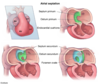
Describe the steps of atrial septation

A
  • (1) Septum primum grow toward endocardial cushion, narrowing foramen (ostium) primum
  • (2) Foramen secundum forms in septum primum (foramen primum disappears)
    • Perforations in the septum primum eventually fuse together to become the foramen secundum
  • (3) Septum secundum develops as foramen secundum maintains R-to-L shunt
  • (4) Septum secundum expands and covers most of the foramen secundum
    • The residual foramen in the septum secundum is the foramen ovale
  • (5) Remaining portion of septum primum form valve of foramen ovale
  • (6) Septum secundum and septum primum fuse to form the atrial septum
  • (7) Foramen ovale usually closes soon after birth because in increased LA pressure
2
Q

Derivatives of the 1st aortic arch

A
  • Part of maxillary artery
3
Q

Derivatives of 2nd aortic arch

A
  • Stapedial artery and hyoid artery
4
Q

Derivatives of 3rd aortic arch

A

Common carotid artery and proximal part of internal carotids

5
Q

Derivatives of 4th aortic arch

A
  • On left – aortic arch
  • On right – proximal part of R subclavian
6
Q

Derivatives of 6th aortic arch

A
  • Proximal part of pulmonary arteries
  • On left – ductus arteriosus
7
Q

What are the 3 shunts involved in fetal circulation + describe fetal circulation

A

Ductus venosus, foramen ovale, ductus arteriosus

  • Umbilical vein carries oxygenated blood from placenta to liver
    • In the liver there is mixing of oxygenated blood from umbilical vein with deoxygenated blood from lower extremities
    • Some blood from umbilical vein goes to liver into hepatic circulation while some blood is shunted directly into the IVC via ductus venosus, bypassing hepatic circulation
  • Blood goes from IVC to RA
    • In the RA, blood can either go into the RV or straight into the LA via foramen ovale
  • Blood that went to the RV then enters the pulmonary artery
    • In the pulmonary artery, blood can either go to the lungs, or go through the ductus arteriosus which will shunt the blood directly into the descending aorta
      • This shunt is due to the high fetal pulmonary artery resistance
8
Q

Describe the transition from fetal circulation to adult circulation (e.g. closure of shunts)

A
  • When the infant takes a breath, this decreases intrathoracic pressure, thus decreasing resistance in pulmonary vasculature
  • Decreased resistance leads to more blood entering the pulmonary artery (less leaving through ductus arteriosus), and thus more blood entering the LA
  • Increased LA pressure causes closure of the foramen ovale
  • Highly oxygenated blood in the aorta causes closure of ductus arteriosus
9
Q

Cardiac disorders associated with Turner syndrome

A
  • Coarctation of aorta
  • Bicuspid aortic valve
10
Q

Cardiac disorders associated with Down Syndrome

A
  • Endocardial cushion defect (ASD and VSD)
    • Complete atrioventricular canal defect
11
Q

Cardiac disorders associated with DiGeorge Syndrome

A
  • Tetralogy of Fallot
  • Truncus arteriosus
12
Q

Cardiac disorders associated with Marfan syndrome

A

Aortic insufficiency due to abnormal aortic valve

13
Q

Defect and heart sound heard in atrial septal defect

A
  • Most commonly due to ostium secundum defect
  • Loud S1; wide, fixed split S2
  • Symptoms range from none to heart failure
14
Q

Heart sounds associated with patent ductus arteriosus

A
  • Due to decreased pulmonary vascular resistance after birth, shunt becomes left to right
  • Continuous “machine-like” murmur
15
Q

What drug do you use to keep the PDA open and to close it?

A
  • Indomethacin closes PDA
  • PGE keeps PDA open
16
Q

Describe the pathogenesis of Eisenmenger syndrome

A
  • Uncorrected L-to-R shunt = increased pulmonary blood flow = remodeling of vasculature = pulmonary arterial HTN = right ventricular hypertrophy = increase RV pressure = shunt become R-to-L
  • Causes late cyanosis, clubbing, and polycythemia
17
Q

Location and presentation of infantile aortic coarctation

A
  • Coarctation is proximal to ductus arteriosus
    • Associated with PDA
  • Presentation:
    • Lower extremity cyanosis in infants – blood going to LE is deoxygenated (from RV to pulmonary artery to PDA)
  • Associated with Turner syndrome
18
Q

Location and presentation of adult aortic coarctation

A
  • Coarctation is distal to ductus arteriosus
  • Presentation:
    • HTN in upper extremities and hypotension with weak pulses in lower extremities
    • “Notching” of ribs on X-ray
      • Collateral arteries form and engorged intercostal erode ribs
19
Q

Complications of aortic coarctation

A
  • Heart failure, increased risk of cerebral hemorrhage, aortic rupture, endocarditis
20
Q

What other defect is often associated with persistent truncus arteriosus

A
  • Failure of truncus arteriosus to divide into pulmonary trunk and aorta
  • Usually associated with VSD
21
Q

What is required in order for life in an infant with transposition of great vessels

A
  • Not compatible with life unless a shunt is present to allow mixing of blood (e.g. VSD, PDA, or patent foramen ovale)
  • Defect = 2 closed loop circuits
    • LV enters into the pulmonary trunk
    • RV enters into the aorta
22
Q

Describe the defect and required shunting in tricuspid atresia

A
  • Absence of tricuspid valve and hypoplastic RV
  • Requires 2 shunts:
    • ASD to get blood from RA to LA
    • VSD to get blood from LV to RV so it can go to the lungs
23
Q

What are the defects in tetralogy of fallot

A
  • 4 defects:
    • (a) Pulmonary valve stenosis
    • (b) RV hypertrophy
    • (c) VSD
    • (d) Overriding aorta (sitting over VSD)
24
Q

Describe presentation of Tetralogy of Fallot

A
  • “Tet spells”
    • Cyanosis exacerbated by increased R-to-L shunt due to crying fever, exercise
  • Symptoms relieved by squatting
    • Increased SVR = R-to-L shunt becomes L-to-R shunt = improved cyanosis
  • Will see a boot-shaped heart on X-ray due to RV hypertrophy
25
Q

Describe the defect in Total anomalous pulmonary venous return (TAPVR)

A
  • Pulmonary veins drain into R heart circulation (e.g. SVC, coronary sinus, RA)
  • Associated with ASD and sometimes PDA to allow for R-to-L shunting
26
Q

Describe the defect in Ebstein anomaly

A
  • Displacement of tricuspid valve leaflets downward into RV, “atrializing” the RV
  • Associated with tricuspid regurgitation and right HF
  • Most patients have a patent foramen ovale with a R to L shunt
  • Can be caused by lithium exposure in utero
27
Q

Equation for Cardiac output

A
  • CO = Stroke volume (SV) x Heart rate (HR)
28
Q

Equation for stroke volume and causes of changes in SV

A
  • SV = end-diastolic volume (EDV) – end-systolic volume (ESV)
  • Change of stroke volume:
    • Contractility = increased SV
    • Increased afterload = decreased SV
    • Increased preload = increased SV
29
Q

What is Fick Principle

A
  • CO = (Rate of O2 consumption) / (arterial O2 content – venous O2 content)
30
Q

Equation for mean arterial pressure

A
  • MAP = CO (Q) x Total peripheral resistance (TPR)
  • MAP = (2/3 diastolic pressure) + (1/3 systolic pressure)
31
Q

Equation for pulse pressure

A
  • PP = Systolic pressure – Diastolic pressure
    • PP is proportional to SV
32
Q

Equation for ejection fraction

A
  • = what your heart can pump out / what your heart can hold
  • EF = (stroke volume) / (end-diastolic volume)
33
Q

How is CO maintained in early and sustained exercise

A
  • Early exercise:
    • CO maintained due to increased HR and increased SV
  • Sustained exercise:
    • CO maintained due to increased HR
    • SV plateaus
34
Q
A