Lecture 14 Flashcards

(61 cards)

1
Q

What bones make up the shoulder girdle and the upper extremity, respectively?

A

Shoulder girdle - scapula and clavicle

Upper extremity - humerus, radius and ulna, carpals, metacarpals, and phalanges

2
Q

Where do both ends of the clavicle attach to?

A

Medial attachment - sternum

Lateral - acromion process of the scapula

3
Q

The ____ of the humerus attaches at the glenoid cavity to form a _____ joint. The lateral projection is called the ____ tubercle, while the medial projection is the ____ tubercle.

A

head, ball and socket, greater, lesser

4
Q

The tendon of the long head of the biceps muscle runs in this groove; found in between the greater and lesser tubercles of the humerus.

A

Intertubercular sulcus/groove

5
Q

A 5 year old child is admitted to the ER for a fracture in his arm. The X-ray shows a fracture in his humerus. What part of the humerus is likely fractured?

A

Surgical neck

6
Q

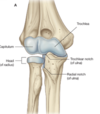
The rounded protuberance at the distal end of the humerus is called a ______. This forms an articulation with another bone.

A

Condyle

7
Q

T/F? The coronoid process is found on the anterior side of the humerus, and the olecranon process is located posteriorly.

A

TRUE

8
Q

List the condyles found within the humerus, and whether they are medial or lateral.

A

Capitulum - lateral; for radial articulation

Trochlea - medial; for ulnar articulation

Epicondyles - lateral and medial

9
Q

Name the joints of the shoulder. Which is capable of flexion, extension, adduction, and abduction (most flexible)?

A

Glenohumoral, sternoclavicular, and acromioclavicular.

The glenohumoral is the most flexible

10
Q

Because the cup of the glenoid cavity has more of a flattened shape, what provides for a better fit for the head of humerus?

A

Glenoid labrum

11
Q

Where are the sternoclavicular and acromioclavicular joints found?

A

Sternoclavicular - between manubrium of the sternum and the medial end of the clavicle

Acromioclavicular - between acromion of the scapula and the lateral end of the clavicle

12
Q

The radius of the forearm will articulate with the _____ in the humerus

A

Capitulum

13
Q

Radial Tuberosity

A

Bumpout point of the radius where the bicep will attach here

14
Q

T/F? The radius and the ulna can both do flexion, extension, and a pivoting motion.

A

FALSE - the ulna and the radius can do both flexion and extension, however only the radius can pivot. The ulna is “locked” into place due to its articulation fashion.

15
Q

Describe whether the ulna and radius are smaller are larger at both the proximal and distal ends.

A

Proximal - radius is smaller, ulna is larger

Distal - radius is larger and the ulna is smaller

16
Q

The ______ _____ is a pen-like projection located at the distal end of both the ___ and ____ and will form the ___ joint.

A

Styloid process, ulna and radius, wrist joint

17
Q

The _____ ______ separates the ulna and the radius; this helps stabilize and separate the forearm into compartments.

A

Interosseous sheath

18
Q

T/F? Pronation and supination occur at both the wrist and the elbow.

A

FALSE - pronation and supination both occur at the ELBOW ONLY.

19
Q

What allows for pronation and supination at the elbow? Hint: it has to do with one of the bones in the forearm!

A

The radius is not locked into place across the humerus; during pronation, the radius will cross the ulna.

20
Q

What are the 3 joints of the elbow?

A

1) trochlear notch of the ulna and trochlea of the humerus
2) Head of the radius and capitulum of humerus
3) Head of the radius and the radial notch of the ulna.

21
Q

List the carpal bones. Out of these bones, which is a sesmoid bone? Which bone will form the joint between the carpal and the metacarpal of the thumb? What type of joint does it form?

A

Scaphoid, lunate, triquetrum, pisiform (bottom) trapezium, trapezoid, capitate, hamate (top)

The pisiform.

Trapezium, saddle joint (only type in the body)

22
Q

Anular Ligament

A

A ligament that goes around the radial head to hold it in place; enables pivoting motion without moving out of its socket.

23
Q

What is the tissue layer that covers the arch created by the carpal bones?

A

Flexor retinacullum

24
Q

Define the carpals, metacarpals, and the metacarpophalalangeal joints, and the interphalangeal joints.

A
  • Carpals - little bones in the wrist
  • Metacarpals - bones in between the carpals and phalanges
  • MCP - joints between the metacarpals and the proximal phalanges
  • Interphalangeal joints - found between the phalanges
25
What muscles lay over the scapula? Which muscles are above and below the scapular spine? What innervates the infraspinatus and supraspinatus muscles?
Infraspinatus, supraspinatus, teres major, teres minor, subscapularis Infraspinatus, teres major, and teres minor are below. supraspinatus is above the scapular spine. Suprascapular nerve
26
The teres minor is innervated by the _____ and the teres major is innervated by the \_\_\_\_\_\_.
minor - axillary major - lower subscapular
27
Describe the location of the subscapularis muscle.
The subscapularis is on the anterior side of the scapula, on the sub scapular fossa.
28
Where does the brachial plexus comes from and travel to? What does it innervate?
Travels from the anterior (ventral) rami of C5-T1 spinal segments and travels into the opening of the axilla, down the arm. Innervates muscles that are in upper limb and that move the upper limb
29
What structures pass through the axilla area (b/w neck and upper extremity)?
Subclavian to axillary (artery and vein), brachial plexus, lymphatics, axillary tail of breast, biceps and coracobrachialis (upper parts of some arm muscles)
30
What defines the axillary inlet?
Bones, specifically clavicle anteriorly, scapulla posteriorly, and rib 1 medially
31
What are the anterior, medial, lateral and posterior walls of the axilla?
anterior: clavipectoral fascia, pec major medial: thoracic wall and serratus anterior lateral: humerus posterior: muscles over scapula and triceps muscle
32
Subclavian artery becomes the axillary artery after passing \_\_\_\_\_\_\_\_\_\_\_\_\_\_\_\_
under the clavicle and lateral to rib 1
33
Axillary artery is divided into 3 parts by\_\_\_\_\_ What are the 3 parts and what are the arteries called in each part called?
pec minor 1. proximal to pec minor: superior thoracic 2. posterior to pec minor: thoraco-acromial, lateral thoracic 3. distal to pec minor: subscapular, anterior and posterior circumflex humeral
34
Axillary artery becomes the _______ artery after passing the \_\_\_\_\_\_\_\_\_\_\_\_\_
brachial artery border of teres major muscle
35
Brachial artery travels in which part of arm? Brachial artery divides into ______ and ______ arteries near the \_\_\_\_\_\_, which travel on what sides of the arm?
anterior part of the arm Divides into radial and ulnar arteries near the elbow Radial travels on lateral side and ulnar travels on medial side of arm
36
What is the Profunda Brachii and what does this travel along with?
artery used to measure blood pressure; It's a major branch off of the brachial artery that supplies the posterior arm; travels with radial nerve
37
axillary vein becomes subclavian vein after passing \_\_\_\_\_\_\_
rib 1
38
Axillary lymph nodes drain what? These nodes are divided into groups based on \_\_\_\_\_\_\_\_\_\_\_\_\_\_
drain the UE, parts of upper back, shoulders, neck, and most of the breast location in the axilla
39
Brachial plexus is divided into --- list from proximal to distal
roots, trunks, divisions, cords
40
What are the 3 trunks? What are the divisions called? What are the cords called?
Trunks: superior, middle and inferior Divisions: Anterior and posterior Cords - lateral, posterior, medial
41
What structure are the brachial plexus divisions oriented around?
Axillary artery
42
Name the branches of the divisions and their innervations
Fool. There are none.
43
Name the branches off the roots and their innervations
Dorsal Scapular - innervates rhomboid Long thoracic nerve - innervates serratus anterior
44
Name the branches of the trunks and their innervations
3 trunks - lateral, medial, posterior suprascapular - innervates supraspinatous and infraspinatous Nerve to subclavius
45
Name the branches of the cords and their innervations
Lateral cord - lateral pectoral nerve, terminal nerve, musculocutaneous, terminal 1/2 of median nerve Posterior cord - Axillary (deltoid muscle and teres minor), terminal 1/2 of radial, thoracodorsal (lattisimus muscle), subscapular (subscapularis muscle and teres major) Medial cord - median, medial pectoral, medial cutaneous nerves of the arm and forearm, ulnar nerve
46
What nerve runs posterior to the axillary artery?
Radial nerve, off of posterior cord of brachial plexus
47
\_\_\_\_\_\_\_\_\_\_\_ nerve and ___________ nerve innervate ONLY skin
medial cutaneous nerve of arm and medial cutaneous nerve of forearm both branch off of medial cord
48
The dorsal scapular nerve and long thoracic nerve branch from which part of the brachial plexus? a) trunks b) divisions c) cords d) roots
D
49
Which is NOT a muscle that make up the posterior wall of the axillary region? a) triceps b) biceps c) suprascapular d) teres major and minor
B - The posterior wall of the axilla is made up of muscles from the scapula and the triceps!
50
What are the nerves that branch off the lateral cord from the brachial plexus?
lateral pectoral, musculocutaneous, and median
51
What are the nerves that branch off the posterior cord from the brachial plexus?
thoracodorsal, superior and inferior subscapular, axillary, and radial
52
What are the nerves that branch off the medial cord from the brachial plexus?
Median, medial pectoral, medial cutaneous nerves of the arm and forearm, and the ulnar nerve
53
T/F? The median nerve branches from both the lateral and median cords of the brachial plexus.
TRUE
54
The coronoid process of the humerus is located above which condyle?
Trochlea of the humerus
55
The glenohumoral joint of the shoulder is cushioned by which structures? a) fatty pads b) bursae c) tendon sheaths d) synovial fluid
B
56
The suprascapular artery and nerve will travel through which foramen? What is this foramen created by?
Suprascapular foramen - created by the suprascapular notch and the clavicle passing over top of it
57
The suprascapular ligament will travel through the ______ \_\_\_\_\_\_.
suprascapular notch of the scapula
58
Which vein passes posterior-laterally in the upper extremity and travels in the delto-pectoral groove?
Cephalic vein
59
Superficial structures of the arm are drained by the ____ vein, which is more medial, and the _____ vein (lateral).
Cephalic (lateral) Basilic (medial)
60
Which vein forms the bulk of the axillary vein and also travels with the axillary artery?
Basilic vein
61
T/F Scapula and clavicle are part of the axial skeleton
FALSE. They are part of the appendicular skeleton