Neuroanatomy Flashcards
(72 cards)
What is the correct term/name for nerve cell bodies in the CNS?
Nucleus
What is the correct term for a bundle of axons in the CNS?
Tract
What is the correct term/name for nerve cell bodies in the PNS?
Ganglion
What is the correct term for a bundle of axons in the PNS?
Nerve
What do spinal nerves supply?
Soma/body wall
Where is the only place spinal nerves are located?
Intervertebral foramina

What is the name given to spinal nerves once they have exited the intervertebral foramina to connect with the body wall?
Rami

What is the name given to the connecting structures attaching the spinal cord and spinal nerves of the same level?
Roots and rootlets

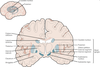
Label these structures


What are nerve plexuses?
Networks of intertwined anterior rami
Which axons are transported in the anterior horn of the spinal cord?
Motor axons
Which axons are transported in the posterior horn of the spinal cord?
Sensory axons
Which segment of the spinal cord has lateral horns and why?
T1 to L2 - transports sympathetic axons
Which nerve is the only sensory nerve not to synapse in the thalamus prior to entering the cortex?
Olfactory
How many layers of scalp are there?
5
What are the layers of the scalp?
S = Skin
C = Connective tissue
A = Aponeurosis
L = Loose connective tissue
P= Pericranium
What is the arterial supply of the scalp?
Scalp branches from the external carotid artery
What is the thinnest part of the skull?
Pterion
Which artery courses over the deep aspect of the pterion?
The middle meningeal artery
What are the three layers of meninges superficial to deep?
Dura mater
Arachnoid mater
Pia mater
What is the function of the dura mater?
Protection - this is the toughest meningeal layer
What is the function of the granulations on the arachnoid mater?
Reabsorbing CSF
What is the function of the pia mater?
Adherence to the brain and all nerves & vessels entering or leaving the brain
What is the dura mater adherent to?
The internal aspect of all bones of the skull