Autoimmune Diseases 2 Flashcards

1
Q

Non-genetic factors in autoimmunity: Infection

  • Some infections have been linked with the subsequent development of autoimmune disease
  • An immunological explanation is known as … …, in which epitopes relevant to the pathogen are shared with host antigens
A
  • Some infections have been linked with the subsequent development of autoimmune disease
  • An immunological explanation is known as molecular mimicry, in which epitopes relevant to the pathogen are shared with host antigens
2
Q

Molecular mimicry

  • Viral infection: presentation of viral peptides to a CD4 T cell via MHC …, causing T cell activation
  • The viral peptides happen to be similar to a host-derived peptide; the T cell would normally recognise these peptides, but would not react to them
  • The activated T cell now reacts strongly to the self-peptide and initiates …
  • The process depends on having the correct MHC molecules to present this critical epitope that is common to both virus and host (inherited)
  • Also depends on having the correct T cell to recognise it (mainly bad luck)
A
  • Viral infection: presentation of viral peptides to a CD4 T cell via MHC 2, causing T cell activation
  • The viral peptides happen to be similar to a host-derived peptide; the T cell would normally recognise these peptides, but would not react to them
  • The activated T cell now reacts strongly to the self-peptide and initiates inflammation
  • The process depends on having the correct MHC molecules to present this critical epitope that is common to both virus and host (inherited)
  • Also depends on having the correct T cell to recognise it (mainly bad luck)
3
Q

Molecular mimicry

  • Viral infection: presentation of viral peptides to a CD4 T cell via MHC 2, causing T cell activation
  • The viral peptides happen to be similar to a host-derived peptide; the T cell would normally recognise these peptides, but would not react to them
  • The activated T cell now reacts strongly to the self-peptide and initiates inflammation
  • The process depends on having the correct MHC molecules to present this critical epitope that is common to both virus and host (…)
  • Also depends on having the … to recognise it (mainly bad luck)
A
  • Viral infection: presentation of viral peptides to a CD4 T cell via MHC 2, causing T cell activation
  • The viral peptides happen to be similar to a host-derived peptide; the T cell would normally recognise these peptides, but would not react to them
  • The activated T cell now reacts strongly to the self-peptide and initiates inflammation
  • The process depends on having the correct MHC molecules to present this critical epitope that is common to both virus and host (inherited)
  • Also depends on having the correct T cell to recognise it (mainly bad luck)
4
Q

Molecular mimicry

  • Viral …: presentation of viral peptides to a CD4 T cell via MHC 2, causing T cell activation
  • The viral peptides happen to be similar to a host-derived peptide; the T cell would normally recognise these peptides, but would not react to them
  • The activated T cell now reacts strongly to the self-peptide and initiates inflammation
  • The process depends on having the correct MHC molecules to present this critical epitope that is common to both virus and host (inherited)
  • Also depends on having the correct T cell to recognise it (mainly bad luck)
A
  • Viral infection: presentation of viral peptides to a CD4 T cell via MHC 2, causing T cell activation
  • The viral peptides happen to be similar to a host-derived peptide; the T cell would normally recognise these peptides, but would not react to them
  • The activated T cell now reacts strongly to the self-peptide and initiates inflammation
  • The process depends on having the correct MHC molecules to present this critical epitope that is common to both virus and host (inherited)
  • Also depends on having the correct T cell to recognise it (mainly bad luck)
5
Q

Molecular mimicry

  • Viral infection: presentation of viral peptides to a CD4 T cell via MHC 2, causing T cell activation
  • The viral peptides happen to be similar to a host-derived peptide; the T cell would normally recognise these peptides, but would not react to them
  • The activated T cell now … strongly to the self-peptide and initiates …
  • The process depends on having the correct MHC molecules to present this critical epitope that is common to both virus and host (inherited)
  • Also depends on having the correct T cell to recognise it (mainly bad luck)
A
  • Viral infection: presentation of viral peptides to a CD4 T cell via MHC 2, causing T cell activation
  • The viral peptides happen to be similar to a host-derived peptide; the T cell would normally recognise these peptides, but would not react to them
  • The activated T cell now reacts strongly to the self-peptide and initiates inflammation
  • The process depends on having the correct MHC molecules to present this critical epitope that is common to both virus and host (inherited)
  • Also depends on having the correct T cell to recognise it (mainly bad luck)
6
Q

Give 2 examples of molecular mimicry

A
  • Autoimmune haemolysis after Mycoplasma pneumoniae
    • Mycoplasma antigen has homology to ‘I’ antigen on red blood cells
    • IgM antibody to mycoplasma may cause transient haemolysis
  • Rheumatic fever: inflammatory disease occurring after streptococcal infection affecting heart, joints, skin and brain
    • Anti-streptococcal antibodies believed to cross-react with connective tissue
  • Even for these ‘best examples’ the target antigens are not well-defined; for other diseases the paradigm remains rather theoretical
7
Q

Autoimmune haemolysis after Mycoplasma pneumoniae is an example of molecular …

A
  • molecular mimicry
    • Autoimmune haemolysis after Mycoplasma pneumoniae
    • Mycoplasma antigen has homology to ‘I’ antigen on red blood cells
    • IgM antibody to mycoplasma may cause transient haemolysis
8
Q

Rheumatic fever is an example of molecular …

A
  • molecular mimicry
    • It is an inflammatory disease occurring after streptococcal infection affecting heart, joints, skin and brain
  • Anti-streptococcal antibodies believed to cross-react with connective tissue
9
Q

Autoimmune haemolysis after Mycoplasma pneumoniae

  • Mycoplasma antigen has homology to ‘I’ antigen on red blood cells
  • Ig… antibody to mycoplasma may cause transient …
A
  • Mycoplasma antigen has homology to ‘I’ antigen on red blood cells
  • IgM antibody to mycoplasma may cause transient haemolysis
10
Q

Rheumatic fever

  • It is an inflammatory disease occurring after … infection affecting heart, joints, skin and brain
    • Anti-… antibodies believed to cross-react with connective tissue
A
  • It is an inflammatory disease occurring after streptococcal infection affecting heart, joints, skin and brain
    • Anti-streptococcal antibodies believed to cross-react with connective tissue
11
Q

Type 1 diabetes

  • Lack of insulin impairs cellular update of glucose, leading to polyuria, polydypsia, polyphagia and weight loss
  • Onset at any age, but typically …
  • Disease prevalence around …%; rising by around 5% per anum
  • Treatment by injection of insulin and diet
  • Important to differentiate from:
    • … diabetes - can present with a similar phenotype but requires different management
    • Type II diabetes mellitus = older onset, insulin secretion, ketoacidosis less likely and insulin not necessarily required
A
  • Lack of insulin impairs cellular update of glucose, leading to polyuria, polydypsia, polyphagia and weight loss
  • Onset at any age, but typically childhood
  • Disease prevalence around 0.8%; rising by around 5% per anum
  • Treatment by injection of insulin and diet
  • Important to differentiate from:
    • Monogenic diabetes - can present with a similar phenotype but requires different management
    • Type II diabetes mellitus = older onset, insulin secretion, ketoacidosis less likely and insulin not necessarily required
12
Q

Type 1 diabetes

  • Lack of insulin impairs cellular update of glucose, leading to polyuria, polydypsia, polyphagia and weight …
  • Onset at any age, but typically childhood
  • Disease prevalence around 0.8%; rising by around …% per anum
  • Treatment by injection of insulin and diet
  • Important to differentiate from:
    • Monogenic diabetes - can present with a similar phenotype but requires different management
    • Type II diabetes mellitus = older onset, insulin secretion, ketoacidosis less likely and insulin not necessarily required
A
  • Lack of insulin impairs cellular update of glucose, leading to polyuria, polydypsia, polyphagia and weight loss
  • Onset at any age, but typically childhood
  • Disease prevalence around 0.8%; rising by around 5% per anum
  • Treatment by injection of insulin and diet
  • Important to differentiate from:
    • Monogenic diabetes - can present with a similar phenotype but requires different management
    • Type II diabetes mellitus = older onset, insulin secretion, ketoacidosis less likely and insulin not necessarily required
13
Q

Immunology of type 1 diabetes

  • Evidence for … disease
    • Islet cell antibodies detectable for months to years before the onset of clinical disease
    • HLA associations
    • Mouse model
    • Early pancreatic biopsy shows … with CD4/ 8 T cells
    • Note that although antibodies present, they do not appear to be directly relevant to destruction of the pancreas
    • By the time patient has established diabetes, generally no active inflammation in pancreatic biopsy
A
  • Evidence for autoimmune disease
    • Islet cell antibodies detectable for months to years before the onset of clinical disease
    • HLA associations
    • Mouse model
    • Early pancreatic biopsy shows infiltration with CD4/ 8 T cells
    • Note that although antibodies present, they do not appear to be directly relevant to destruction of the pancreas
    • By the time patient has established diabetes, generally no active inflammation in pancreatic biopsy
14
Q

Immunology of type 1 diabetes

  • Is there evidence for autoimmune disease?
A
  • Yes - Evidence for autoimmune disease
    • Islet cell antibodies detectable for months to years before the onset of clinical disease
    • HLA associations
    • Mouse model
    • Early pancreatic biopsy shows infiltration with CD4/ 8 T cells
    • Note that although antibodies present, they do not appear to be directly relevant to destruction of the pancreas
    • By the time patient has established diabetes, generally no active inflammation in pancreatic biopsy
15
Q

This is a pancreatic biopsy showing what?

A

pre-diabetic pancreatic biopsy showing infiltration with CD8 T cells

16
Q

Progression to type 1 diabetes

  • Fill in the blanks
A
17
Q

Progression to Type 1 diabetes

  • By the time overt diabetes has developed, over …% of the pancreas has been destroyed
A
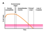
  • By the time overt diabetes has developed, over 90% of the pancreas has been destroyed​
18
Q

Genetics and type 1 diabetes

  • Genetic background definitely important – conconcordance in monozygotic twins is close to 100% if they are observed for long enough (nb older texts underestimate this risk)
  • … class II alleles are the major defined genetic risk factor
    • DR3 or DR4 relative risk is 6
    • DR3 and DR4 relative risk is 15
  • Rather like coeliac disease, believed that these molecules are required to present relevant islet cell antigens to CD4 T cells
  • Autoimmune response may occur if appropriate T cell receptors are present, together with other … and … co-factors
A
  • Genetic background definitely important – conconcordance in monozygotic twins is close to 100% if they are observed for long enough (nb older texts underestimate this risk)
  • HLA class II alleles are the major defined genetic risk factor
    • DR3 or DR4 relative risk is 6
    • DR3 and DR4 relative risk is 15
  • Rather like coeliac disease, believed that these molecules are required to present relevant islet cell antigens to CD4 T cells
  • Autoimmune response may occur if appropriate T cell receptors are present, together with other genetic and environmental co-factors
19
Q

Genetics and type 1 diabetes

  • Is Genetic background definitely important?
A
  • yes - Genetic background definitely important – conconcordance in monozygotic twins is close to 100% if they are observed for long enough (nb older texts underestimate this risk)
  • HLA class II alleles are the major defined genetic risk factor
    • DR3 or DR4 relative risk is 6
    • DR3 and DR4 relative risk is 15
  • Rather like coeliac disease, believed that these molecules are required to present relevant islet cell antigens to CD4 T cells
  • Autoimmune response may occur if appropriate T cell receptors are present, together with other genetic and environmental co-factors
20
Q

… class II alleles are the major defined genetic risk factor for T1 diabetes

A
  • HLA class II alleles are the major defined genetic risk factor
    • –DR3 or DR4 relative risk is 6
    • –DR3 and DR4 relative risk is 15
21
Q

What is the major defined genetic risk factor for T1 diabetes?

A

HLA class II alleles are the major defined genetic risk factor for T1 diabetes

22
Q

Precipitating events - T1 diabetes

  • … to islet cell antigens present for months-years before onset of clinical disease
  • Gap between initiation of disease and its presentation makes identification of triggers difficult
  • Much of the data is epidemiological
  • Some evidence for … virus
    • Stronger immune response to virus in cases compared to controls
    • Viral infection can cause pancreatitis in mice and humans, and precipitate autoimmune diabetes in mouse models
    • Protein 2C from Coxsackie virus has homology with islet cell antigen glutamic acid decarboxylase (GAD) (?molecular mimicry mechanism)
A
  • Autoantibodies to islet cell antigens present for months-years before onset of clinical disease
  • Gap between initiation of disease and its presentation makes identification of triggers difficult
  • Much of the data is epidemiological
  • Some evidence for Coxsackie virus
    • Stronger immune response to virus in cases compared to controls
    • Viral infection can cause pancreatitis in mice and humans, and precipitate autoimmune diabetes in mouse models
    • Protein 2C from Coxsackie virus has homology with islet cell antigen glutamic acid decarboxylase (GAD) (?molecular mimicry mechanism)
23
Q

Precipitating events - T1 diabetes

  • Autoantibodies to islet cell antigens present for months-years before onset of clinical disease
  • Gap between … of disease and its … makes identification of triggers difficult
  • Much of the data is epidemiological
  • Some evidence for Coxsackie virus
    • Stronger immune response to virus in cases compared to controls
    • Viral infection can cause pancreatitis in mice and humans, and precipitate autoimmune diabetes in mouse models
    • Protein 2C from Coxsackie virus has homology with islet cell antigen glutamic acid decarboxylase (GAD) (?molecular mimicry mechanism)
A
  • Autoantibodies to islet cell antigens present for months-years before onset of clinical disease
  • Gap between initiation of disease and its presentation makes identification of triggers difficult
  • Much of the data is epidemiological
  • Some evidence for Coxsackie virus
    • Stronger immune response to virus in cases compared to controls
    • Viral infection can cause pancreatitis in mice and humans, and precipitate autoimmune diabetes in mouse models
    • Protein 2C from Coxsackie virus has homology with islet cell antigen glutamic acid decarboxylase (GAD) (?molecular mimicry mechanism)
24
Q

Summary: The development of AID is multi-step

  • … background: Critical for some diseases in determining which peptides can be presented
  • T cell receptor …: critical in determining whether the peptide-MHC complex can be recognised. Note: the gene segments are inherited, but the receptors are produced randomly and will differ even in identical twins
  • Infection: may influence the activation of T cells and B cells that are potentially auto reactive
  • Likely to be myriad other genetic and environmental factors
A
  • MHC background: Critical for some diseases in determining which peptides can be presented
  • T cell receptor repertoire: critical in determining whether the peptide-MHC complex can be recognised. Note: the gene segments are inherited, but the receptors are produced randomly and will differ even in identical twins
  • Infection: may influence the activation of T cells and B cells that are potentially auto reactive
  • Likely to be myriad other genetic and environmental factors
25
Q

Summary: The development of AID is multi-step

  • MHC background: Critical for some diseases in determining which peptides can be presented
  • T cell receptor repertoire: critical in determining whether the peptide-MHC complex can be recognised. Note: the gene segments are inherited, but the receptors are produced randomly and will differ even in identical twins
  • …: may influence the activation of T cells and B cells that are potentially auto reactive
  • Likely to be myriad other … and … factors
A
  • MHC background: Critical for some diseases in determining which peptides can be presented
  • T cell receptor repertoire: critical in determining whether the peptide-MHC complex can be recognised. Note: the gene segments are inherited, but the receptors are produced randomly and will differ even in identical twins
  • Infection: may influence the activation of T cells and B cells that are potentially auto reactive
  • Likely to be myriad other genetic and environmental factors
26
Q

Multi-system autoimmune diseases

  • What are some examples? (3)
A
  • Rheumatoid arthritis – predominantly a joint disease, but also many extra-articular features; see symposium for more discussion
  • SLE
  • Sjogrens syndrome
27
Q

Rheumatoid arthritis(predominantly a joint disease but also many extra-articular features), SLE and Sjogrens syndrome are all examples of what?

A

multi-system autoimmune diseases

28
Q

Systemic lupus erythematosis (SLE)

  • A true multi-system autoimmune disorder
  • Skin
    • Butterfly (‘…’ rash)
    • Photosensitivity
    • Hives
  • Serositis
    • Pleurisy, pleural …
    • Pericarditis
  • Renal
  • Nephritis
  • … fibrosis
  • Joint pain
  • Autoimmune cytopenias
A
  • A true multi-system autoimmune disorder
  • Skin
    • Butterfly (‘lupine’ rash)
    • Photosensitivity
    • Hives
  • Serositis
    • Pleurisy, pleural effusion
    • Pericarditis
  • Renal
  • Nephritis
  • Pulmonary fibrosis
  • Joint pain
  • Autoimmune cytopenias
29
Q

Systemic lupus erythematosis (SLE)

  • A true …-system autoimmune disorder
  • Skin
    • … (‘lupine’ rash)
    • Photosensitivity
    • Hives
  • Serositis
    • Pleurisy, pleural effusion
    • Peri…
  • Renal
  • Nephritis
  • Pulmonary fibrosis
  • Joint pain
  • Autoimmune cytopenias
A
  • A true multi-system autoimmune disorder
  • Skin
    • Butterfly (‘lupine’ rash)
    • Photosensitivity
    • Hives
  • Serositis
    • Pleurisy, pleural effusion
    • Pericarditis
  • Renal
  • Nephritis
  • Pulmonary fibrosis
  • Joint pain
  • Autoimmune cytopenias
30
Q

Systemic lupus erythematosis (SLE)

  • A true multi-system autoimmune disorder
  • Skin
    • Butterfly (‘lupine’ rash)
    • Photosensitivity
    • Hives
  • S…
    • Pleurisy, pleural effusion
    • Pericarditis
  • Renal
  • Nephritis
  • Pulmonary fibrosis
  • Joint pain
  • Autoimmune …
A
  • A true multi-system autoimmune disorder
  • Skin
    • Butterfly (‘lupine’ rash)
    • Photosensitivity
    • Hives
  • Serositis
    • Pleurisy, pleural effusion
    • Pericarditis
  • Renal
  • Nephritis
  • Pulmonary fibrosis
  • Joint pain
  • Autoimmune cytopenias
31
Q

Systemic lupus erythematosis (SLE)

  • What gender usually?
  • More commin in people of … and … decent
  • Associated with presence of what antibodies?
A
  • Mostly women of reproductive age, rare in men
  • More common in people of Asian and African decent
  • Associated with presence of anti-nuclear antibodies – a collection of antibodies that react with cell nuclei and cell division apparatus; however, these don’t seem to directly cause disease and are probably an epi-phenomon
  • Some elements of disease probably caused by immune complex deposition; others may be explained by disordered apoptosis
  • Some patients have deficiency of classical complement components (C1, C4, C2)
32
Q

Systemic lupus erythematosis (SLE) is more common in who?

A
  • women of reproductive age - rare in men
  • also people of asian and african decent
33
Q

SLE is associated with presence of what antibodies?

A

anti-nuclear antibodies - a collection of antibodies that react with cell nuclei and cell division apparatus; however, these don’t seem to directly cause disease and are probably an epi-phenomon

34
Q

Monogenic disorders and autoimmunity: classical complement deficiency

  • Immune complexes are cleared by phagocytes; process enhanced by phagocyte Fc receptors and … receptors
  • Deficiency of C1q/ C2/ C4 predispose to …, presumably because immune complexes cannot be cleared effectively
A
  • Immune complexes are cleared by phagocytes; process enhanced by phagocyte Fc receptors and C3b receptors
  • Deficiency of C1q/ C2/ C4 predispose to lupus, presumably because immune complexes cannot be cleared effectively
35
Q

Deficiency of C1q/ C2/ C4 predispose to …, presumably because immune complexes cannot be cleared effectively

A

Deficiency of C1q/ C2/ C4 predispose to lupus, presumably because immune complexes cannot be cleared effectively

36
Q

… protein is the most important and abundant protein in the complement system

A

C3 protein is the most important and abundant protein in the complement system

37
Q

Autoimmune serology for diagnosis

  • Some autoantibodies have diagnostic value
    • In some cases the antibodies are …
    • In others, they are simply a … effect
  • Broadly three methods for detection:
    • Indirect immunofluorescence
    • Solid-phase immunoassay
    • Direct immunofluorescence
A
  • Some autoantibodies have diagnostic value
    • In some cases the antibodies are pathogenic
    • In others, they are simply a bystander effect
  • Broadly three methods for detection:
    • Indirect immunofluorescence
    • Solid-phase immunoassay
    • Direct immunofluorescence
38
Q

Autoimmune serology for diagnosis

  • Some autoantibodies have diagnostic value
    • In some cases the antibodies are pathogenic
    • In others, they are simply a bystander effect
  • Broadly three methods for detection:
    • Indirect …
    • …-phase immunoassay
    • Direct …
A
  • Some autoantibodies have diagnostic value
    • In some cases the antibodies are pathogenic
    • In others, they are simply a bystander effect
  • Broadly three methods for detection:
    • Indirect immunofluorescence
    • Solid-phase immunoassay
    • Direct immunofluorescence
39
Q

Detecting antibodies in blood: indirect immunofluorescence

A
40
Q

Detecting antibodies in blood: … …

A

Detecting antibodies in blood: indirect immunofluorescence

41
Q

Detecting antibodies in blood: indirect immunofluorescence

  • Glass slide with tissue of interest, harvested from animal source
  • Patient serum containing (or not) relevant … onto glass slide
  • Add detection antibody labelled with … …
  • Look for … under microscope
A
  • Glass slide with tissue of interest, harvested from animal source
  • Patient serum containing (or not) relevant antibodies onto glass slide
  • Add detection antibody labelled with fluorescent marker
  • Look for fluorescence under microscope
42
Q

Type 1 diabetes - Indirect Immunofluorescence

  • Indirect immunofluorescence with monkey pancreas – … staining of pancreatic islets demonstrates the presence of islet cell autoantibodies
A
  • Indirect immunofluorescence with monkey pancreas – positive staining of pancreatic islets demonstrates the presence of islet cell autoantibodies
43
Q

Why is it important to identify type 1 DM?

  • Risk of …
  • Requires …
  • … diabetes and type 2 diabetes require a different approach
A
  • Risk of ketoacidosis
  • Requires insulin
  • Monongenic diabetes and type 2 diabetes require a different approach
44
Q

Distinction between type 1/ type 2 and … diabetes not as straightforward as once thought on clinical grounds

A

Distinction between type 1/ type 2 and monogenic diabetes not as straightforward as once thought on clinical grounds

45
Q

Detecting antibodies in blood by immunoassay

  • What is this experiment?
A
  • ELISA
    • …..being replaced by newer methods that are more automated eg particle bead suspension
46
Q

ELISA

  • Antigen put onto well
  • Then add the serum to well - specific antibody binds to antigen (if it’s present)
  • …-linked antibody binds to specific antibody
  • Substrate added and converted by enzyme into coloured product - rate of colour formation is proportional to the amount of specific antibody
  • Must … in between steps to ensure things that unbound
A
  • Antigen put onto well
  • Wash - Then add the serum to well - specific antibody binds to antigen (if it’s present)
  • Enzyme-linked antibody binds to specific antibody
  • Substrate added and converted by enzyme into coloured product - rate of colour formation is proportional to the amount of specific antibody
  • Must wash in between steps to ensure things that unbound
47
Q

ELISA

  • Antigen put onto well
  • Wash - Then add the serum to well - specific antibody binds to antigen (if it’s present)
  • Enzyme-linked antibody binds to specific antibody
  • Substrate added and converted by enzyme into coloured product - … of … formation is … to the amount of specific antibody
  • Must wash in between steps to ensure things that unbound
A
  • Antigen put onto well
  • Wash - Then add the serum to well - specific antibody binds to antigen (if it’s present)
  • Enzyme-linked antibody binds to specific antibody
  • Substrate added and converted by enzyme into coloured product - rate of colour formation is proportional to the amount of specific antibody
  • Must wash in between steps to ensure things that unbound
48
Q

Introduction to ELISA tutorial

  • A classic ELISA system uses a … well plastic plate
  • The first step is to coat each well with the … of interest - for this example, let’s say that we’re looking for tTG antibody to diagnose coeliac disease
  • To do coat the plate, tTG solution is simply added to the wells; because it’s a protein, it sticks to the plastic. Excess tTG is then washed off
A
  • A classic ELISA system uses a 96 well plastic plate
  • The first step is to coat each well with the antigen of interest - for this example, let’s say that we’re looking for tTG antibody to diagnose coeliac disease
  • To do coat the plate, tTG solution is simply added to the wells; because it’s a protein, it sticks to the plastic. Excess tTG is then washed off
49
Q

Introduction to ELISA tutorial

  • A classic ELISA system uses a … well plastic plate
  • The first step is to coat each well with the antigen of interest - for this example, let’s say that we’re looking for tTG antibody to diagnose coeliac disease
  • To do coat the plate, tTG solution is simply added to the wells; because it’s a protein, it sticks to the plastic. Excess tTG is then …
A
  • A classic ELISA system uses a 96 well plastic plate
  • The first step is to coat each well with the antigen of interest - for this example, let’s say that we’re looking for tTG antibody to diagnose coeliac disease
  • To do coat the plate, tTG solution is simply added to the wells; because it’s a protein, it sticks to the plastic. Excess tTG is then washed off
50
Q

A classic … system uses a 96 well plastic plate

A

A classic ELISA system uses a 96 well plastic plate

51
Q

Introduction to ELISA tutorial

  • The first step is to coat each well with the … of interest (e.g tTG)
  • tTG Solution is simply added to the wells; because it’s a protein, it sticks to the plastic. Excess is then …
  • .. samples are added to the wells
  • The samples are …, to allow time for tTG antibody in the samples to react with the tTG antibody
  • Excess antibody is…
  • If there was tTG antibody in the sample, it will now be captured by the immobilised tTG antigen (…)
  • A … antibody is added
  • The secondary antibody is an anti-IgA antibody, which binds to IgA Fc regions
  • It’s covalently linked to an enzyme such as horse radish peroxidase, indicated as a red circle on the figure below
  • Excess secondary antibody is washed away
  • A trigger is now added, which reacts with the horse radish peroxidase that is bound to the secondary antibody, which in turn is bound to antibody tTG antibody that was present in the patient sample
  • This produces a colour change
  • The amount of colour change can be measured using a photocell
A
  • The first step is to coat each well with the antigen of interest (e.g tTG)
  • tTG Solution is simply added to the wells; because it’s a protein, it sticks to the plastic. Excess is then washed off
  • Patient samples are added to the wells
  • The samples are incubated, to allow time for tTG antibody in the samples to react with the tTG antibody
  • Excess antibody is washed off
  • If there was tTG antibody in the sample, it will now be captured by the immobilised tTG antigen (invisible)
  • A secondary antibody is added
  • The secondary antibody is an anti-IgA antibody, which binds to IgA Fc regions
  • It’s covalently linked to an enzyme such as horse radish peroxidase, indicated as a red circle on the figure below
  • Excess secondary antibody is washed away
  • A trigger is now added, which reacts with the horse radish peroxidase that is bound to the secondary antibody, which in turn is bound to antibody tTG antibody that was present in the patient sample
  • This produces a colour change
  • The amount of colour change can be measured using a photocell
52
Q

Introduction to ELISA tutorial

  • The first step is to coat each well with the antigen of interest (e.g tTG)
  • tTG Solution is simply added to the wells; because it’s a protein, it sticks to the plastic. Excess is then washed off
  • Patient samples are added to the wells
  • The samples are incubated, to allow time for tTG antibody in the samples to react with the tTG antibody
  • Excess antibody is washed off
  • If there was tTG antibody in the sample, it will now be captured by the immobilised tTG antigen (invisible)
  • A … … is added
  • The … is an anti-IgA antibody, which binds to IgA … regions
  • It’s covalently linked to an enzyme such as horse radish peroxidase, indicated as a red circle on the figure below
  • Excess secondary antibody is washed away
  • A … is now added, which reacts with the horse radish peroxidase that is bound to the secondary antibody, which in turn is bound to antibody tTG antibody that was present in the patient sample
  • This produces a colour change
  • The amount of colour change can be measured using a photocell
A
  • The first step is to coat each well with the antigen of interest (e.g tTG)
  • tTG Solution is simply added to the wells; because it’s a protein, it sticks to the plastic. Excess is then washed off
  • Patient samples are added to the wells
  • The samples are incubated, to allow time for tTG antibody in the samples to react with the tTG antibody
  • Excess antibody is washed off
  • If there was tTG antibody in the sample, it will now be captured by the immobilised tTG antigen (invisible)
  • A secondary antibody is added
  • The secondary antibody is an anti-IgA antibody, which binds to IgA Fc regions
  • It’s covalently linked to an enzyme such as horse radish peroxidase, indicated as a red circle on the figure below
  • Excess secondary antibody is washed away
  • A trigger is now added, which reacts with the horse radish peroxidase that is bound to the secondary antibody, which in turn is bound to antibody tTG antibody that was present in the patient sample
  • This produces a colour change
  • The amount of colour change can be measured using a photocell
53
Q

Introduction to ELISA tutorial

  • The first step is to coat each well with the antigen of interest (e.g tTG)
  • tTG Solution is simply added to the wells; because it’s a protein, it sticks to the plastic. Excess is then washed off
  • Patient samples are added to the wells
  • The samples are incubated, to allow time for tTG antibody in the samples to react with the tTG antibody
  • Excess antibody is washed off
  • If there was tTG antibody in the sample, it will now be captured by the immobilised tTG antigen (invisible)
  • A secondary antibody is added
  • The secondary antibody is an anti-IgA antibody, which binds to IgA Fc regions
  • It’s covalently linked to an enzyme such as horse radish peroxidase, indicated as a red circle on the figure below
  • Excess secondary antibody is washed away
  • A trigger is now added, which reacts with the horse radish peroxidase that is bound to the secondary antibody, which in turn is bound to antibody tTG antibody that was present in the patient sample
  • This produces a … change
  • The amount of … change can be measured using a …
A
  • The first step is to coat each well with the antigen of interest (e.g tTG)
  • tTG Solution is simply added to the wells; because it’s a protein, it sticks to the plastic. Excess is then washed off
  • Patient samples are added to the wells
  • The samples are incubated, to allow time for tTG antibody in the samples to react with the tTG antibody
  • Excess antibody is washed off
  • If there was tTG antibody in the sample, it will now be captured by the immobilised tTG antigen (invisible)
  • A secondary antibody is added
  • The secondary antibody is an anti-IgA antibody, which binds to IgA Fc regions
  • It’s covalently linked to an enzyme such as horse radish peroxidase, indicated as a red circle on the figure below
  • Excess secondary antibody is washed away
  • A trigger is now added, which reacts with the horse radish peroxidase that is bound to the secondary antibody, which in turn is bound to antibody tTG antibody that was present in the patient sample
  • This produces a colour change
  • The amount of colour change can be measured using a photocell
54
Q

What must you do in between steps of ELISA?

A

Washings! ensures anything unbound is washed away

55
Q

In the ELISA test, what are we looking for? (what does it tell us)

A

colour change - meausred using a photocell - rate of colour formation is proportional to the amount of specific antibody

56
Q

Photocell - ELISA

  • To provide a value for the samples, they need to be compared with something
  • This involves setting up wells containing known … of tTG antibody
  • The photocell readings from each well are used to create a standard …
  • Positive and negative controls are also run to show that the assay has worked correctly
A
  • To provide a value for the samples, they need to be compared with something
  • This involves setting up wells containing known concentrations of tTG antibody
  • The photocell readings from each well are used to create a standard curve
  • Positive and negative controls are also run to show that the assay has worked correctly
57
Q

Photocell - ELISA

  • To provide a value for the samples, they need to be compared with something
  • This involves setting up wells containing known concentrations of tTG antibody
  • The photocell readings from each well are used to create a standard curve
  • … and … controls are also run to show that the assay has worked correctly
A
  • To provide a value for the samples, they need to be compared with something
  • This involves setting up wells containing known concentrations of tTG antibody
  • The photocell readings from each well are used to create a standard curve
  • Positive and negative controls are also run to show that the assay has worked correctly
58
Q

Detecting antibodies bound to tissue: direct immunofluorescence

A
59
Q

This is showing … immunofluorescence

A

Detecting antibodies bound to tissue: direct immunofluorescence

60
Q

Bullous skin disease: pemphigoid

  • …-walled bullae, rarely on mucus membranes
  • Fulfils criteria for …-… disease
  • Target is antigen at dermo-epidermal junction
  • Linear deposition of antibody, which activates complement producing skin dehiscence and tense blister
A
  • Thick-walled bullae, rarely on mucus membranes
  • Fulfils criteria for antibody-mediated disease
  • Target is antigen at dermo-epidermal junction
  • Linear deposition of antibody, which activates complement producing skin dehiscence and tense blister
61
Q

Bullous skin disease: pemphigoid

  • Thick-walled bullae, rarely on … membranes
  • Fulfils criteria for antibody-mediated disease
  • Target is antigen at dermo-epidermal junction
  • Linear deposition of antibody, which activates … producing skin dehiscence and tense blister
A
  • Thick-walled bullae, rarely on mucus membranes
  • Fulfils criteria for antibody-mediated disease
  • Target is antigen at dermo-epidermal junction
  • Linear deposition of antibody, which activates complement producing skin dehiscence and tense blister
62
Q

What bullous skin diseases fulfil criteria for antibody-mediated disease?

A

pemphigus and pemphigoid

63
Q

Bullous skin disease: pemphigus

  • …-walled bullae on skin and mucus membranes, rupture easily
  • Fulfils critiera for antibody-mediated disease
  • Target is the intercellular cement protein desmoglein 3 in superficial skin layers
A
  • Thin-walled bullae on skin and mucus membranes, rupture easily
  • Fulfils critiera for antibody-mediated disease
  • Target is the intercellular cement protein desmoglein 3 in superficial skin layers
64
Q

Bullous skin disease: pemphigus

  • …-walled bullae on skin and mucus membranes, … easily
  • Fulfils critiera for antibody-mediated disease
  • Target is the intercellular cement protein desmoglein 3 in superficial skin layers
A
  • Thin-walled bullae on skin and mucus membranes, rupture easily
  • Fulfils critiera for antibody-mediated disease
  • Target is the intercellular cement protein desmoglein 3 in superficial skin layers
65
Q

pemphigus vs pemphigoid - which is most severe?

A
  • pemphigus
  • thin walled blisters - affects mucus membranes - nutrition and hydration difficult and also large areas of opened skin
66
Q

Coeliac disease diagnosis

  • … binding to the … of smooth muscle fibres found to have a strong disease association with coeliac disease in the 1990s.
  • Subsequently target antigen found to be tissue … (tTG), which is now expressed in recombinant systems to provide antigen for modern immunoassays
  • HLA typing also increasing utilised – absence of HLA DQ-2/ 8 makes coeliac disease very unlikely (ie high negative predictive value)
A
  • Antibodies binding to the endomysium of smooth muscle fibres found to have a strong disease association with coeliac disease in the 1990s.
  • Subsequently target antigen found to be tissue tranglutaminase (tTG), which is now expressed in recombinant systems to provide antigen for modern immunoassays
  • HLA typing also increasing utilised – absence of HLA DQ-2/ 8 makes coeliac disease very unlikely (ie high negative predictive value)
67
Q

Coeliac disease diagnosis

  • Antibodies binding to the … of smooth muscle fibres found to have a strong disease association with coeliac disease in the 1990s.
  • Subsequently target antigen found to be tissue … (tTG), which is now expressed in recombinant systems to provide antigen for modern immunoassays
  • … typing also increasing utilised – absence of … DQ-2/ 8 makes coeliac disease very unlikely (ie high negative predictive value)
A
  • Antibodies binding to the endomysium of smooth muscle fibres found to have a strong disease association with coeliac disease in the 1990s.
  • Subsequently target antigen found to be tissue tranglutaminase (tTG), which is now expressed in recombinant systems to provide antigen for modern immunoassays
  • HLA typing also increasing utilised – absence of HLA DQ-2/ 8 makes coeliac disease very unlikely (ie high negative predictive value)
68
Q

Pernicious anaemia

  • Vitamin … absorbed in terminal ileum
  • Absorption requires a co-factor called … FACTOR which is secreted by the gastric parietal cells
  • Pernicious anaemia describes … destruction of the gastric parietal cells
  • Loss of intrinsic factor abrogates … absorption
  • Liver stores around 2 years supply of …
  • Once depleted, multiple possible manifestations:
    • Anaemia
    • Neurological
    • Subfertility
A
  • Vitamin B12 absorbed in terminal ileum
  • Absorption requires a co-factor called INTRINSIC FACTOR which is secreted by the gastric parietal cells
  • Pernicious anaemia describes autoimmune destruction of the gastric parietal cells
  • Loss of intrinsic factor abrogates B12 absorption
  • Liver stores around 2 years supply of B12
  • Once depleted, multiple possible manifestations:
    • Anaemia
    • Neurological
    • Subfertility
69
Q

Pernicious anaemia

  • Vitamin B12 absorbed in terminal …
  • Absorption requires a co-factor called INTRINSIC FACTOR which is secreted by the … … cells
  • Pernicious anaemia describes autoimmune destruction of the … … cells
  • Loss of intrinsic factor abrogates B12 absorption
  • Liver stores around … yearssupply of B12
  • Once depleted, multiple possible manifestations:
    • Anaemia
    • Neurological
    • Subfertility
A
  • Vitamin B12 absorbed in terminal ileum
  • Absorption requires a co-factor called INTRINSIC FACTOR which is secreted by the gastric parietal cells
  • Pernicious anaemia describes autoimmune destruction of the gastric parietal cells
  • Loss of intrinsic factor abrogates B12 absorption
  • Liver stores around 2 years supply of B12
  • Once depleted, multiple possible manifestations:
    • Anaemia
    • Neurological
    • Subfertility
70
Q

… anaemia describes autoimmune destruction of the gastric parietal cells

A

Pernicious anaemia describes autoimmune destruction of the gastric parietal cells

71
Q

Vitamin B12 absorbed in … …, absorption requires a co-factor called … FACTOR which is secreted by the gastric parietal cells

A

Vitamin B12 absorbed in terminal ileum, absorption requires a co-factor called INTRINSIC FACTOR which is secreted by the gastric parietal cells

72
Q

Vitamin B12 absorbed in terminal ileum, absorption requires a co-factor called INTRINSIC FACTOR which is secreted by the … … cells

A

Vitamin B12 absorbed in terminal ileum, absorption requires a co-factor called INTRINSIC FACTOR which is secreted by the gastric parietal cells

73
Q

Liver stores around … years supply of B12

A

Liver stores around 2 years supply of B12

74
Q

Treatment of AID: manage the consequences

  • Often preferable to treating the immunology
    • … drugs are toxic
    • By the time the disease is overt, the damage may already have been done and … may be unhelpful
  • Examples:
    • Thyroxine for under-active thyroid
    • Carbimazole, surgery or drugs for thyrotoxicosis
    • Insulin for diabetes
    • B12 for pernicious anaemia
  • However, in some AID, particularly non organ-specific, treatment of the immune system is the best option
A
  • Often preferable to treating the immunology
    • Immunosuppressive drugs are toxic
    • By the time the disease is overt, the damage may already have been done and immunosuppression may be unhelpful
  • Examples:
    • Thyroxine for under-active thyroid
    • Carbimazole, surgery or drugs for thyrotoxicosis
    • Insulin for diabetes
    • B12 for pernicious anaemia
  • However, in some AID, particularly non organ-specific, treatment of the immune system is the best option
75
Q

Treatment of AID: manage the consequences

  • Often preferable to treating the immunology
    • Immunosuppressive drugs are toxic
    • By the time the disease is overt, the damage may already have been done and immunosuppression may be unhelpful
  • Examples:
    • … for under-active thyroid
    • Carbimazole, surgery or drugs for thyrotoxicosis
    • … for diabetes
    • … for pernicious anaemia
  • However, in some AID, particularly non organ-specific, treatment of the immune system is the best option
A
  • Often preferable to treating the immunology
    • Immunosuppressive drugs are toxic
    • By the time the disease is overt, the damage may already have been done and immunosuppression may be unhelpful
  • Examples:
    • Thyroxine for under-active thyroid
    • Carbimazole, surgery or drugs for thyrotoxicosis
    • Insulin for diabetes
    • B12 for pernicious anaemia
  • However, in some AID, particularly non organ-specific, treatment of the immune system is the best option
76
Q

Treatment of AID: manage the consequences

  • Often … to treating the immunology
    • Immunosuppressive drugs are …
    • By the time the disease is …, the damage may already have been done and immunosuppression may be unhelpful
  • Examples:
    • Thyroxine for under-active thyroid
    • Carbimazole, surgery or drugs for thyrotoxicosis
    • Insulin for diabetes
    • B12 for pernicious anaemia
  • However, in some AID, particularly non organ-specific, treatment of the immune system is the best option
A
  • Often preferable to treating the immunology
    • Immunosuppressive drugs are toxic
    • By the time the disease is overt, the damage may already have been done and immunosuppression may be unhelpful
  • Examples:
    • Thyroxine for under-active thyroid
    • Carbimazole, surgery or drugs for thyrotoxicosis
    • Insulin for diabetes
    • B12 for pernicious anaemia
  • However, in some AID, particularly non organ-specific, treatment of the immune system is the best option
77
Q

Treatment of AID: manage the consequences

  • Often preferable to treating the immunology
    • Immunosuppressive drugs are toxic
    • By the time the disease is overt, the damage may already have been done and immunosuppression may be unhelpful
  • Examples:
    • Thyroxine for under-active thyroid
    • Carbimazole, surgery or drugs for thyrotoxicosis
    • Insulin for diabetes
    • B12 for pernicious anaemia
  • However, in some AID, particularly … …-specific, treatment of the immune system is the best option
A
  • Often preferable to treating the immunology
    • Immunosuppressive drugs are toxic
    • By the time the disease is overt, the damage may already have been done and immunosuppression may be unhelpful
  • Examples:
    • Thyroxine for under-active thyroid
    • Carbimazole, surgery or drugs for thyrotoxicosis
    • Insulin for diabetes
    • B12 for pernicious anaemia
  • However, in some AID, particularly non organ-specific, treatment of the immune system is the best option
78
Q

Some drugs used for immunomodulation

  • Used particularly for ‘…-system’ autoimmune diseases
    • Systemic …
    • Small molecule immunosuppressive drugs (eg methotrexate, azathioprine, ciclosporin)
    • Increasing interest in ‘biologics’
A
  • Used particularly for ‘multi-system’ autoimmune diseases
    • Systemic corticosteroids
    • Small molecule immunosuppressive drugs (eg methotrexate, azathioprine, ciclosporin)
    • Increasing interest in ‘biologics’
79
Q

Some drugs used for immunomodulation

  • Used particularly for ‘multi-system’ autoimmune diseases
    • Systemic corticosteroids
    • Small molecule … drugs (eg methotrexate, azathioprine, ciclosporin)
    • Increasing interest in ‘…’
A
  • Used particularly for ‘multi-system’ autoimmune diseases
    • Systemic corticosteroids
    • Small molecule immunosuppressive drugs (eg methotrexate, azathioprine, ciclosporin)
    • Increasing interest in ‘biologics
80
Q

Plasmapharesis

  • Plasmapharesis removes … from the … therefore may be useful in …-mediated diseases
A
  • Plasmapharesis removes antibodies from the bloodstream therefore may be useful in antibody-mediated diseases
81
Q

What is plasmapharesis?

A

Plasmapheresis is a process in which the liquid part of the blood, or plasma, is separated from the blood cells (Plasmapharesis removes antibodies from the bloodstream therefore may be useful in antibody-mediated diseases)